. 2003 Oct 23;425(6960):851-6.
doi: 10.1038/nature02009. Epub 2003 Sep 14.

Hedgehog is an early and late mediator of pancreatic cancer tumorigenesis

Affiliations

Hedgehog is an early and late mediator of pancreatic cancer tumorigenesis

Sarah P Thayer et al. Nature. .

Abstract

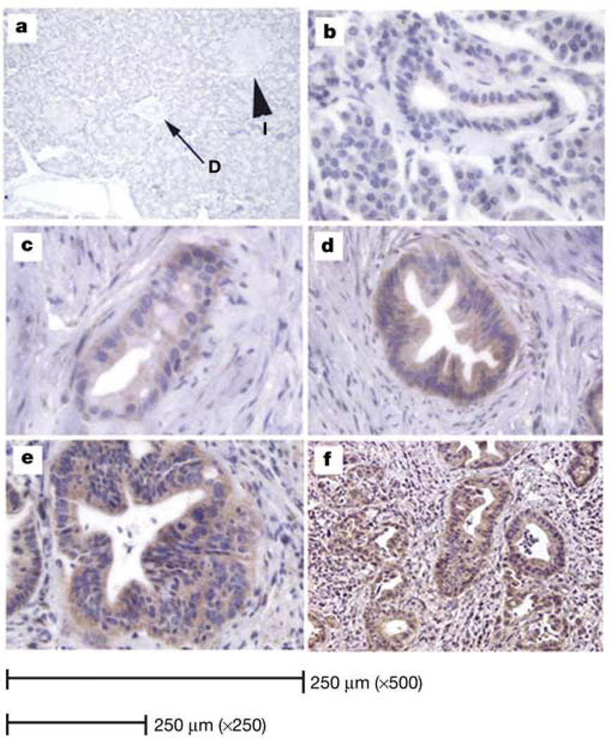
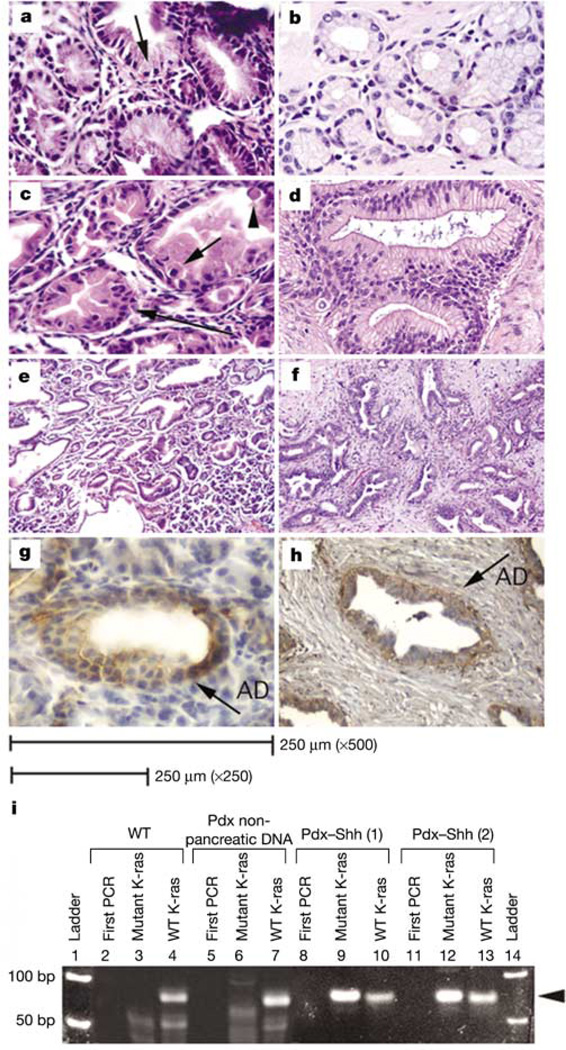
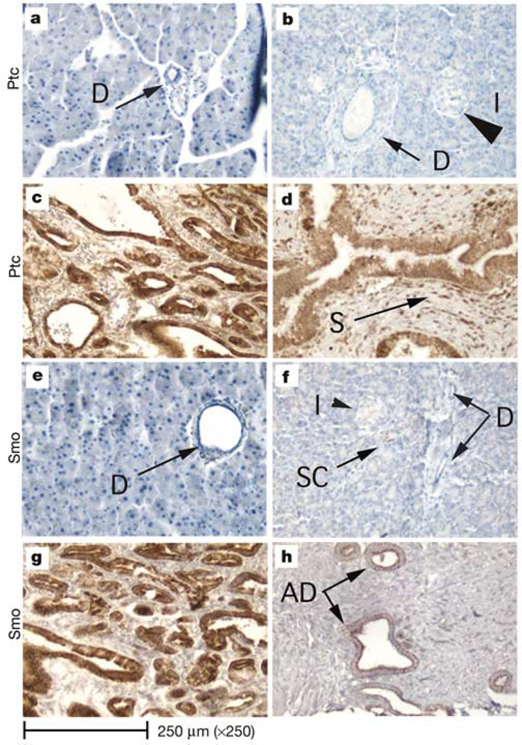
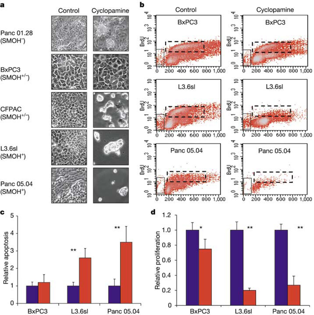
Hedgehog signalling--an essential pathway during embryonic pancreatic development, the misregulation of which has been implicated in several forms of cancer--may also be an important mediator in human pancreatic carcinoma. Here we report that sonic hedgehog, a secreted hedgehog ligand, is abnormally expressed in pancreatic adenocarcinoma and its precursor lesions: pancreatic intraepithelial neoplasia (PanIN). Pancreata of Pdx-Shh mice (in which Shh is misexpressed in the pancreatic endoderm) develop abnormal tubular structures, a phenocopy of human PanIN-1 and -2. Moreover, these PanIN-like lesions also contain mutations in K-ras and overexpress HER-2/neu, which are genetic mutations found early in the progression of human pancreatic cancer. Furthermore, hedgehog signalling remains active in cell lines established from primary and metastatic pancreatic adenocarcinomas. Notably, inhibition of hedgehog signalling by cyclopamine induced apoptosis and blocked proliferation in a subset of the pancreatic cancer cell lines both in vitro and in vivo. These data suggest that this pathway may have an early and critical role in the genesis of this cancer, and that maintenance of hedgehog signalling is important for aberrant proliferation and tumorigenesis.


Figures

Figure 1
Figure 1
Immunohistochemical identification of SHH. a, Normal human pancreas. No specific staining for SHH protein was identified in acini, islets (I) or ducts (D) (×125 magnification). b, No SHH expression is detected in normal ductal epithelium (×500 magnification). c, PanIN-1 expresses minimal amounts of SHH (×500). d, PanIN-2 expresses moderate levels of SHH (×250). e, PanIN-3 (×250) and f, invasive cancer (×125): moderate to high levels of SHH.
Figure 2
Figure 2
Histological comparison of human PanINs and Pdx–Shh mouse pancreata. a, Pdx–Shh pancreata display abnormal intestinal epithelial changes (arrow identifies abundant supranuclear mucin) that resemble human PanIN-1 (b) (×500). c, Pdx–Shh pancreata (×500) also exhibit a higher degree of atypia, with nuclear disarray and stratification (long arrow), mitosis (short arrow) and apoptosis (arrowhead), resembling human PanIN-2 (d) (×250). e, f, Low-magnification (×125) view of Pdx–Shh pancreata (e) and tubular complexes seen in human adenocarcinoma (f). g, Pdx–Shh mice (×500) and human PanINs (h) (×250) overexpress HER-2/neu, identified by a brown stain (AD; abnormal ductal epithelium). i, Mutant-allele-specific PCR amplification using mismatched primers (mutant K-ras) to detect codon 12 K-ras mutation. Wild-type primer (WT K-ras) is used as control. Lanes 1 and 14, ladder; 2, 5, 8, 11, first round of PCR (first PCR) reveals no detectable band, as expected. Wild-type mice express only wild-type K-ras (lanes 3, 4). Abnormal pancreatic epithelium of two Pdx–Shh mice expresses a TGT codon 12 mutation of K-ras (lanes 9, 12) and wild-type K-ras (lanes 10, 13), whereas the extrapancreatic tissues of Pdx–Shh mice outside the Shh expression domain express only wild-type K-ras (lanes 6, 7). The arrowhead identifies the 70-bp amplicon.
Figure 3
Figure 3
Hedgehog signalling pathway. ad, Immunohistochemical identification of Ptc1 receptor. The Ptc1 receptor is not identified in normal acini, ducts or islets in either mouse (a) or human (b) pancreata. Ptc1 (brown stain) is markedly overexpressed in the abnormal pancreatic epithelium of both Pdx–Shh mice (c) and neoplastic human pancreata (d). In humans, PTC1 is also overexpressed throughout the reactive stroma (S) that surrounds the epithelium. eh, Immunohistochemical identification of smoothened (Smo). Smoothened is not detected in normal duct epithelium (D) of normal mouse (e) or human (f) pancreata. Very infrequently Smo is detected in scattered cells (SC) in human pancreata; however, Smo (brown stain) is detected in the abnormal epithelium of Pdx–Shh mice (g) and neoplastic human pancreata (h) (arrows). The magnification is ×250 for all images.
Figure 4
Figure 4
Effects of cyclopamine treatment on pancreatic adenocarcinoma cells. a, Cell cultures of five pancreatic adenocarcinoma cell lines untreated (control) or treated with 10 µM cyclopamine for 7 days. b, Representative FACS histograms of three cell lines after BrdU incorporation (n = 4). The x axes show DNA content whereas the y axes show BrdU level; vertical boxes mark apoptotic cells; dashed horizontal boxes mark proliferating cells. c, Quantification of apoptotic cells measured in b (n = 4). To compare treated and untreated cells, the number of apoptotic cells in control samples was adjusted to 1. d, Quantification of proliferating cells measured in b (n = 4). To compare treated and untreated cells, the number of proliferating cells in control samples was adjusted to 1. Error bars indicate standard deviation. Asterisk, P < 0.05; double asterisks, P < 0.01. SMOH, no expression; SMOH+/−, low level expression; SMOH+, strong expression.
Figure 5
Figure 5
Cyclopamine treatment blocks tumour formation of human pancreatic adenocarcinoma cells after transplantation into nude mice. a, Schematic indicating sites of tumour cell and cyclopamine/vehicle injections. b, Isolated tumours derived from control or cyclopamine-treated L3.6sl and Panc 05.04 adenocarcinoma cells. Cyclopamine/vehicle injections were initiated either after palpable tumours had formed (delayed) or simultaneously with injection of tumour cells (concurrent). All pictures are shown at the same magnification. c, Weight of isolated tumours. Untreated control tumours of each cell line were adjusted to 1 to allow comparison of relative change in tumour mass. For delayed cyclopamine/vehicle injections, the values are: BxPC3, control 1 (n = 6), cyclopamine 1.1 (n = 5); Panc 05.04, control 1 (n = 5), cyclopamine 0.48 (n = 4); L3.6sl, control 1 (n = 5), cyclopamine 0.39 (n = 4). The values for concurrent cyclopamine/vehicle injections are: L3.6sl, control 1 (n = 4), cyclopamine 0.16 (n = 4). Error bars indicate standard deviation. Double asterisks, P < 0.01. dh, Histological analysis of the effect of cyclopamine treatment on L3.6sl-derived tumours. d, e, Haematoxylin/eosin staining of sections through the peripheral tumour regions. f, g, TUNEL staining of apoptotic cells in control (f) and cyclopamine-treated (g) tumours. h, Quantification of TUNEL-positive cells in control (blue) and cyclopamine-treated (red) tumours. Error bars indicate s.e.m. Double asterisks, P < 0.01.

Comment in

References

    1. Capdevila J, Estrada MP, Sanchez-Herrero E, Guerrero I. The Drosophila segment polarity gene patched interacts with decapentaplegic in wing development. EMBO J. 1994;13:71–82. - PMC - PubMed
    1. Hardcastle Z, Mo R, Hui C-C, Sharpe PT. The Shh signalling pathway in tooth development: defects in Gli2 and Gli3 mutants. Development. 1998;125:2803–2811. - PubMed
    1. Marigo V, Tabin C. Regulation of patched by sonic hedgehog in the developing neural tube. Proc. Natl Acad. Sci. USA. 1996;93:9346–9351. - PMC - PubMed
    1. Riddle RD, Johnson RL, Laufer E, Tabin C. Sonic hedgehog mediates the polarizing activity of the ZPA. Cell. 1993;75:1401–1416. - PubMed
    1. St-Jacques B, et al. Sonic hedgehog signaling is essential for hair development. Curr. Biol. 1998;8:1058–1068. - PubMed

Publication types

MeSH terms