Review
. 2003 Oct;36 Suppl 1(Suppl 1):17-32.
doi: 10.1046/j.1365-2184.36.s.1.3.x.

Isolation and characterization of functional mammary gland stem cells

Affiliations
Review

Isolation and characterization of functional mammary gland stem cells

Bryan Welm et al. Cell Prolif. 2003 Oct.

Abstract

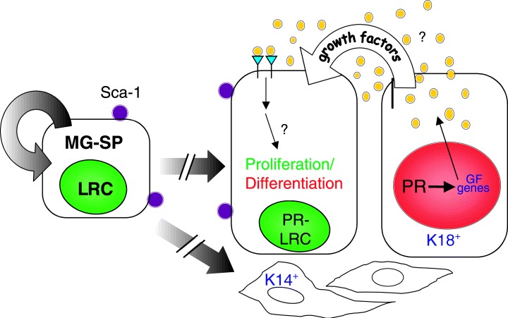
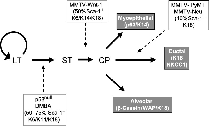
Significant advances in the stem-cell biology of several tissues, including the mammary gland, have occurred over the past several years. Recent progress on stem-cell fate determination, molecular markers, signalling pathways and niche interactions in haematopoietic, neuronal and muscle tissue may provide parallel insight into the biology of mammary epithelial stem cells. Taking advantage of approaches similar to those employed to isolate and characterize haematopoietic and epidermal stem cells, we have identified a mammary epithelial cell population with several stem/progenitor cell qualities. In this article, we review some recent data on mammary epithelial stem/progenitor cells in genetically engineered mouse models. We also discuss several potential molecular markers, including stem-cell antigen-1 (Sca-1), which may be useful for both the isolation of functional mammary epithelial stem/progenitor cells and the analysis of tumour aetiology and phenotype in genetically engineered mouse models. In different transgenic mammary tumour models, Sca-1 expression levels, as well as several other putative markers of progenitors including keratin-6, possess dramatically altered expression profiles. These data suggest that the heterogeneity of mouse models of breast cancer may partially reflect the selection or expansion of different progenitors.


Figures

Figure 1
Figure 1
A simplified mammary gland cell lineage model. Mammary gland side population (MG‐SP) cells, defined by their ability to efflux Hoechst dye, are estimated to represent approximately 0.5–3% of the epithelial cells in the virgin mammary gland. These cells also have been identified by their ability to retain BrdU during a longterm labelling and chase protocol, and are therefore designated as label‐retention cells (LRC). By FACS analysis, approximately 75% of the SP cells are stem‐cell antigen (Sca‐1) positive. The SP/Sca‐1/LRC‐positive mammary stem cells have the capability to either self‐renew to generate more stem cells (symmetric proliferation) or to undergo asymmetric proliferation to generate differentiated cells including keratin‐18 progesterone receptor (K18‐PR)‐positive mammary luminal epithelial and keratin 14 (K14)‐positive myoepithelial cells. The broken arrows indicate the presence of a number of potential intermediate progenitors for these cell lineages. The luminal epithelial cells are organized in the mature mammary ducts into nondividing steroid receptor‐positive cells (red nucleus, designated PR+) representing approximately 25% of the ductal epithelium, and proliferative cells (green nucleus, designated PR, LRC). Local growth factors including IGF‐II, amphiregulin, Wnt 4 and RankL, induced by oestrogen, progesterone and prolactin, act to regulate proliferation of adjacent cells via a paracrine/juxtacrine mechanism. Co‐localization of oestrogen receptor‐alpha, the progesterone receptor and prolactin receptor has been observed in the cells designated PR+. The long‐term labelling and chase experiments suggest that the majority of the stem/progentior cells are steroid receptor‐negative and that the proliferative cells may then give rise to the steroid receptor‐positive cells.
Figure 2
Figure 2
Cellular heterogeneity of mouse models of breast cancer may partially reflect the selection of different progenitors. Analysis of different tumour models has indicated the differential expression of Sca‐1 and K6 as well as K14 in hyperplasias and tumours of differing aetiologies. By analogy to haematopoietic stem cells, LT refers to a long‐term stem‐cell progenitor population, which is capable of self‐renewal, and ST to a short‐term progenitor, which can differentiate into a committed progenitor (CP). The dashed lines indicate the putative origins of mouse mammary tumours induced by the loss of p53, treatment with the carcinogen, DMBA, or overexpression of Wnt‐1, Neu and PyMT under the control of the MMTV‐LTR (Li et al. submitted for publication).

Similar articles

Cited by

References

    1. Al‐Hajj M, Wicha M, Benito‐Hernandez A, Morrison SJ, Clarke MF (2003) Prospective identification of tumorigenic breast cancer cells. Proc. Natl Acad. Sci. USA 100, 6980. - PMC - PubMed
    1. Allen JD, Brinkhuis RF, Wijnholds J, Schinkel AH (1999) The mouse Bcrp1/Mxr/Abcp gene: amplification and overexpression in cell lines selected for resistance to topotecan, mitoxantrone, or doxorubicin. Cancer Res. 59, 4237. - PubMed
    1. Alvi AJ, Clayton H, Joshi C, Enver T, Ashworth A, Vivanco MDM, Dale TC, Smalley MJ (2002) Functional and molecular characterisation of mammary side population cells. Breast Cancer Res. 5, R1. - PMC - PubMed
    1. Artavanis‐Tsakonas S, Rand MD, Lake RJ (1999) Notch signaling: cell fate control and signal integration in development. Science. 284, 770. - PubMed
    1. Attisano L, Wrana JL, Lopez‐Casillas F, Massague J (1994) TGF‐beta receptors and actions. Biochim. Biophys. Acta 1222, 71. - PubMed

Publication types