

Epithelial progenitor cell lines as models of normal breast morphogenesis and neoplasia
- PMID: 14521514
- PMCID: PMC2933221
- DOI: 10.1046/j.1365-2184.36.s.1.4.x
Epithelial progenitor cell lines as models of normal breast morphogenesis and neoplasia
Abstract
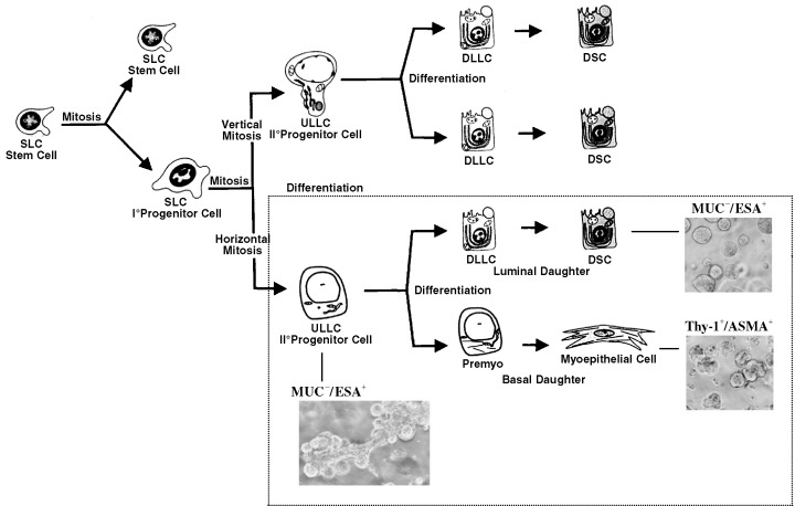
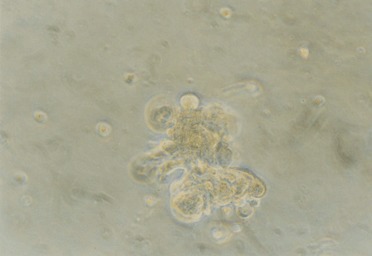
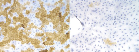
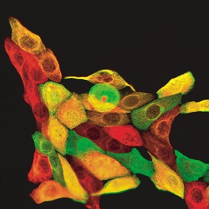
The majority of human breast carcinomas exhibit luminal characteristics and as such, are most probably derived from progenitor cells within the luminal epithelial compartment. This has been subdivided recently into at least three luminal subtypes based on gene expression patterns. The value of knowing the cellular origin of individual tumours is clear and should aid in designing effective therapies. To do this, however, we need strategies aimed at defining the nature of stem and progenitor cell populations in the normal breast. In this review, we will discuss our technical approach for delineating the origin of the epithelial cell types. A major step forward was the purification of each cell type by the application of immunomagnetic cell sorting based on expression of lineage-specific surface antigens. We then developed chemically defined media that could support either the luminal epithelial or the myoepithelial cell phenotype in primary cultures. Having succeeded in continuous propagation presumably without loss of markers, we could show that a subset of the luminal epithelial cells could convert to myoepithelial cells, signifying the possible existence of a progenitor cell population. By combining the information on marker expression and in situ localization with immunomagnetic sorting and subsequent immortalization, we have identified and isolated a cytokeratin 19-positive suprabasal putative precursor cell in the luminal epithelial compartment and established representative cell lines. This suprabasal-derived epithelial cell line is able to generate both itself and differentiated luminal epithelial and myoepithelial cells, and in addition, is able to form elaborate terminal duct lobular unit (TDLU)-like structures within a reconstituted basement membrane. As more than 90% of breast cancers arise in TDLUs and more than 90% are also cytokeratin 19-positive, we suggest that this cell population contains a breast-cancer progenitor.
Figures
References
-
- Aggeler J, Ward J, Blackie LM, Barcellos‐Hoff MH, Streuli CH, Bissell MJ (1991) Cytodifferentiation of mouse mammary epithelial cells cultured on a reconstituted basement membrane reveals striking similarities to development in vivo . J. Cell Sci. 99, 407. - PubMed
-
- Band V (1998) The role of retinoblastoma and p53 tumor suppressor pathways in human mammary epithelial cell immortalization. Int. J. Oncol. 12, 499. - PubMed
-
- Bartek J, Durban EM, Hallowes RC, Taylor‐Papadimitriou J (1985) A subclass of luminal epithelial cells in the human mammary gland, defined by antibodies to cytokeratins. J. Cell Sci. 75, 17. - PubMed
Publication types
MeSH terms
Grants and funding
LinkOut - more resources
Full Text Sources
Other Literature Sources
Medical
Research Materials
