. 2003 Oct;140(3):547-57.
doi: 10.1038/sj.bjp.0705464.

Tetrahydrocannabinol-induced neurotoxicity depends on CB1 receptor-mediated c-Jun N-terminal kinase activation in cultured cortical neurons

Affiliations

Tetrahydrocannabinol-induced neurotoxicity depends on CB1 receptor-mediated c-Jun N-terminal kinase activation in cultured cortical neurons

Eric J Downer et al. Br J Pharmacol. 2003 Oct.

Abstract

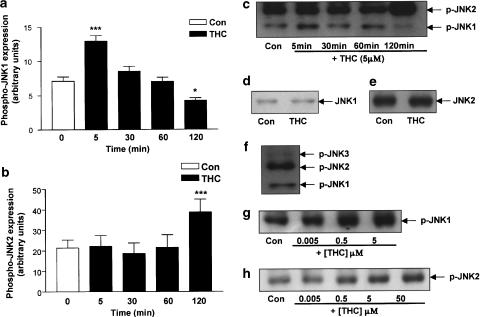
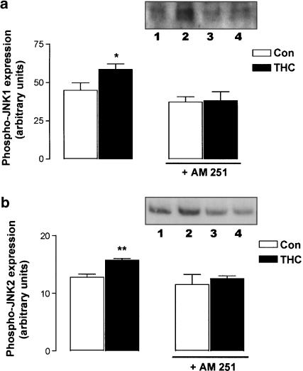
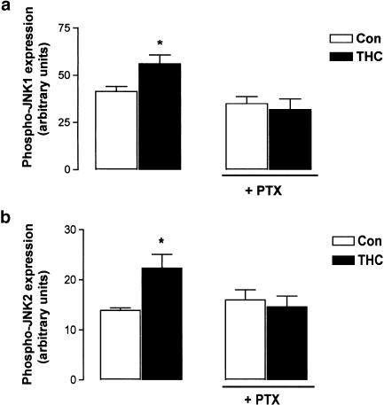
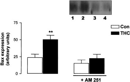
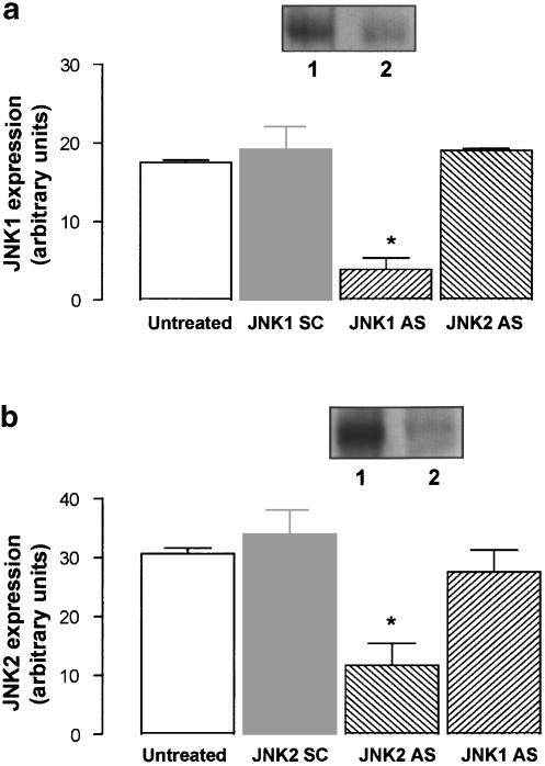
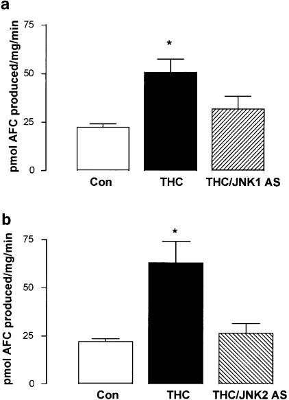
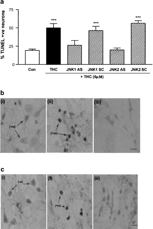
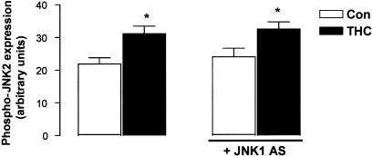
Delta9-Tetrahydrocannabinol (THC), the main psychoactive ingredient of marijuana, induces apoptosis in cultured cortical neurons. THC exerts its apoptotic effects in cortical neurons by binding to the CB1 cannabinoid receptor. The CB1 receptor has been shown to couple to the stress-activated protein kinase, c-Jun N-terminal kinase (JNK). However, the involvement of specific JNK isoforms in the neurotoxic properties of THC remains to be established. The present study involved treatment of rat cultured cortical neurons with THC (0.005-50 microM), and combinations of THC with the CB1 receptor antagonist, AM 251 (10 microM) and pertussis toxin (PTX; 200 ng ml-1). Antisense oligonucleotides (AS) were used to deplete neurons of JNK1 and JNK2 in order to elucidate their respective roles in THC signalling. Here we report that THC induces the activation of JNK via the CB1 receptor and its associated G-protein, Gi/o. Treatment of cultured cortical neurons with THC resulted in a differential timeframe of activation of the JNK1 and JNK2 isoforms. Use of specific JNK1 and JNK2 AS identified activation of caspase-3 and DNA fragmentation as downstream consequences of JNK1 and JNK2 activation. The results from this study demonstrate that activation of the CB1 receptor induces JNK and caspase-3 activation, an increase in Bax expression and DNA fragmentation. The data demonstrate that the activation of both JNK1 and JNK2 isoforms is central to the THC-induced activation of the apoptotic pathway in cortical neurons.


Figures

Figure 1
Figure 1
Time course of THC-induced activation of JNK. Cortical neurons were exposed to THC for 5–120 min, then cells were harvested and the cytosolic fractions analysed for the expression levels of the phosphorylated active forms of JNK 1 and JNK2 using Western immunoblot. (a) A significant increase in phospho-JNK1 expression was found following treatment with THC (5 μM) for 5 min. Exposure to THC for 30 and 60 min had no effect on phospho-JNK1 expression. However, treatment with THC for 120 min significantly decreased phospho-JNK1 expression. Results are expressed as mean±s.e.m. for seven observations, ***P<0.001, *P<0.05. (b) Phospho-JNK2 expression was significantly increased when neurons were treated with THC (5 μM) for 120 min. Exposure to THC for 5, 30 and 60 min had no effect on phospho-JNK2 expression. Results are expressed as mean±s.e.m. for seven observations, ***P<0.001. (c) A sample Western immunoblot demonstrating the increase in phospho-JNK1 expression at 5 min of THC treatment and the corresponding increase in phospho-JNK2 expression following treatment with THC for 120 min. (d) Neurons were treated with THC (5 μM) for 5 min, then cells were harvested in lysis buffer and the cytosolic fractions analysed for total JNK1 protein expression using Western immunoblot. THC had no effect on JNK1 protein expression at 5 min. Inset: A sample Western immunoblot showing that THC treatment for 5 min had no effect on JNK1 protein expression as compared to vehicle-treated neurons. (e) Cortical neurons were treated with THC (5 μM) for 2 h, then cells were harvested and analysed for total JNK2 protein expression by Western blot analysis. Incubation of cells with THC for 2 h did not affect JNK2 protein expression. Inset: A sample Western immunoblot demonstrating total JNK2 protein expression in vehicle-treated neurons and neurons treated with THC for 2 h. (f) Untreated cortical neurons were harvested and analysed for phosphorylated JNK1, JNK2 and JNK3 expression. JNK3 expression was moderate compared to JNK1/2 expression. (g) A sample Western immunoblot showing that a 5 min treatment with THC increases phospho-JNK1 expression in a dose-dependent manner. Similar results were observed in five separate experiments. (h) A sample Western immunoblot showing that a 120 min treatment with THC increases phospho-JNK2 expression in a dose-dependent manner. Similar results were observed in five separate experiments.
Figure 2
Figure 2
THC-induced activation of JNK is mediated by the CB1 receptor. (a) Treatment of neurons with THC (5 μM) for 5 min evoked a significant increase in phospho-JNK1 expression. Preincubation of cortical neurons with the CB1 receptor antagonist AM 251 (10 μM) for 30 min abolished the THC-induced increase in phospho-JNK1 expression. Results are expressed as mean±s.e.m. for six observations, *P<0.05. Inset: A sample Western immunoblot demonstrating the increase in phospho-JNK1 expression after 5 min of THC treatment (lane 2) compared to control cells (lane 1). While AM 251 had no effect on phospho-JNK1 expression (lane 3), it abolished the ability of THC to increase phospho-JNK1 expression (lane 4). (b) Treatment of neurons with THC (5 μM) for 120 min evoked a significant increase in phospho-JNK2 expression. Preincubation of cortical neurons with the CB1 receptor antagonist AM 251 (10 μM) for 30 min abolished the THC-induced increase in phospho-JNK2 expression. Results are expressed as mean±s.e.m. for six observations, **P<0.01. Inset: A sample Western immunoblot demonstrating the increase in phospho-JNK2 expression after 5 min of THC treatment (lane 2) compared to control cells (lane 1). While AM 251 had no effect on phospho-JNK2 expression (lane 3), it abolished the ability of THC to increase phospho-JNK2 expression (lane 4).
Figure 3
Figure 3
THC-induced JNK activation is mediated by G-protein subtypes Gi/o. (a) THC (5 μM; 5 min) significantly increased phospho-JNK1 expression and this was prevented by pretreatment with PTX (200 ng ml−1; 24 h). Results are expressed as mean±s.e.m. for five observations, *P<0.05. (b) THC (5 μM; 120 min) significantly increased phospho-JNK2 expression and this was prevented by pretreatment with PTX (200 ng ml−1; 24 h). Results are expressed as mean±s.e.m. for five observations, *P<0.05.
Figure 4
Figure 4
THC increases Bax expression via the CB1 receptor. Neurons were preincubated with AM 251 (10 μM) for 30 min, treated with THC (5 μM) for 2 h and analysed for the expression levels of Bax using Western immunoblotting. THC significantly increased Bax expression and this was abolished by AM 251. Results are expressed as mean±s.e.m. for five observations, **P<0.01. Inset: A sample Western immunoblot showing that THC increases Bax expression (lane 2) compared to vehicle-treated neurons (lane 1). The stimulatory effect of THC on Bax expression was prevented by AM 251 (lane 4). Exposure of cells to AM 251 alone had no effect on Bax expression (lane 3).
Figure 5
Figure 5
JNK AS effectively reduces JNK protein expression. Neurons were incubated with 2 μM JNK1/JNK2 antisense (AS) or JNK1/JNK2 scrambled control (SC) for 48 h. Cells were harvested in lysis buffer and the total expression of JNK1 and JNK2 protein was examined by Western blot analysis. (a) Treatment with JNK1 AS significantly decreased JNK1 protein expression in comparison to untreated cells and cells treated with JNK1 SC. JNK2 AS had no effect on JNK1 protein expression. Results are expressed as mean±s.e.m. for four experiments, *P<0.05. Inset: A sample immunoblot demonstrating that JNK1 AS significantly reduces JNK1 protein expression (lane 2) compared to JNK1 SC-treated neurons (lane 1). (b) JNK2 AS significantly reduced JNK2 protein expression. JNK1 AS had no effect on JNK2 protein expression. Results are expressed as mean±s.e.m. for four experiments, *P<0.05. Inset: A sample immunoblot demonstrating the significant reduction in JNK2 protein expression in JNK2 AS-treated neurons (lane 2) compared to JNK2 SC-treated cells (lane 1).
Figure 6
Figure 6
JNK is required for caspase-3 activity in THC-induced apoptosis. (a) Treatment of cortical neurons with THC (5 μM) for 2 h significantly increased caspase-3 activity, as assessed by the cleavage of the fluorogenic DEVD substrate. The stimulatory effect of THC on caspase-3 activity was prevented by pretreatment with JNK1 AS (2 μM; 48 h). Results are expressed as mean±s.e.m. for eight observations, *P<0.05. (b) Similarly, pretreatment of cells with JNK2 AS (2 μM; 48 h) prior to THC treatment abolished the stimulatory effect of THC on caspase-3 activity. Results are expressed as mean±s.e.m. for seven observations, *P<0.05.
Figure 7
Figure 7
JNK activity is required for THC-induced DNA fragmentation. (a) Exposure of neurons to THC (5 μM) for 3 h significantly increased the percentage of cells with DNA fragmentation. Treatment with JNK1 AS or JNK2 AS (2 μM; 48 h) prior to THC treatment prevented the THC-induced increase in DNA fragmentation. In contrast, in neurons treated with JNK1 SC or JNK2 SC (2 μM; 48 h), THC still evoked a significant increase in DNA fragmentation. Results are expressed as mean±s.e.m. for five observations, ***P<0.001. (b) Representative images of (i) control, (ii) THC-treated and (iii) THC/JNK1 AS-treated cortical neurons following TUNEL staining for DNA fragmentation. Arrows indicate TUNEL-negative (−ve) and TUNEL-positive (+ve) cells. Scale bar is 50 μM. (c) Representative images of TUNEL staining of (i) control, (ii) THC-treated and (iii) THC/JNK2 AS-treated cortical neurons. Scale bar is 50 μM.
Figure 8
Figure 8
JNK1 is not upstream of JNK2 in THC-induced apoptosis. Neurons were preincubated with JNK1 AS (2 μM) for 48 h, treated with THC (5 μM) for 2 h and analysed for phosphorylated JNK2 expression using Western immunoblot. THC evoked a significant increase in phospho-JNK2 expression. In cells exposed to JNK1 AS, THC was still capable of inducing a significant increase in phospho-JNK2. Results are expressed as mean±s.e.m. for four observations, *P<0.05.

Comment in

  • Neurons on cannabinoids: dead or alive?
    Guzman M. Guzman M. Br J Pharmacol. 2003 Oct;140(3):439-40. doi: 10.1038/sj.bjp.0705465. Br J Pharmacol. 2003. PMID: 14522839 Free PMC article. Review. No abstract available.

References

    1. ABOOD M.E., MARTIN B.R. Neurobiology of marijuana abuse. Trends Pharmacol. Sci. 1992;13:201–206. - PubMed
    1. ABOOD M.E., RIZVI G., SALLAPUDI N., MCALLISTER S. Activation of the CB1 cannabinoid receptor protects cultured mouse spinal neurons against excitotoxicity. Neurosci. Lett. 2001;309:197–201. - PubMed
    1. BOUABOULA M., POINOT-CHAZEL C., BOURRIE B., CANAT X., CALANDRA B., RINALDI-CARMONA M., LE FUR G., CASELLAS P. Activation of mitogen-activated protein kinases by stimulation of the central cannabinoid receptor CB1. Biochem. J. 1995;312:637–641. - PMC - PubMed
    1. BOUABOULA M., POINOT-CHAZEL C., MARCHAND J., CANAT X., BOURRIE B., RINALDI-CARMONA M., CALANDRA B., LE FUR G., CASELLAS P. Signaling pathway associated with stimulation of CB2 peripheral cannabinoid receptor: involvement of both mitogen-activated protein kinase and induction of Krox-24 expression. Eur. J. Biochem. 1996;237:704–711. - PubMed
    1. BOZYCZKO-COYNE D., O'KANE T.M., WU Z.L., DOBRZANSKI P., MURTHY S., VAUGHT J.L., SCOTT R.W. CEP-1347/KT-7515, an inhibitor of SAPK/JNK pathway activation, promotes survival and blocks multiple events associated with Aβ-induced cortical neuron apoptosis. J. Neurochem. 2001;77:849–863. - PubMed

Publication types

Substances

LinkOut - more resources