. 2003 Oct 1;17(19):2396-405.
doi: 10.1101/gad.1109403.

mSin3-associated protein, mSds3, is essential for pericentric heterochromatin formation and chromosome segregation in mammalian cells

Affiliations

mSin3-associated protein, mSds3, is essential for pericentric heterochromatin formation and chromosome segregation in mammalian cells

Gregory David et al. Genes Dev. .

Abstract

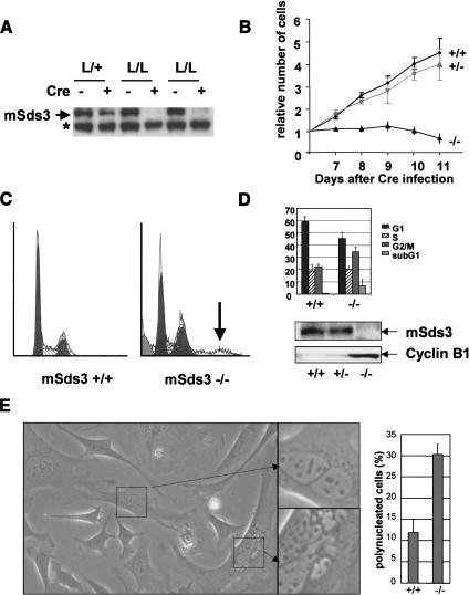
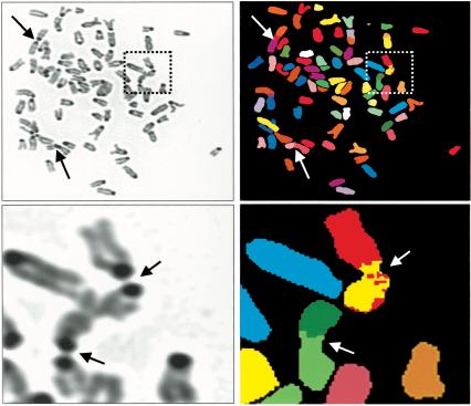
The histone code guides many aspects of chromosome biology including the equal distribution of chromosomes during cell division. In the chromatin domains surrounding the centromere, known as pericentric heterochromatin, histone modifications, particularly deacetylation and methylation, appear to be essential for proper chromosome segregation. However, the specific factors and their precise roles in this highly orchestrated process remain under active investigation. Here, we report that germ-line or somatic deletion of mSds3, an essential component of the functional mSin3/HDAC corepressor complex, generates a cell-lethal condition associated with rampant aneuploidy, defective karyokinesis, and consequently, a failure of cytokinesis. mSds3-deficient cells fail to deacetylate and methylate pericentric heterochromatin histones and to recruit essential heterochromatin-associated proteins, resulting in aberrant associations among heterologous chromosomes via centromeric regions and consequent failure to properly segregate chromosomes. Mutant mSds3 molecules that are defective in mSin3 binding fail to rescue the mSds3 null phenotypes. On the basis of these findings, we propose that mSds3 and its associated mSin3/HDAC components play a central role in initiating the cascade of pericentric heterochromatin-specific modifications necessary for the proper distribution of chromosomes during cell division in mammalian cells.


Figures

Figure 1.
Figure 1.
mSds3 deficiency leads to early embryonic lethality. (A) Targeting strategy and mutant alleles of mSds3. The neomycin resistance cassette is flanked by FRT sites (diamonds), and the neomycin cassette plus the exon 6 region to be deleted (hatched bars) are flanked by LoxP sites (triangles). The external probe used for Southern screening is depicted in gray. Restriction sites used are EcoRI (E) and SalI (S). Exon 5 corresponds to nucleotides 341-360 starting at the initiating ATG; exon 6, nucleotides 361-517; exon 7, nucleotides 518-613. (B) Southern analysis of ES cell clones showing targeting of the mSds3 locus. (C) Genotyping by PCR of mice bearing a lox allele (top) or a null allele (bottom) for mSds3. (D) Genotype distribution of offspring and embryos resulting from mSds3 heterozygote mice intercrosses. (R) Resorbed. (E) In vitro outgrowth of blastocysts from mSds3 heterozygote intercrosses harvested at 3.5 dpc (=d0). The two bottom panels show putative mSds3 null blastocysts undergoing death. (ICM) Inner cell mass; (TE) trophectoderm.
Figure 2.
Figure 2.
mSds3 deficiency results in cell cycle defects. (A) Western blot analysis with an anti-mSds3 antibody of MEFs, 8 d after Cre recombinase infection. The asterisk marks a nonspecific band. (B) Growth curve of MEFs wild-type, heterozygous or null for mSds3 after Cre recombinase infection. For each genotype, six independent cell lines were tested for this assay in duplicate. Empty vector-infected mSds3L/L MEFs grew similar to wild-type MEFs (data not shown). (C) Flow cytometry analysis of mSds3 MEFs 8 d after Cre recombinase infection. Shown is a representative result obtained for mSds3+/+ and mSds3-/- cells. The arrow points to polyploid cells in mSds3-/- cell population. (D, top) Cell cycle repartition average on two independent cell lines for each genotype in duplicate 8 d after Cre recombinase infection. (Bottom) Western Blots performed on the indicated cell lines with an anti-mSds3 antibody or an anti-cyclin-B1 antibody 8 d after Cre recombinase infection. (E, left) Bright field picture of representative mSds3-/- MEFs culture 8 d after Cre recombinase infection. (Right) Quantification of polynucleated cells in three independent primary MEFs of each genotype (at least 200 cells were counted for each cell line).
Figure 3.
Figure 3.
mSds3 deficiency impairs pericentric heterochromatin structure. (a) Immunofluorescence performed on mSds3+/+ (panels A,C,E) or mSds3L/L (panels B,D,F) immortalized cells 8 d after Cre recombinase infection. Primary MEFs null for mSds3 exhibited the same pattern for each of the antibodies used in this study (data not shown). Left column is immunofluorescence performed using an anti-acetylated H4 on Lys 12 antibody (panels A,B), an anti-methylated H3 on Lys 9 antibody (panels C,D) or an anti-HP1α antibody (panels E,F). DAPI is visualized in the middle column and merge is shown in the right column. Shown is a representative cell for each genotype and antibody used. No differences were observed between mSds3+/+ and mSds3+/- cells. Images were acquired using a deconvolution microscope. Bar, 10 μm. (b) Western blot performed on matched cell lines to those shown in a, with the indicated antibodies. The bottom right gel represent a Comassie stain of the relevant molecular weight region.
Figure 4.
Figure 4.
mSds3 binding to mSin3A is necessary to rescue cell viability and pericentric heterochromatin defects. (A) Schematic representation of mSds3 and mSds3 mutants. The coiled coil is encompassed by amino acids 59-170, and the SID (mSin3 interaction domain) by amino acids 188-226. The region deleted in each of the mutants is depicted as a white area. Δ1 corresponds to mSds3Δ161-170, Δ2 to mSds3Δ171-180, Δ3 to mSds3Δ181-190, Δ4 to mSds3Δ191-200, Δ5 to mSds3Δ201-210, Δ6 to mSds3Δ211-220, and Δ7 to mSds3Δ221-230. (B) Western blot documenting the interaction between mSin3A and mSds3 or mSds3 deletion mutants in 293T cells. (Top) Western blot with an anti-Flag antibody detecting transfected Flag-tagged mSds3 or Flag-tagged mSds3 deletion mutants in whole-cell extracts. Ten percent of the amount of proteins used in the immunoprecipitation were loaded. (Bottom) Western blot with an anti-Flag antibody detecting Flag-tagged mSds3 or Flag-tagged mSds3 deletion mutants pulled down with an anti-mSin3A antibody. As reported previously, mSds3 runs as a doublet when overexpressed. mSin3AFlag was transfected to increase the amount of pulled-down mSds3. (C) Growth curve of immortalized mSds3L/- MEFS transduced with retrovirus encoding the mSds3 mutants, and subsequently infected with a retrovirus encoding Cre recombinase. For each genotype, two independent cell lines were tested. The growth of mSds3L/+ cell lines expressing the different mutants was not affected after Cre infection (data not shown). (D) Table representing the percentage of colocalization of HP1α with pericentric heterochromatin identified as DAPI-bright domains in the cell lines described in C, 8 d after Cre-mediated infection.
Figure 5.
Figure 5.
mSds3 deficiency results in genomic instability. (A) Chromosomal counts of metaphases from mSds3 wild-type primary MEFs (left) or mSds3 null primary MEFs (right), 8 d after Cre infection. Each bar represents one metaphase. Ten metaphase were counted on three independent cell lines for each genotype. (B) DAPI (blue) and centromeric-specific FISH (red) staining of one representative metaphase from mSds3+/+ primary MEFs (top) or mSds3-/- primary MEFs (bottom). The arrows point to associated centromeric regions. (C) Chromosomes from mSds3 null primary MEFs metaphase stained with DAPI (blue) and centromeric-specific FISH (red). (D) Double FISH with centromeric-specific probe (red) and telomeric-specific probe (green) of a portion of a representative metaphase from mSds3 null primary MEFs. Arrows indicate associated centromeric regions.
Figure 6.
Figure 6.
DAPI-stained representation (left) and corresponding spectral karyotyping (SKY) analysis (right) of a representative metaphase from primary mSds3-/- MEFs 4 d after Cre-mediated deletion. The arrows point to the associated chromosomes. (Bottom) A close-up of the aberrant chromosomal structures designated by the dotted windows at top.

References

    1. Alland L., David, G., Shen-Li, H., Potes, J., Muhle, R., Lee, H.C., Hou Jr., H., Chen, K., and DePinho, R.A. 2002. Identification of mammalian Sds3 as an integral component of the Sin3/histone deacetylase corepressor complex. Mol. Cell. Biol. 22: 2743-2750. - PMC - PubMed
    1. Ayer D.E., Lawrence, Q.A., and Eisenman, R.N. 1995. Mad-Max transcriptional repression is mediated by ternary complex formation with mammalian homologs of yeast repressor Sin3. Cell 80: 767-776. - PubMed
    1. Bannister A.J., Zegerman, P., Partridge, J.F., Miska, E.A., Thomas, J.O., Allshire, R.C., and Kouzarides, T. 2001. Selective recognition of methylated lysine 9 on histone H3 by the HP1 chromo domain. Nature 410: 120-124. - PubMed
    1. Brown D.C., Grace, E., Sumner, A.T., Edmunds, A.T., and Ellis, P.M. 1995. ICF syndrome (immunodeficiency, centromeric instability and facial anomalies): Investigation of heterochromatin abnormalities and review of clinical outcome. Hum. Genet. 96: 411-416. - PubMed
    1. Dobie K.W., Hari, K.L., Maggert, K.A., and Karpen, G.H. 1999. Centromere proteins and chromosome inheritance: A complex affair. Curr. Opin. Genet. Dev. 9: 206-217. - PubMed

Publication types

MeSH terms