. 2003 Oct 1;17(19):2421-35.
doi: 10.1101/gad.1126303.

KNL-1 directs assembly of the microtubule-binding interface of the kinetochore in C. elegans

Affiliations

KNL-1 directs assembly of the microtubule-binding interface of the kinetochore in C. elegans

Arshad Desai et al. Genes Dev. .

Abstract

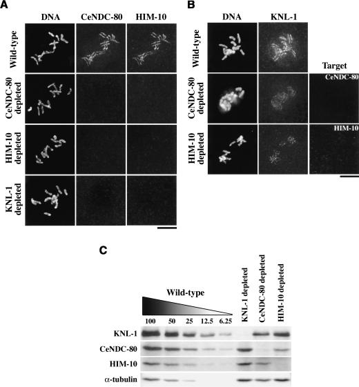
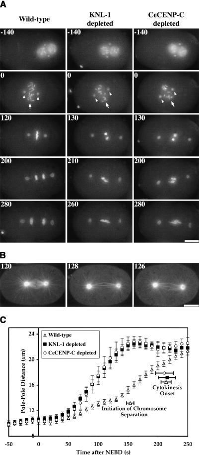
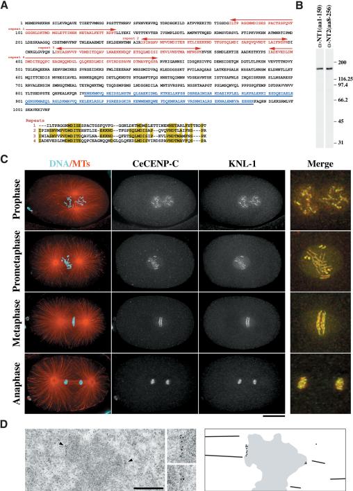
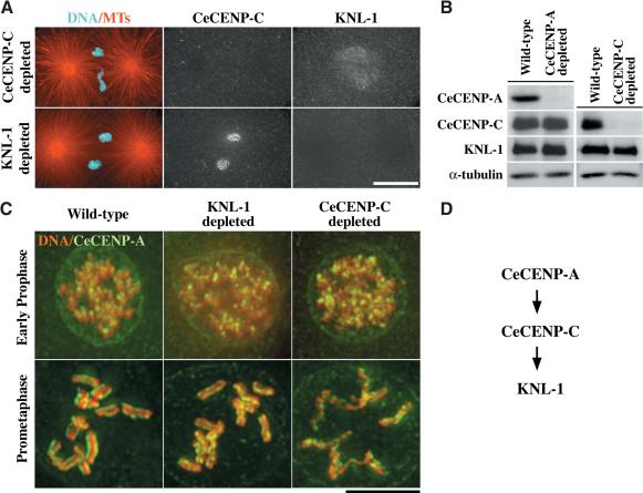
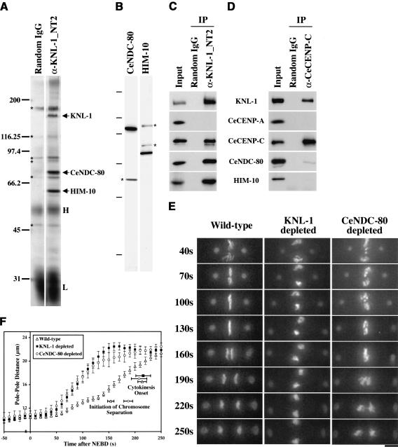
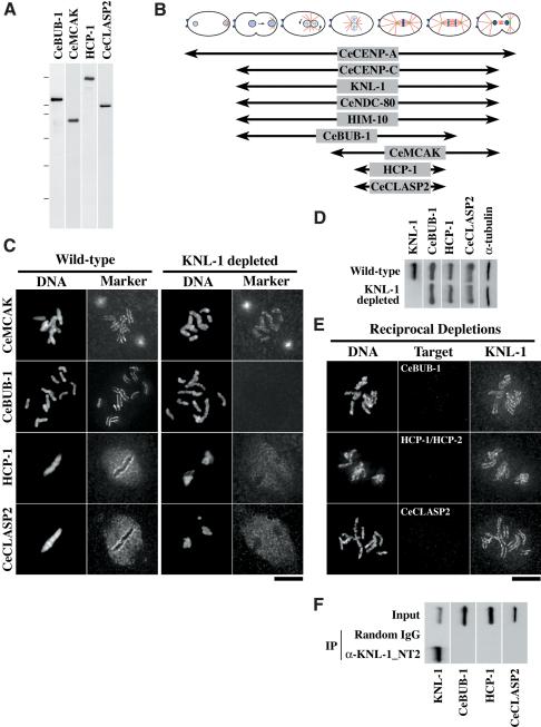
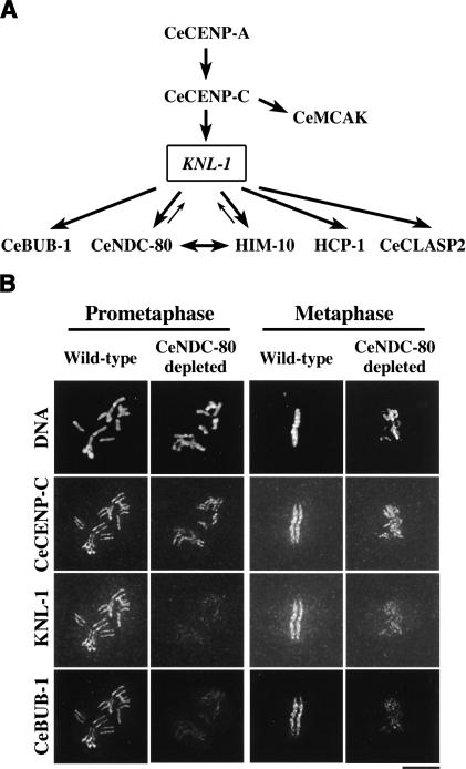
Segregation of the replicated genome during cell division requires kinetochores, mechanochemical organelles that assemble on mitotic chromosomes to connect them to spindle microtubules. CENP-A, a histone H3 variant, and CENP-C, a conserved structural protein, form the DNA-proximal foundation for kinetochore assembly. Using RNA interference-based genomics in Caenorhabditis elegans, we identified KNL-1, a novel kinetochore protein whose depletion, like that of CeCENP-A or CeCENP-C, leads to a "kinetochore-null" phenotype. KNL-1 is downstream of CeCENP-A and CeCENP-C in a linear assembly hierarchy. In embryonic extracts, KNL-1 exhibits substoichiometric interactions with CeCENP-C and forms a near-stoichiometric complex with CeNDC-80 and HIM-10, the C. elegans homologs of Ndc80p/HEC1p and Nuf2p-two widely conserved outer kinetochore components. However, CeNDC-80 and HIM-10 are not functionally equivalent to KNL-1 because their inhibition, although preventing formation of a mechanically stable kinetochore-microtubule interface and causing chromosome missegregation, does not result in a kinetochore-null phenotype. The greater functional importance of KNL-1 may be due to its requirement for targeting multiple components of the outer kinetochore, including CeNDC-80 and HIM-10. Thus, KNL-1 plays a central role in translating the initiation of kinetochore assembly by CeCENP-A and CeCENP-C into the formation of a functional microtubule-binding interface.


Figures

Figure 5.
Figure 5.
KNL-1, CeNDC-80, and HIM-10 exhibit asymmetric dependencies during kinetochore assembly. (A) Wild-type, CeNDC-80-depleted (n = 33), HIM-10-depleted (n = 36), and KNL-1-depleted (n = 67) embryos were stained for DNA (left panels), CeNDC-80 (middle panels), and HIM-10 (right panels). Chromosomes from prometaphase-stage one-cell embryos are shown. (B) Wild-type, CeNDC-80-depleted (n = 37), and HIM-10-depleted (n = 40) embryos were stained for DNA (left panels), KNL-1 (middle panels), and the depletion target (right panels). (C) Western blot of extracts prepared from wild-type, KNL-1-depleted, CeNDC-80-depleted, and HIM-10-depleted worms. Serial dilutions of wild-type extract were loaded to quantify depletion levels.
Figure 1.
Figure 1.
KNL-1 depletion results in a “kinetochorenull” phenotype. Selected panels from time-lapse sequences of wild-type (left column), KNL-1 depleted (middle column), and CeCENP-C depleted (right column) embryos are shown. Time after NEBD (in seconds) is indicated in the upper left corner of each panel. (A) Chromosome segregation was monitored in embryos coexpressing GFP-histone H2B and GFP-γ-tubulin. The 3D widefield data sets were acquired every 10 sec for wild-type (n = 27), KNL-1-depleted (n = 24), and CeCENP-C-depleted (n = 18) embryos. Time-aligned projections of representative 3D movies are shown (see also Supplementary Videos 1-3). Chromosomes (arrows in 0-sec panels) and centrosomes (arrowheads in 0-sec panels) are easily distinguished in these images. (B) Spindle morphology was monitored by single-section spinning disk confocal imaging of embryos expressing GFP-β-tubulin. Images were acquired every 6-8 sec for wild-type (n = 27), KNL-1-depleted (n = 32), and CeCENP-C-depleted (n = 16) embryos (see also Supplementary Videos 4-6). A single image, concurrent with metaphase in wild type, is shown for each type of embryo. (C) The distance between spindle poles was tracked for 14 wild-type, 15 KNL-1-depleted, and 12 CeCENP-C-depleted embryos filmed as in A. The plot shows average pole-to-pole distance versus time after NEBD. Error bars represent the S.E.M. with a confidence interval of 0.95. The average times for the onset of cytokinesis (for all three conditions) and the initiation of chromosome separation (in wild type) are also indicated. Bars: A,B, 10 μm.
Figure 2.
Figure 2.
KNL-1 localizes to kinetochores throughout mitosis. (A) KNL-1 is a 1010-amino-acid novel protein with a predicted molecular mass of 113 kD. The N terminus contains four repeats of a loosely conserved sequence motif (indicated in red and aligned beneath the sequence). The C terminus of KNL-1 has a region predicted to form a coiled coil (underlined and lettered in blue). (B) Western blots of embryo extracts probed using affinity-purified antibodies raised against two overlapping regions of KNL-1 (amino acids 1-150 and amino acids 8-256). (C) Projected 3D data sets of fixed embryos at different cell cycle stages stained for microtubules and DNA (red and cyan; left column), the kinetochore marker CeCENP-C (second column), and KNL-1 (third column). The merged images on the far right show CeCENP-C (red) and KNL-1 (green) at 2.5× higher magnification. (D) Immuno-EM of KNL-1 in multicellular embryos prepared by high-pressure freezing/freeze substitution. Insets were magnified 2×. The schematic to the right illustrates the positions of the gold particles in the field of view. Bars: C, 10 μm; D, 500 nm.
Figure 3.
Figure 3.
KNL-1 is downstream of CeCENP-A and CeCENP-C in the kinetochore assembly hierarchy. (A) Embryos were fixed and stained for microtubules and DNA (red and cyan; left panels), CeCENP-C (middle panels), and KNL-1 (right panels). In CeCENP-C-depleted embryos (n = 39), KNL-1 did not localize to chromosomes, although the weaker “matrix” staining in the region of the spindle was still observed. In KNL-1-depleted embryos, CeCENP-A (n = 45; not shown) and CeCENP-C (n = 31) localized normally to kinetochores. Both CeCENP-C (Oegema et al. 2001) and KNL-1 (n = 31 embryos; not shown) failed to target to chromosomes in embryos depleted of CeCENP-A. (B) Western blots of wild-type, CeCENP-A-depleted, and CeCENP-C-depleted worms. α-Tubulin was used as a loading control. (C) Wild-type (left panels), KNL-1-depleted (middle panels), and CeCENP-C-depleted (right panels) embryos were fixed and stained for DNA (red) and CeCENP-A (green). Representative prophase (top row) and prometaphase (bottom row) embryos are shown. (D) Schematic illustrating the linear assembly hierarchy for CeCENP-A, CeCENP-C, and KNL-1. The relationship between CeCENP-A and CeCENP-C was established previously (Moore and Roth 2001; Oegema et al. 2001). Bars: A, 10 μm; C, 5 μm.
Figure 4.
Figure 4.
KNL-1 forms a complex with two widely conserved outer kinetochore proteins. (A) Silver-stained gel of immunoprecipitates from embryo extracts. The three bands consistently detected in KNL-1 (α-KNL-1_NT2) but not control (Random IgG) immunoprecipitates (indicated by arrows; n = 4 extracts) were identified as KNL-1, CeNDC-80, and HIM-10 by mass spectrometry. The same bands were detected in immunoprecipitates using a second KNL-1 antibody (data not shown). Contaminating antibody heavy (H) and light (L) chains and background bands (*) are also indicated. (B) Western blots of embryo extracts probed with affinity-purified antibodies against CeNDC-80 and HIM-10. Asterisks indicate background bands that were not eliminated by RNAi of the corresponding genes. The molecular mass markers were 200, 97.4, 66.2, 45, 31, and 14.4 kD. (C) Western blots of KNL-1 immunoprecipitates probed with the indicated antibodies. Identical results were obtained in immunoprecipitations performed on four independent extracts. (D) Western blots of CeCENP-C immunoprecipitates probed with the indicated antibodies. Identical results were obtained in immunoprecipitations performed on two independent extracts. CeCENP-C was not detectable by silver staining in either KNL-1 or CeCENP-C immunoprecipitates. (E) 3D widefield data sets were acquired every 10 sec for wild-type (n = 27), KNL-1-depleted (n = 24), CeNDC-80-depleted (n = 21), and HIM-10-depleted (n = 22; not shown; see Supplementary Videos 11, 12) embryos. Time-aligned projections of representative 3D movies are shown (see also Supplementary Videos 7-14). The times after NEBD are indicated on the left in seconds. Bar, 5 μm. (F) The distance between spindle poles was tracked for 11 CeNDC-80-depleted embryos filmed as in E. The plot shows average pole-to-pole distance versus time after NEBD. Wild-type and KNL-1-depleted tracking data are replotted from Figure 1C. Error bars represent the S.E.M. with a confidence interval of 0.95. The average times of the onset of cytokinesis (all three conditions) and the average time when chromosome separation was first visible (wild-type and CeNDC-80-depleted embryos) are also indicated.
Figure 6.
Figure 6.
KNL-1 is required to target multiple outer kinetochore components. (A) Western blots of embryo extracts probed with affinity-purified antibodies against the indicated C. elegans kinetochore components. The molecular mass markers were 200, 116.25, 97.4, 66.2, 45, and 31 kD. (B) Schematic summarizing when during the first embryonic mitosis the nine proteins analyzed in this study localize to kinetochores (Supplementary Fig. 1; Oegema et al. 2001). (C) Wild-type and KNL-1-depleted embryos were stained for DNA (left panels in each pair), KNL-1 (not shown), and one of four kinetochore markers (right panels in each pair) as indicated. KNL-1 was not detectable by immunofluorescence in all cases where it was depleted (data not shown). CeMCAK targets normally to kinetochores in KNL-1-depleted embryos (n = 63), but CeBUB-1 (n = 50), HCP-1 (n = 21), and CeCLASP2 (n = 19) fail to target. (D) Westerns blots of wild-type and KNL-1-depleted worms were probed with antibodies to the three proteins that require KNL-1 for kinetochore targeting. α-Tubulin blotting was used as a loading control. (E) Embryos depleted of CeBUB-1 (n = 55), HCP-1 and HCP-2 (n = 37), or CeCLASP2 (n = 33) were stained for DNA (left panels), the depleted component (middle panels), and KNL-1 (right panels). KNL-1 targeted normally to kinetochores in all cases. (F) Western blots of KNL-1 immunoprecipitates probed with antibodies against the three proteins (CeBUB-1, HCP-1, and CeCLASP2) that require KNL-1 for kinetochore targeting. The control blot panel for KNL-1 is the same as in Figure 4C.
Figure 7.
Figure 7.
The architectural logic of a mitotic metazoan kinetochore. (A) Schematic of the kinetochore assembly hierarchy in C. elegans based on phenotypic analysis, targeting dependencies, and biochemical data. KNL-1 plays a central role in propagating the initiation of kinetochore assembly by CeCENP-A and CeCENP-C into the formation of a functional microtubule-binding interface. (B) A test of the proposed kinetochore assembly hierarchy. Wild-type and CeNDC-80-depleted embryos were fixed and stained for DNA (top row), CeCENP-C (second row), KNL-1 (third row), and CeBUB-1 (bottom row). Exposures for each kinetochore marker were identical for wild-type and CeNDC-80-depleted embryos. Images were equivalently processed to facilitate comparisons of staining intensities. Similar diminution of KNL-1 and CeBUB-1 staining, but not of CeCENP-C staining, was observed in 16 prophase and 20 prometaphase-early anaphase CeNDC-80-depleted one-cell-stage embryos. Bar, 5 μm.

References

    1. Akhmanova A., Hoogenraad, C.C., Drabek, K., Stepanova, T., Dortland, B., Verkerk, T., Vermeulen, W., Burgering, B.M., De Zeeuw, C.I., Grosveld, F., et al. 2001. Clasps are CLIP-115 and -170 associating proteins involved in the regional regulation of microtubule dynamics in motile fibroblasts. Cell 104: 923-935. - PubMed
    1. Cheeseman I.M., Anderson, S., Jwa, M., Green, E.M., Kang, J., Yates III, J.R., Chan, C.S., Drubin, D.G., and Barnes, G. 2002a. Phospho-regulation of kinetochore-microtubule attachments by the Aurora kinase Ipl1p. Cell 111: 163-172. - PubMed
    1. Chen Y., Riley, D.J., Chen, P.L., and Lee, W.H. 1997. HEC, a novel nuclear protein rich in leucine heptad repeats specifically involved in mitosis. Mol. Cell. Biol. 17: 6049-6056. - PMC - PubMed
    1. Cleveland D.W., Mao, Y., and Sullivan, K.F. 2003. Centromeres and kinetochores: From epigenetics to mitotic checkpoint signaling. Cell 112: 407-421. - PubMed
    1. DeLuca J.G., Moree, B., Hickey, J.M., Kilmartin, J.V., and Salmon, E.D. 2002. hNuf2 inhibition blocks stable kinetochore-microtubule attachment and induces mitotic cell death in HeLa cells. J. Cell Biol. 159: 549-555. - PMC - PubMed

Publication types

MeSH terms

LinkOut - more resources