. 2003 Oct;112(7):989-98.
doi: 10.1172/JCI17970.

Stat3 protects against Fas-induced liver injury by redox-dependent and -independent mechanisms

Affiliations

Stat3 protects against Fas-induced liver injury by redox-dependent and -independent mechanisms

Sanae Haga et al. J Clin Invest. 2003 Oct.

Abstract

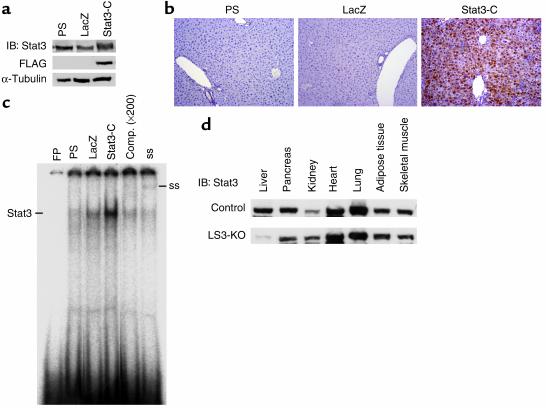
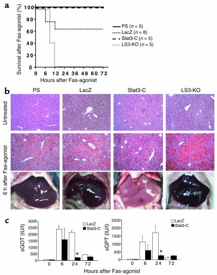
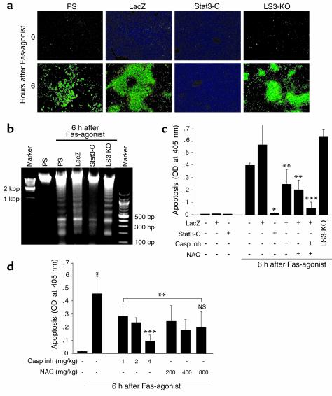
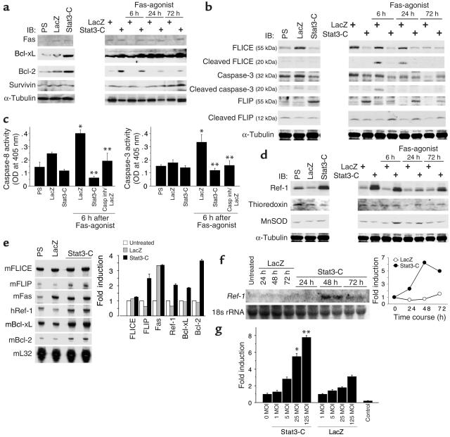
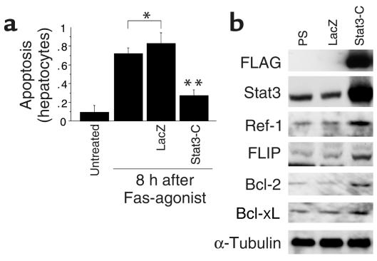
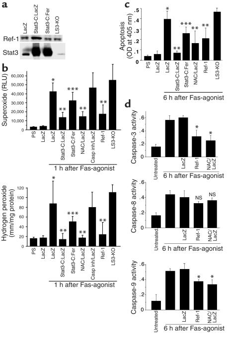
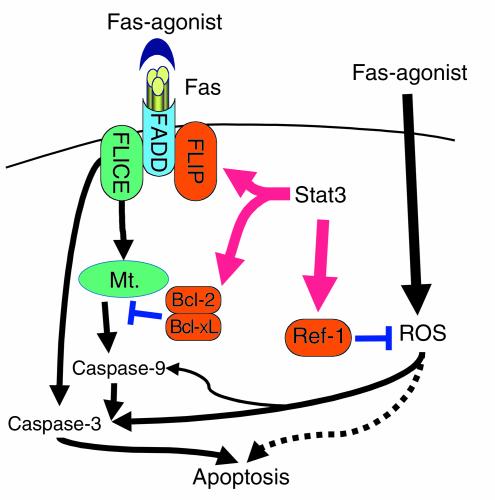
Signal transducer and activator of transcription-3 (Stat3) is one of the most important molecules involved in the initiation of liver development and regeneration. In order to investigate the hepatoprotective effects of Stat3, we examined whether Stat3 protects against Fas-mediated liver injury in the mouse. A constitutively activated form of Stat3 (Stat3-C) was adenovirally overexpressed in mouse liver by intravenous injection, and then a nonlethal dose of Fas agonist (Jo2) was injected intraperitoneally into the mouse (0.3 microg/g body wt). Stat3-C dramatically suppressed both apoptosis and necrosis induced by Jo2. In contrast, liver-specific Stat3-knockout mice failed to survive following Jo2 injection. Stat3-C upregulated expression of FLICE inhibitor protein (FLIP), Bcl-xL, and Bcl-2, and accordingly downregulated activities of FLICE and caspase-3 that were redox-independent. Interestingly, Stat3-C also upregulated the redox-associated protein redox factor-1 (Ref-1) and reduced apoptosis in liver following Jo2 injection by suppressing oxidative stress and redox-sensitive caspase-3 activity. These findings indicate that Stat3 activation protects against Fas-mediated liver injury by inhibiting caspase activities in redox-dependent and -independent mechanisms.


Figures

Figure 1
Figure 1
Stat3-C protein is successfully overexpressed in mouse liver and activates Stat3 DNA binding. (a) Anti-Stat3 and anti-FLAG immunoblots were performed to determine total and exogenous Stat3 protein in liver tissue, respectively, 3 days after adenovirus infection. (b) Immunohistochemical study using anti-Stat3 showed that Stat3-C protein is expressed in nuclei and cytoplasm of the liver in Stat3-C mice. Original magnification, ×200. (c) Electrophoretic mobility shift assay (EMSA) analysis showed elevated Stat3 DNA binding in the liver of Stat3-C mice. Five micrograms of nuclear extract was incubated with SIE-m67 probe. In order to confirm specific Stat3 DNA binding, competition (comp.) and supershift assays were performed using cold SIE-m67 probe and anti-Stat3. (d) Western blot analysis of Stat3 protein in various tissues of control and LS3-KO mice. These data are representative of at least three independent experiments. FP, free probe; ss, supershift; IB, immunoblot.
Figure 2
Figure 2
Stat3-C prevents liver injury induced by Fas agonist. (a) Survival after Fas agonist treatment. Mice were injected intraperitoneally with Jo2 (0.3 μg/g body wt) and followed for 72 hours. (b) Liver appearance and microscopic change 6 hours after Fas agonist. H&E staining; original magnification, ×100. (c) Serum levels of GOT and GPT (sGot and sGPT) until 72 hours after Fas agonist treatment. *P ≤ 0.01 vs. LacZ 24 hours after treatment with Fas agonist. Data are expressed as mean ± SEM (n = 5).
Figure 3
Figure 3
Stat3-C suppresses Fas-induced apoptotic cell death in liver. (a) Fluoromicroscopic photographs of liver sections with TUNEL staining from PS, LacZ, Stat3-C, and LS3-KO mice treated with Fas agonist (formalin-fixed, paraffin-embedded sections; original magnification, ×100). (b) The DNA ladder pattern was analyzed 6 hours after Fas agonist. Each photo is representative of at least three independent experiments. (c) Apoptotic cell death was quantitatively measured by ELISA. Pretreatment with the general caspase inhibitor Z-VAD-fmk (1 μg/g body wt) or the antioxidant NAC (200 mg/kg body wt) was performed 1 hour prior to the experiments. *P < 0.01 vs. LacZ/Fas agonist, **P < 0.05 vs. LacZ/Fas agonist and Stat3-C/Fas agonist, ***P < 0.05 vs. LacZ/Fas agonist + caspase inhibitor and LacZ/Fas agonist + NAC. (d) Dose-dependent effects of caspase inhibitor (Z-VAD-fmk) or antioxidant (NAC) upon hepatic apoptosis after Fas agonist. *P < 0.01 vs. untreated control, **P < 0.05 vs. Fas agonist, ***P < 0.05 vs. Fas agonist + caspase inhibitor (1 and 2 mg/kg, respectively). All data are expressed as mean ± SEM (n = 5). Casp inh, caspase inhibitor.
Figure 4
Figure 4
Stat3-C induces apoptosis-related proteins and redox-related protein. Immunoblot, Northern blot, and multiprobe ribonuclease protection assay were performed on liver tissues from PS, LacZ, and Stat3-C mice. Sequential changes of the protein amounts after Fas agonist treatment were analyzed in LacZ and Stat3-C mice. Each blot is representative of three independent experiments. (a) Expression of Fas, Bcl-related proteins, and Survivin. (b) Expression of caspase-related proteins. (c) Enzyme activity assays of caspase-3 and caspase-8. *P < 0.05 vs. LacZ, **P < 0.01 vs. LacZ/Fas agonist. (d) Protein expression of redox-related proteins Ref-1, thioredoxin, and MnSOD. (e) Multiprobe ribonuclease protection assay for apoptosis-related genes and redox-related genes 48 hours after adenoviral gene transfer. m, mouse; h, human. (f) Northern blot analysis for Ref-1 mRNA after adenoviral gene transfer of Stat3-C. Results are from a single representative experiment that was reproduced once (df). (g) Transcriptional activity of Ref-1 promoter by Stat3-C. Luciferase assay for Ref-1 promoter activity stimulated by AxCAS3-C or AdLacZ. Control was pGL3 without Ref-1 promoter. *P ≤ 0.01 vs. uninfected (0 MOI, 0) and LacZ (25 MOI), **P ≤ 0.01 vs. uninfected (0 MOI) and LacZ (125 MOI). All data are expressed as mean ± SEM (n = 5).
Figure 5
Figure 5
Stat3-C protects primary hepatocytes from Jo2-induced apoptosis with significant expression of apoptosis- and antioxidant-related proteins in hepatocytes. (a) Apoptotic cell death of primary hepatocytes was quantitatively measured by ELISA. *P < 0.05 vs. untreated, **P < 0.01 vs. LacZ/Fas agonist. All data are expressed as mean ± SEM (n = 5). (b) Protein expression by Stat3-C in primary hepatocytes.
Figure 6
Figure 6
Hepatic ROS production and apoptosis induced by Fas agonist are suppressed by Stat3-C via Ref-1. LacZ, Stat3-C:LacZ, and Stat3-C:AdFer (Stat3-C:Fer) mice were infected with AdLacZ only, AxCAS3-C:AdLacZ (1:1), and AxCAS3-C:AdFer (1:1), respectively. Total amounts of adenovirus vectors were adjusted to 3 × 10⁸ PFU/mouse in all experiments. (a) Immunoblots of Ref-1 and Stat3 proteins showing that AxCAS3-C increased Stat3 and Ref-1 proteins, but AdFer reduced them. Ref-1 protein was, however, expressed even in the liver of LS3-KO mice. (b) Superoxide generation assay and hydrogen peroxide assay in liver tissue*P < 0.05 vs. LacZ, **P < 0.05 vs. LacZ/Fas agonist, ***P < 0.05 vs. Stat3-C:LacZ/Fas agonist. Casp inh; i.e. Z-VAD-fmk. (c) Apoptotic cell death was quantitatively analyzed by ELISA. *P < 0.01 vs. LacZ, **P < 0.05 vs. LacZ/Fas agonist, ***P < 0.05 vs. Stat3-C:LacZ/Fas agonist. (d) Enzyme activity assays of caspase-3 and caspase-8. *P < 0.05 vs. LacZ/Fas agonist. All data are expressed as mean ± SEM (n = 5). RLU, relative light unit.
Figure 7
Figure 7
Schematic view of the protective mechanisms of Stat3 against Fas-mediated liver injury. Activation of Stat3 upregulates caspase-related proteins (such as FLIP, Bcl-XL, and Bcl-2) and redox-related protein Ref-1. These proteins collaboratively prevent Fas-mediated liver injury in redox-dependent and/or independent ways. Mt., mitochondria.

Comment in

References

    1. Darnell JE., Jr STATs and gene regulation. Science. 1997;277:1630–1635. - PubMed
    1. Schuringa JJ, Jonk LJ, Dokter WH, Vellenga E, Kruijer W. Interleukin-6-induced STAT3 transactivation and Ser727 phosphorylation involves Vav, Rac-1 and the kinase SEK-1/MKK-4 as signal transduction components. Biochem. J. 2000;347:89–96. - PMC - PubMed
    1. Leu JI, Crissey MA, Leu JP, Ciliberto G, Taub R. Interleukin-6-induced STAT3 and AP-1 amplify hepatocyte nuclear factor 1-mediated transactivation of hepatic genes, an adaptive response to liver injury. Mol. Cell. Biol. 2001;21:414–424. - PMC - PubMed
    1. Schuringa JJ, van der Schaaf S, Vellenga E, Eggen BJ, Kruijer W. LIF-induced STAT3 signaling in murine versus human embryonal carcinoma (EC) cells. Exp. Cell Res. 2002;274:119–129. - PubMed
    1. Levy DE, Lee CK. What does Stat3 do? J. Clin. Invest. 2002;109:1143–1148. doi:10.1172/JCI200215650. - PMC - PubMed

MeSH terms

Substances