Comparative Study
. 2004 Jan;91(1):78-91.
doi: 10.1152/jn.00741.2003. Epub 2003 Oct 1.

Long-lasting activation of rhythmic neuronal activity by a novel mechanosensory system in the crustacean stomatogastric nervous system

Affiliations
Comparative Study

Long-lasting activation of rhythmic neuronal activity by a novel mechanosensory system in the crustacean stomatogastric nervous system

Mark P Beenhakker et al. J Neurophysiol. 2004 Jan.

Abstract

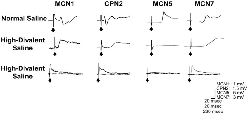
Sensory neurons enable neural circuits to generate behaviors appropriate for the current environmental situation. Here, we characterize the actions of a population (about 60) of bilaterally symmetric bipolar neurons identified within the inner wall of the cardiac gutter, a foregut structure in the crab Cancer borealis. These neurons, called the ventral cardiac neurons (VCNs), project their axons through the crab stomatogastric nervous system to influence neural circuits associated with feeding. Brief pressure application to the cardiac gutter transiently modulated the filtering motor pattern (pyloric rhythm) generated by the pyloric circuit within the stomatogastric ganglion (STG). This modulation included an increased speed of the pyloric rhythm and a concomitant decrease in the activity of the lateral pyloric neuron. Furthermore, 2 min of rhythmic pressure application to the cardiac gutter elicited a chewing motor pattern (gastric mill rhythm) generated by the gastric mill circuit in the STG that persisted for < or =30 min. These sensory actions on the pyloric and gastric mill circuits were mimicked by either ventral cardiac nerve or dorsal posterior esophageal nerve stimulation. VCN actions on the STG circuits required the activation of projection neurons in the commissural ganglia. A subset of the VCN actions on these projection neurons appeared to be direct and cholinergic. We propose that the VCN neurons are mechanoreceptors that are activated when food stored in the foregut applies an outward force, leading to the long-lasting activation of projection neurons required to initiate chewing and modify the filtering of chewed food.


Figures

FIG. 1.
FIG. 1.
Crab foregut and stomatogastric nervous system. A: schematic of crab foregut with innervating stomatogastric nervous system (STNS) in black. Arrows with full lines point to foregut regions. Arrows with dotted lines point to STNS elements. B: schematic of the isolated STNS. Included are the 4 ganglia, connecting nerves, peripheral nerves, and 4 identified CoG projection neurons. Each of these projection neurons occurs as a single copy in each CoG. C: motor patterns generated by the pyloric and gastric mill circuits in the STG. The top 3 intracellular recordings correspond to pyloric neurons and the bottom 3 intracellular recordings correspond to gastric mill neurons. Extracellular recording shows the activity of the MCN1 projection neuron. Abbreviations: Ganglia: CoG, commissural ganglion; OG, esophageal ganglion; STG, stomatogastric ganglion. Nerves: dgn, dorsal gastric nerve; dpon, dorsal posterior esophageal nerve; gpn, gastropyloric nerve; ion, inferior esophageal nerve; lgn, lateral gastric nerve; lvn, lateral ventricular nerve; mgn, medial gastric nerve; mvn, medial ventricular nerve; pdn, pyloric dilator nerve; pyn, pyloric nerve; son, superior esophageal nerve; stn, stomatogastric nerve; vcn, ventral cardiac nerve. Neurons: MCN1, 5, 7, modulatory commissural neuron 1, 5, 7; CPN2, commissural projection neuron 2; LP, lateral pyloric; PY, pyloric; PD, pyloric dilator; LG, lateral gastric; GM, gastric mill; DG, dorsal gastric.
FIG. 2.
FIG. 2.
Location and anatomy of VCN neurons. A: region of the foregut innervated by the vcn nerve. This schematic represents the ventral aspect of the foregut. vcn branches from the dpon and projects ventrally to innervate a region of the cardiac sac that lies on/near the ventral anteroposterior midline, near the gastric mill compartment (box). B: schematic representation of an enlarged view of the interior of the foregut region within the box of A. Here is located the cv3 muscle, between the paired lateral cardiac ossicles. vcn innervates (from the outside) the ipsilateral invagination (i.e., cardiac gutter) lying slightly anterior to the lateral cardiac ossicles. C: schematic of region found in the box of B. D: preparation consisting of the region found in the box of B stained with the vital dye methylene blue. One of 2 VCN axon bundles plus about 60 bipolar neurons (region within and near box) is shown. Dotted black lines indicate the location of exiting VCN axons. Cal. bar: 500 μm. E: higher-magnification view of region found in box of D, showing 4 VCN somata with proximal neurites. Cal. bar: 20 μm. CG: cardiac gutter.
FIG. 3.
FIG. 3.
Physiological evidence for a population of VCN neurons. Shown is an intracellular recording of an esophageal system motor neuron (OMN) located in the CoG. Single vcn nerve stimuli at voltages sufficient to activate the VCN pathway elicited apparently monosynaptic excitatory postsynaptic potentials (EPSPs) in the OMN. As the stimulus voltage was increased from threshold (4.2 V) to maximal response (7.2 V) in 0.2-V increments, the EPSP amplitude increased with no change in latency and no additional peaks evident. Shown are all 17 averaged EPSPs (black and gray) evoked by vcn stimulation. Representative EPSPs demonstrating that EPSP amplitude increases with increased vcn stimulation voltage are shown in thick black. This result indicates that more presynaptic neurons of the type responsible for the low-amplitude stimulus-evoked EPSP were likely being recruited. Each EPSP is the average of 10–12 individual responses.
FIG. 4.
FIG. 4.
Mechanical stimulation of the cardiac gutter modulates an ongoing pyloric rhythm. Slight pressure to the cardiac gutter (see Fig. 2), on an excised portion of the cardiac sac innervated by the vcn (see Fig. 2) still connected to the isolated STNS, inhibited the LP neuron and increased the frequency of occurrence of PD neuron bursts (pyloric cycle frequency).
FIG. 5.
FIG. 5.
Effects of brief mechanical stimulation of the ventral cardiac gutter are mimicked by either vcn or dpon nerve stimulation. A: effects of mechanical, vcn, and dpon stimulation in the same preparation. In each case, the stimulus inhibited LP neuron activity and increased the frequency of the pyloric rhythm (as measured by PD neuron burst frequency). BE: quantification of the pyloric rhythm in response to mechanical, vcn, or dpon stimulation. Data represented come from 6 preparations in which all 3 modes of stimulation were delivered. Events occurring during VCN stimulation are labeled “Stim.” Bars labeled “−2” and “−1” correspond to 2 consecutive epochs immediately before VCN stimulation, and those labeled “+1” and “+2” correspond to 2 consecutive epochs immediately after stimulation. Data in each epoch represents the average of 5 consecutive pyloric cycles. Pyloric cycle frequency, number of LP spikes/burst, and LP burst duration are significantly different during the stimulus vs. prestimulus epochs (n = 6, P < 0.05, one-way repeated-measures ANOVA, Student–Newman–Keuls method).
FIG. 6.
FIG. 6.
Repetitive mechanical stimulation of the ventral cardiac gutter activates the gastric mill rhythm. Left: before stimulation, there was no gastric mill rhythm (lgn, dgn, mvn) but there was a pyloric rhythm (mvn). Anterior gastric receptor (AGR) neuron is a Golgi tendon organlike sensory neuron that fires tonically in the isolated STNS (Combes et al. 1995). Right: rhythmic applications of pressure to the cardiac gutter (see Fig. 2), in an excised portion of the cardiac sac inner-vated by the vcn (see Fig. 2) still connected to the isolated STNS, activated the gastric mill rhythm.
FIG. 7.
FIG. 7.
Effects of repetitive mechanical stimulation are mimicked by either repetitive vcn or dpon nerve stimulation. A: gastric mill rhythms elicited by repetitive mechanical, vcn, or dpon stimulation in the same preparation. In each case, the stimulus activated rhythmic bursting in the LG (n = 6), DG (n = 6), and GM (n = 4) neurons, as well as gastric mill rhythm-timed inhibition of the IC and VD neurons (n = 3). BG: quantification of several gastric mill rhythm parameters in response to mechanical, vcn, or dpon stimulation. Regardless of the stimulus, the characteristics of the resulting motor pattern were similar (P > 0.05, one-way repeated-measures ANOVA), except for DG neuron firing frequency (P < 0.05).
FIG. 8.
FIG. 8.
Different dpon nerve stimulus patterns have the same influence on the gastric mill rhythm. A: types of stimulus protocols used to activate VCN pathway. Using the dpon, the VCN pathway was stimulated with a protocol reminiscent of either the gastric mill/cardiac sac (GM/CS) or pyloric rhythm, or was stimulated tonically. In each case, 900 individual stimuli were delivered. B: resulting gastric mill rhythm was assessed 1 min after each stimulation. Based on the 2 parameters tested, gastric mill cycle period (n = 5, P = 0.42) and LG neuron duty cycle (n = 5, P = 0.74), the gastric mill rhythms generated by the 3 protocols outlined in a are comparable (one-way ANOVA).
FIG. 9.
FIG. 9.
Activation of the gastric mill rhythm by VCN stimulation requires communication between the commissural ganglia (CoGs) and the STG. A: in the complete STNS, activating the VCN pathway by dpon stimulation initiated the gastric mill rhythm. B: after removing the CoGs from the STNS, stimulating the VCNs no longer activated the gastric mill rhythm. C: after removing the CoGs, tonic extracellular stimulation (25 Hz) of MCN1 (ion, see Fig. 1) still activated the gastric mill rhythm, indicating that the gastric mill circuit was not compromised. Thickened baseline results from tonic barrage of EPSPs from MCN1 (Coleman et al. 1995). All 3 panels are from the same STNS.
FIG. 10.
FIG. 10.
Extracellular dpon stimulation influences CoG projection neurons. Shown are intracellular recordings of each identified CoG projection neuron in response to a train of 10 dpon stimuli (train frequency: 0.06 Hz; stimulus duration: 6 s; stimulus frequency: 15 Hz). A: MCN1. Initially, stimulating the dpon caused MCN1 to hyperpolarize slightly, followed by a mild rebound after stimulation (1st Stim). During the course of the train delivery, VCN excitation of MCN1 built up until MCN1 exhibited strong activity. Nonetheless, MCN1 continued to hyper-polarize in response to dpon stimulation (Middle Stims). MCN1 exhibited strong activity 1 min after the dpon train. B: CPN2. Response of CPN2 to a dpon stimulus train was similar to that of MCN1. Initial CPN2 inhibition was gradually overcome by strong excitation. After the dpon train, CPN2 was highly active. C: MCN5. MCN5 was activated by dpon stimulation. This level of activation built up during the course of the dpon train. After the stimulus train, MCN5 was weakly or not active. D: MCN7. MCN7 is only weakly excited by a dpon stimulus. After the delivery of dpon train, MCN7 was weakly or not active.
FIG. 11.
FIG. 11.
Synaptic actions on CoG projection neurons resulting from dpon stimulation. In normal saline, MCN1, CPN2, and MCN7 received complex PSPs from dpon stimulation. This PSP consisted of a relatively long-duration EPSP interrupted by a shorter-duration IPSP. Only the EPSP persisted in the presence of high-divalent cation saline in MCN1, CPN2, and MCN7, suggesting a monosynaptic action. Furthermore, this presumed monosynaptic component included a long-lasting action (bottom traces). Stimulating the dpon also elicited an EPSP in MCN5. This postsynaptic event did not persist in high-divalent cation saline. Each PSP is the average of 10 individual postsynaptic responses. Arrows indicate stimulus artifact. VmRest: MCN1, −67 mV; CPN2, −57 mV; MCN5, −62 mV; MCN7, −72 mV.
FIG. 12.
FIG. 12.
VCN neurons are cholinergic. A, B: intracellular recordings of CoG neurons (OMN and MCN1) in high-divalent cation saline while single stimuli were delivered to the dpon revealed presumably monosynaptic EPSPs. Amplitude of these EPSPs was reversibly reduced by about 40% when the cholinergic antagonist decamethonium bromide (10−3 M) was superfused. Each PSP is the average of about 30 postsynaptic events. VmRest: OMN: −54 mV; MCN1, −75 mV.
FIG. 13.
FIG. 13.
Working model of the VCN pathway. During rest (i.e., cardiac sac is empty), the VCN pathway is not activated and, consequently, it does not activate CoG projection neurons (PNs). Thus the gastric mill circuit is off. Because the VCN pathway is activated by mechanical pressure, we postulate that the introduction of food to the food-storing cardiac sac distends this foregut compartment and activates the VCN pathway. Because inconsistent or no gastric mill rhythm is recorded during VCN stimulation, the circuit is not coherently activated during cardiac sac distention. Later, once food has moved from the cardiac sac to the gastric mill for chewing, particular CoG projection neurons show regular and persistent activity and therefore the gastric mill rhythm is elicited.

References

    1. Akay T, Bassler U, Gerharz P, and Buschges A. The role of sensory signals from the insect coxa-trochanta joint in controlling motor activity of the femur-tibia joint. J Neurophysiol 85: 594–604, 2001. - PubMed
    1. Alexandrowitcz JS. Muscle receptor organs in the abdomen of Homarus vulgaris and Palinurus vulgaris. Q J Microscop Sci 92: 163–203, 1951.
    1. Andersson O and Grillner S. Peripheral control of the cat’s step cycle. I. Phase dependent effects of ramp-movements of the hip during “fictive locomotion.” Acta Physiol Scand 113: 89–101, 1981. - PubMed
    1. Beenhakker MP, Hertzberg S, and Nusbaum MP. Neural network modulation by mechanosensory activation. Soc Neurosci Abstr 26: 449, 2000.
    1. Bennet-Clark HC. Abdominal stretch and inhibition of moulting in Rhodnius prolixus (hemiptera). J Insect Physiol 12: 1019–1028, 1966.

Publication types

MeSH terms

Substances

LinkOut - more resources