. 2003 Oct 1;23(26):8911-20.
doi: 10.1523/JNEUROSCI.23-26-08911.2003.

Lobster (Panulirus interruptus) pyloric muscles express the motor patterns of three neural networks, only one of which innervates the muscles

Affiliations

Lobster (Panulirus interruptus) pyloric muscles express the motor patterns of three neural networks, only one of which innervates the muscles

Jeff B Thuma et al. J Neurosci. .

Abstract

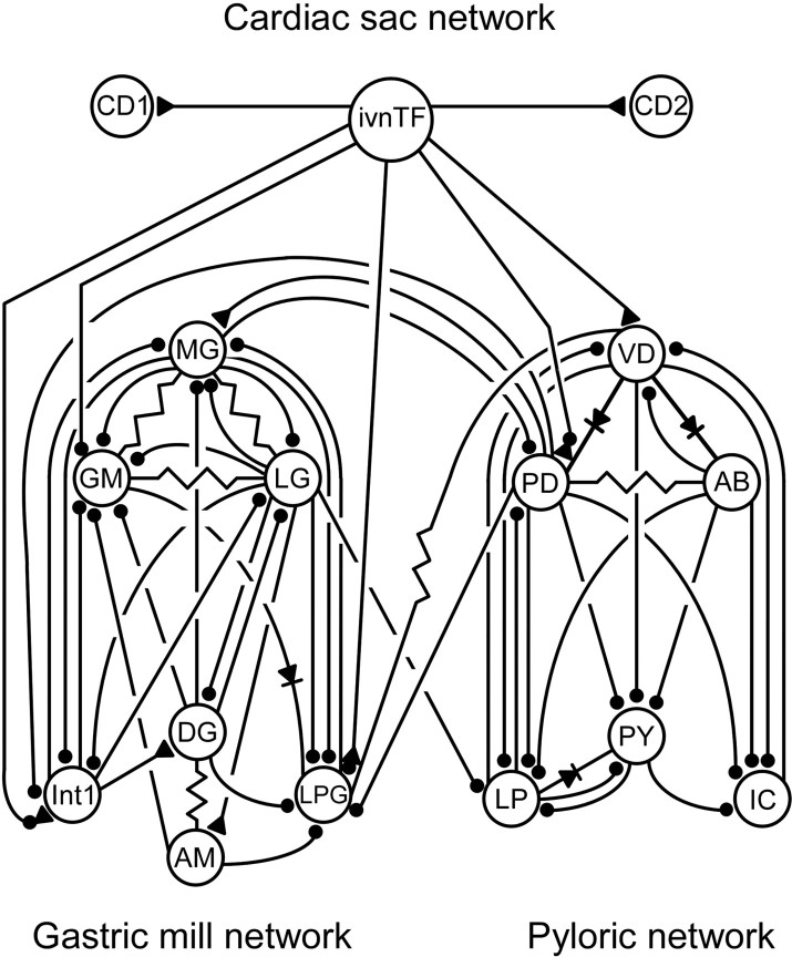
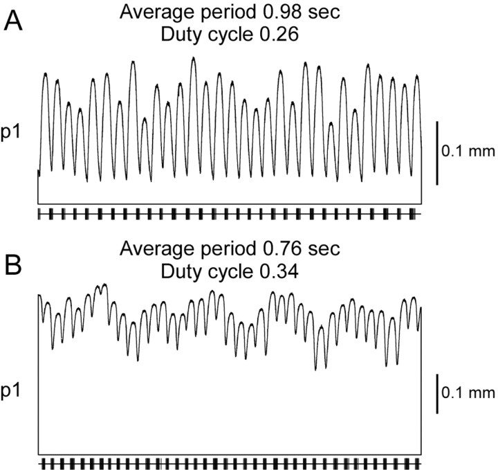
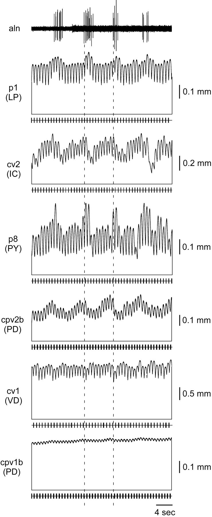
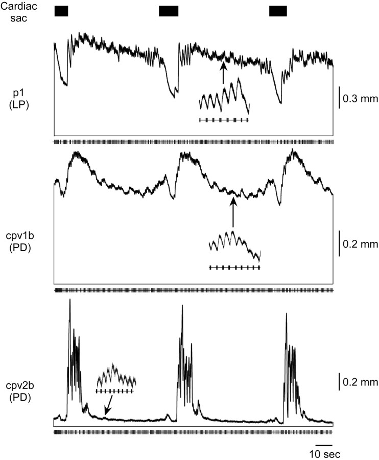
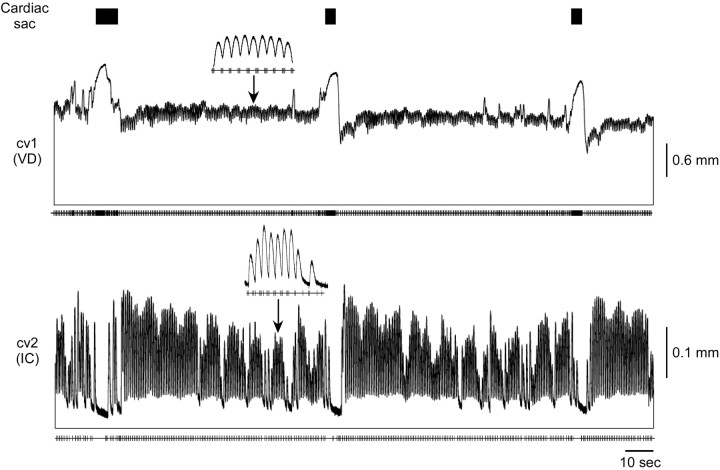
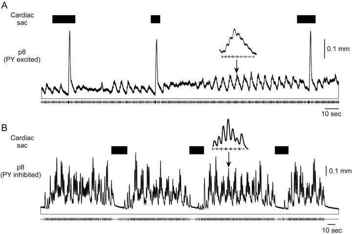
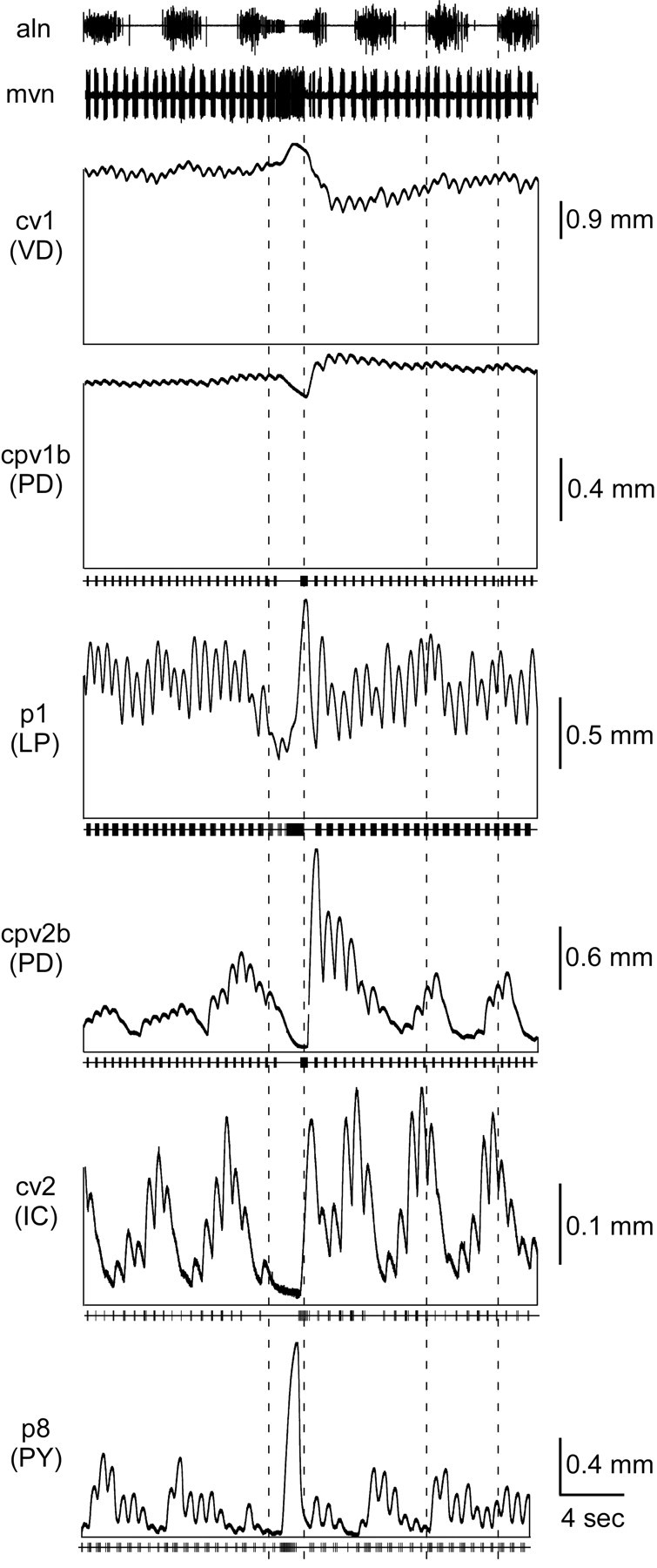
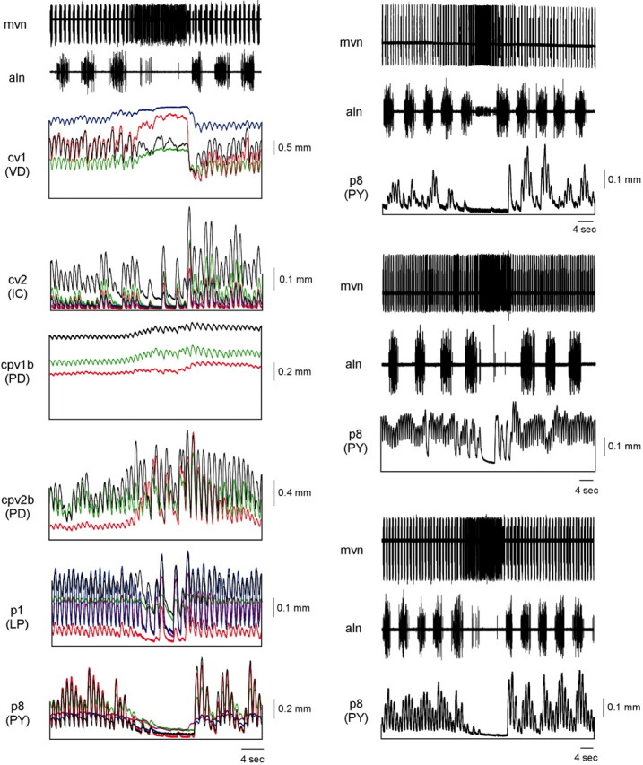
In several systems, including some well studied invertebrate "model" preparations, rapid, rhythmic inputs drive slow muscles. In this situation muscle contractions can summate temporally between motor neuron bursts, tonically contract, and low-pass filter broad-band input. We have investigated how the muscles innervated by each motor neuron type of the rapid, rhythmic (cycle period, approximately 1 sec) lobster pyloric network respond when driven by previously recorded in vitro pyloric network activity from intact stomatogastric nervous systems. Under these conditions the much slower gastric mill and cardiac sac networks of the stomatogastric nervous system are also active and modify pyloric activity. All of the muscles show pyloric timed phasic contractions that ride on a sustained tonic contraction; muscle activity can range from being almost completely phasic to almost completely tonic. The modifications of pyloric neuron activity induced by gastric mill (cycle period, approximately 10 sec) activity result in some pyloric muscles showing prominent, gastric mill-timed, changes in either phasic or tonic contraction amplitude. The strong modification of pyloric neuron activity induced by cardiac sac (cycle period, approximately 60 sec) activity alters the contractions of all pyloric muscles. These changes are sufficient that for some muscles, in some preparations, the primary muscle output is cardiac sac-timed. This is the first work to examine the motor responses of all pyloric muscle classes to spontaneous stomatogastric activity and shows that the pyloric motor pattern is a complex combination of the activities of three neural networks, although only one (the pyloric) innervates the muscles.


Figures

Figure 1.
Figure 1.
Synaptic connectivity diagram for the cardiac sac, gastric mill, and pyloric networks. Ball and stick connections represent ionotropic inhibitory synapses; triangle and stick connections, ionotropic excitatory synapses; resistors, electrical coupling; diodes, rectifying electrical coupling. The ivnTF fibers inhibit the GM neurons, excite the VD neuron, and make combined inhibitory/excitatory synapses onto the PD and LPG neurons and interneuron 1. The LG neuron inhibits the LP neuron. The VD neuron is coupled electrically to the LPG neurons, and the PD neurons inhibit the LPG neurons. CD1, CD2, Cardiac sac dilator neurons 1 and 2; ivnTF, inferior ventricular nerve through fibers; MG, median gastric; GM, gastric mill; LG, lateral gastric; DG, dorsal gastric; LPG, lateral posterior gastric; AM, anterior median; Int1, interneuron 1.
Figure 2.
Figure 2.
Muscle locations. cpv1b and cpv2b are innervated by the two PD neurons, cv1 by the VD neuron, cv2 by the IC neuron, p1 by the LP neuron, and p8 by six to eight PY neurons.
Figure 3.
Figure 3.
Temporal summation in the muscles. A, When stimulated with a train of spikes (vertical lines under muscle trace) mimicking real neural input, the cpv1b muscle relaxes too slowly to relax fully before the next burst of spikes would be received. B, Because of this, when stimulated with rhythmic spike bursts, the muscle contractions temporally summate, creating a sustained contraction (tonic component) on top of which ride phasic contractions corresponding to each spike burst.
Figure 4.
Figure 4.
Muscle responses when the gastric mill and cardiac sac are not modifying pyloric network activity. The LP (p1), PY (p8), VD (cv1), and one PD (cpv2b) neuron-innervated muscle relax relatively quickly, and only approximately one-half of their total contraction amplitude is tonic. The IC neuron-innervated muscle (cv2) relaxes quickly, and its contractions are almost entirely phasic. The other PD neuron-innervated muscle (cpv1b) relaxes extremely slowly, and its contraction is almost completely tonic. In this and Figures 5, 6, 7, 8, 9, 10 the horizontal line under each muscle trace shows muscle rest length; the vertical lines joining this line to the muscle trace are placed to accentuate that the muscle contraction consists of a phasic and a tonic component. The small vertical lines on the bottom horizontal line in each panel indicate motor neuron spikes.
Figure 11.
Figure 11.
Muscle tonic contraction amplitude depends on cycle period and duty cycle. The same p1 muscle was stimulated with neural recordings with different cycle periods and duty cycles. The amount of tonic contraction depends on how much time the muscle has to relax between bursts (i.e., interburst interval). With a longer cycle period and shorter duty cycle (A) the muscle has more time to relax; therefore, the tonic contraction is smaller than for a shorter cycle period and longer duty cycle (B).
Figure 5.
Figure 5.
Muscle responses when the gastric mill (aln) is active. The phasic and tonic contraction amplitudes of the LP (p1), IC (cv2), PY (p8), and one PD (cpv2b) neuron-innervated muscles vary in gastric mill time (dashed lines mark one gastric mill cycle period). The VD (cv1) and other PD neuron-innervated muscle (cpv1b) are affected little by gastric mill activity.
Figure 6.
Figure 6.
p1 (LP), cpv1b (PD), and cpv2b (PD) response when the cardiac sac (black rectangles) and gastric mill are active. The LP neuron is inhibited during cardiac sac activity; thus p1 shows a large relaxation during that time. Between cardiac sac bursts small gastric mill-timed variations are present (insets). cpv1b (PD) and cpv2b (PD) contractions show an increase just before, a decrease during, and a large sustained increase after cardiac sac activity.
Figure 7.
Figure 7.
cv1 (VD) and cv2 (IC) response when cardiac sac (black rectangles) and gastric mill are active. cv1 shows a large increase in contraction amplitude during cardiac sac activity, followed by a decrease below and gradual return to the tonic contraction amplitude present before cardiac sac activity. Very little gastric-timed amplitude variation is seen in cv1 (inset). The IC neuron is inhibited during cardiac sac activity; hence cv2 shows a large relaxation followed by a sustained increase in phasic amplitude. cv2 shows very prominent gastric mill-timed variations in contraction amplitude (inset).
Figure 8.
Figure 8.
Two responses of p8 (PY) when the cardiac sac (black rectangles) and gastric mill networks are active. A, When stimulated by a PY neuron excited during cardiac sac activity, the muscle shows a very large contraction toward the end of cardiac sac activity. B, Stimulation with a PY neuron inhibited by cardiac sac activity causes almost full muscle relaxation during the cardiac sac burst. In both cases gastric mill-timed variations (insets) in amplitude are apparent.
Figure 9.
Figure 9.
Summary of each pyloric muscle type. Dashed lines show cardiac sac (mvn) burst duration and gastric mill (aln) cycle period. The cv1 (VD), cpv1b (PD), and p1 (LP) muscles show a large tonic contraction that persists throughout gastric mill and cardiac sac activity. cv1 shows no gastric mill-timed variations and an increased amplitude during and sustained decrease after cardiac sac activity. cpv1b shows very small gastric mill-timed variations and a small increase in amplitude before, a decrease during, and a large sustained increase after cardiac sac activity. p1 shows little gastric mill-timed variation in this experiment and a decrease in amplitude during cardiac sac activity. cpv2b (PD) shows gastric mill-timed variations in amplitude and an increase before, a decrease during, and a large increase after cardiac sac activity. cv2 (IC) shows large gastric mill variation and a large relaxation during cardiac sac activity. p8 (PY) shows large gastric mill variation and, because in this experiment a PY neuron excited during cardiac sac activity was used to stimulate the muscle, a large contraction during cardiac sac activity.
Figure 10.
Figure 10.
Variation in muscle response to identical driving input and in neuron activity across preparations prevents averaging of muscle response to gastric mill (aln) and cardiac sac (mvn) timed variations in pyloric neuron activity. Left, Five cv1 (VD), five cv2 (IC), three cpv1b (PD), three cpv2b (PD), five p1 (LP), and five p8 (PY) muscles driven by identical neural input containing six gastric mill bursts (aln) and one cardiac sac burst (mvn). Note that in all cases the muscle responses are qualitatively similar but quantitatively different. Right, One p8 (PY) muscle driven by three different neural preparations again containing multiple gastric mill cycles (aln) and one cardiac sac burst (mvn). Note again the qualitative similarity of p8 contractions but quantitative differences caused by differences in the PY neuron activity in the three preparations.

References

    1. Atwood HL ( 1973) An attempt to account for the diversity of crustacean muscles. Am Zool 13: 357-378.
    1. Brezina V, Weiss KR ( 2000) The neuromuscular transform constrains the production of functional rhythmic behaviors. J Neurophysiol 83: 232-259. - PubMed
    1. Brezina V, Orekhova IV, Weiss KR ( 2000) The neuromuscular transform: the dynamic non-linear link between motor neuron firing patterns and muscle contraction in rhythmic behaviors. J Neurophysiol 83: 207-231. - PubMed
    1. Carrier DR ( 1989) Ventilatory action of the hypaxial muscles of the lizard, Iguana iguana: a function of slow muscle. J Exp Biol 143: 435-457. - PubMed
    1. Claiborne BJ, Selverston AI ( 1984) Histamine as a neurotransmitter in the stomatogastric nervous system of the spiny lobster. J Neurosci 4: 708-721. - PMC - PubMed

Publication types

LinkOut - more resources