. 2003 Oct 14;100(21):12480-5.
doi: 10.1073/pnas.2032100100. Epub 2003 Oct 1.

Protein kinase C phosphorylation sensitizes but does not activate the capsaicin receptor transient receptor potential vanilloid 1 (TRPV1)

Affiliations

Protein kinase C phosphorylation sensitizes but does not activate the capsaicin receptor transient receptor potential vanilloid 1 (TRPV1)

Gautam Bhave et al. Proc Natl Acad Sci U S A. .

Abstract

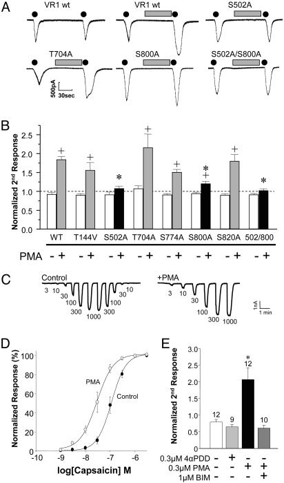
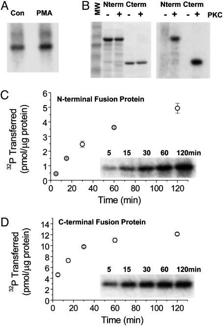
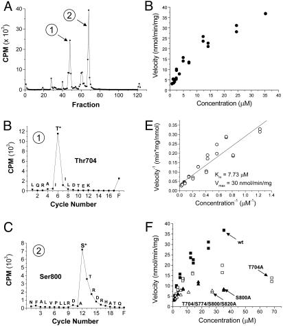
Protein kinase C (PKC) modulates the function of the capsaicin receptor transient receptor potential vanilloid 1 (TRPV1). This modulation manifests as increased current when the channel is activated by capsaicin. In addition, studies have suggested that phosphorylation by PKC might directly gate the channel, because PKC-activating phorbol esters induce TRPV1 currents in the absence of applied ligands. To test whether PKC both modulates and gates the TRPV1 function by direct phosphorylation, we used direct sequencing to determine the major sites of PKC phosphorylation on TRPV1 intracellular domains. We then tested the ability of the PKC-activating phorbol 12-myristate 13-acetate (PMA) to potentiate capsaicin-induced currents and to directly gate TRPV1. We found that mutation of S800 to alanine significantly reduced the PMA-induced enhancement of capsaicin-evoked currents and the direct activation of TRPV1 by PMA. Mutation of S502 to alanine reduced PMA enhancement of capsaicin-evoked currents, but had no effect on direct activation of TRPV1 by PMA. Conversely, mutation of T704 to alanine had no effect on PMA enhancement of capsaicin-evoked currents but dramatically reduced direct activation of TRPV1 by PMA. These results, combined with pharmacological studies showing that inactive phorbol esters also weakly activate TRPV1, suggest that PKC-mediated phosphorylation modulates TRPV1 but does not directly gate the channel. Rather, currents induced by phorbol esters result from the combination of a weak direct ligand-like activation of TRPV1 and the phosphorylation-induced enhancement of the TRPV1 function. Furthermore, modulation of the TRPV1 function by PKC appears to involve distinct phosphorylation sites depending on the mechanism of channel activation.


Figures

Fig. 4.
Fig. 4.
S502 and S800 mediate PKC modulation of capsaicin-evoked currents. (A) Representative current responses to consecutive applications of 20 nM capsaicin (black dots; 10 s) without or with an intervening 1-min application of PMA (100 nM, gray bars) in transiently transfected COS7 cells. (B) Ratio of the second to the initial peak capsaicin response (mean ± SEM) is plotted for wild-type TRPV1 and TRPV1 PKC site mutants in the absence (-) or presence (+) of an intervening PMA application. None of the mutants show a significant difference compared with wild type without PMA (ANOVA, P = 0.20). Only S502A, S800A, and the double mutant S502A/S800A show a decrease in PMA potentiation compared with wild type (ANOVA followed by post hoc Dunnett's test; *, P < 0.05), whereas all of the constructs, except S502A and S502/800A, exhibit some form of PMA potentiation (-PMA versus +PMA, unpaired t test; +, P < 0.05). (C and D) Dose–response relationships for capsaicin are shown under control conditions (•) and after a 3-min pretreatment with the PKC activator, PMA (300 nM, ○). Data are plotted as a percentage of the maximal response elicited by capsaicin (n = 6). C shows representative traces of two cells showing the currents induced by various doses of capsaicin (indicated below the traces, in nM). (E) PMA enhancement of capsaicin-evoked currents is PKC-mediated. Two successive applications of 30 nM capsaicin were performed. The second response is potentiated by the PKC-activating phorbol ester PMA but not by the inactive phorbol ester 4α-phorbol 12,13-didecanoate (4αPDD; 300 nM each). The PMA modulation of capsaicin responses was blocked by the PKC inhibitor bisindolylmaleimide (BIM; 1 μM).
Fig. 1.
Fig. 1.
TRPV1 acts as a PKC substrate in cultured cells and in vitro.(A) Bands represent TRPV1 immunoprecipitated from transiently transfected, metabolically 32P-labeled COS7 cells. PMA treatment (right lane) increases TRPV1 32P incorporation compared with control (left lane). (B) Coomassie blue staining of GST N-terminal (Nterm) and C-terminal (Cterm) TRPV1 fusion proteins without (-) or with (+) added PKC (Left) and the corresponding phosphorimage (Right) showing 32P incorporation into fusion proteins only with added PKC. Reactions were incubated at 30°C for 1 h. (C) Time course of phosphorylation for GST TRPV1 N-terminal fusion protein. Phosphorylation stoichiometry is at least 5 pmol/μg fusion protein or ≈40%. (D) GST TRPV1 C-terminal fusion protein phosphorylation time course showing saturating stoichiometry of ≈12 pmol/μg fusion protein or ≈55%.
Fig. 2.
Fig. 2.
T704 and S800 identified as in vitro PKC phosphorylation sites on the TRPV1 C terminus. (A) Cerenkov counting of HPLC fractions from an in-gel Lys-C digest of PKC-phosphorylated TRPV1 C-terminal fusion protein reveals two major phosphopeptides. (B) Edman sequencing and scintillation counting of the sequencing cycles from the first phosphopeptide fraction delineates T704 as a phosphorylated site. (C) Scintillation counting of the Edman sequencing cycles from the second phosphopeptide fraction reveals S800 as an in vitro PKC phosphorylation site. (D) PKC phosphorylation velocity of the TRPV1 C-terminal fusion protein plotted against protein concentration, showing a relatively strong PKC substrate. (E) Lineweaver–Burk plot (inverse velocity versus inverse concentration) and linear regression delineates a Km of 7.73 μM and Vmax of 30 nmol·min-1·mg-1. (F) Kinetic analysis of various mutants plotted alongside wild-type kinetics from D. The decrement observed in the S800 to alanine mutant is similar to that seen with a mutant fusion protein with all identified PKC phosphorylation sites (T704, S774, S800, and S820) converted to alanine, suggesting that S800 is the predominant substrate.
Fig. 3.
Fig. 3.
Identification of phosphorylation sites on the TRPV1 N-terminal fusion protein and first intracellular loop. (A) Cerenkov counting of the HPLC fractions after an in-gel endoproteinase Lys-C digestion of the PKC-phosphorylated N-terminal fusion protein reveals one major phosphopeptide. (B) Edman sequencing correlated with scintillation counting pinpoints T144 as the phosphorylated site. (C) Cerenkov counting of the HPLC fractions for the T144A mutant TRPV1 N-terminal fusion protein. The major phosphopeptide is eliminated, indicating correct identification of the major site in the wild-type protein, but the radioactivity peak in the flow-through (fraction 83) is dramatically increased. (D) Time course of 32P transfer to TRPV1 first intracellular loop peptide indicates a saturating stoichiometry of ≈50%. (E) Edman sequencing correlated with scintillation counting clearly demarcates S502 as the phosphorylated site rather than S505. (F) Phosphorylation kinetics of the loop peptide with Lineweaver–Burk analysis (Inset) reveals a highly efficacious PKC substrate with a Km of 2.77 μM and Vmax of 7.26 μmol·min-1·mg-1.
Fig. 5.
Fig. 5.
PKC-dependent and -independent effects of phorbol esters on TRPV1. (A) Averaged traces of wild-type TRPV1 responses to 4α-PMA (1 μM) and PMA (1 μM) under control conditions or in the presence of Ro31-8220 (1 μM, present in all perfusion solutions) or of ruthenium red (10 μM). Lower trace shows the response of vector-transfected cells to the same concentrations of 4α-PMA and PMA. (B) Population data showing the average peak responses to 4α-PMA and PMA under control conditions and in the presence of Ro31-8220 [n = 189 (control) and 145 (Ro31-8220)]. (C) Mutation of T704 and S800 to alanine residues reduces the response of TRPV1 to PMA to the level of the 4α-PMA response, whereas mutation of S502A statistically increases the responses to both 4α-PMA and PMA (n = 103–233 cells). *, P < 0.05. Asterisks above the bars indicate a significant difference between the 4α-PMA and PMA response for a given construct. Asterisks within the bars indicate a significant difference for the response to 4α-PMA or PMA compared with the response of wild-type TRPV1 to the same compound. (D) Capsaicin responses of wild-type and TRPV1 PKC-phosphorylation site mutants are not statistically different (n = 38–161). A wild-type control was done on each experimental day to eliminate variability resulting from day-to-day changes in expression. The responses of each mutant were then normalized to the size of the wild-type 4α-PMA response within each day's experiment. Statistical analyses were performed by using a one-way ANOVA followed by a Tukey's post hoc test.

References

    1. Cesare, P. & McNaughton, P. (1996) Proc. Natl. Acad. Sci. USA 93, 15435-15439. - PMC - PubMed
    1. Barber, L. A. & Vasko, M. R. (1996) J. Neurochem. 67, 72-80. - PubMed
    1. Leng, S., Mizumura, K., Koda, H. & Kumazawa, T. (1996) Neurosci. Lett. 206, 13-16. - PubMed
    1. Schepelmann, K., Messlinger, K. & Schmidt, R. F. (1993) Exp. Brain Res. 92, 391-398. - PubMed
    1. Ahlgren, S. C. & Levine, J. D. (1994) J. Neurophysiol. 72, 684-692. - PubMed

Publication types