. 2003 Oct;185(20):6032-41.
doi: 10.1128/JB.185.20.6032-6041.2003.

The iron-responsive regulator fur is transcriptionally autoregulated and not essential in Neisseria meningitidis

Affiliations

The iron-responsive regulator fur is transcriptionally autoregulated and not essential in Neisseria meningitidis

Isabel Delany et al. J Bacteriol. 2003 Oct.

Abstract

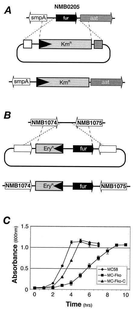
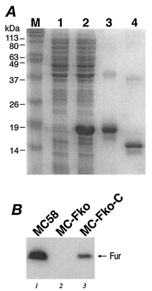
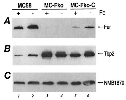
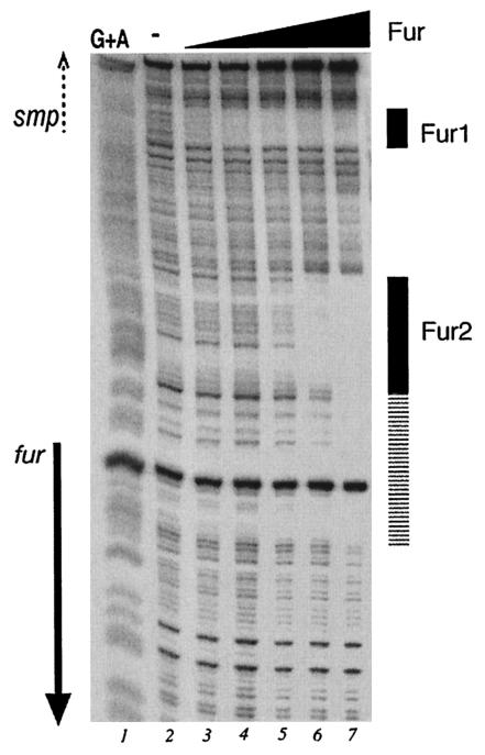
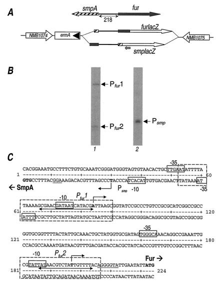
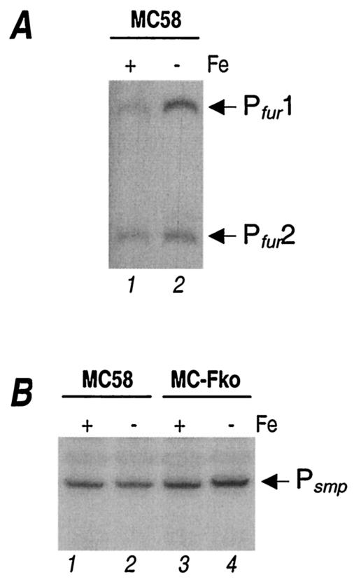
Fur is a well-known iron-responsive repressor of gene transcription, which is used by many bacteria to respond to the low-iron environment that pathogens encounter during infection. The fur gene in Neisseria meningitidis has been described as an essential gene that may regulate a broad array of genes. We succeeded in obtaining an N. meningitidis mutant with the fur gene knocked out and used it to undertake studies of fur-mediated iron regulation. We show that expression of both Fur and the transferrin binding protein Tbp2 is iron regulated and demonstrate that this regulation is Fur mediated for the Tbp2 protein. Footprinting analysis revealed that Fur binds to two distinct sites upstream of its coding region with different affinities and that these binding sites overlap two promoters that differentially control transcription of the fur gene in response to iron. The presence of two independently regulated fur promoters may allow meningococcus to fine-tune expression of this regulator controlling iron homeostasis, possibly during infection.


Figures

FIG. 1.
FIG. 1.
Mutation and complementation of the fur gene of N. meningitidis. (A) Schematic representation of the strategy used to construct a fur mutant; (B) schematic representation of the strategy used for complementation of Fur; (C) growth curves of the wild-type MC58 strain, the fur MC-Fko mutant, and the MC-Fko-C complemented mutant in supplemented GC medium.
FIG. 2.
FIG. 2.
(A) Expression and purification of the Fur protein. Lane 1, protein extracts from noninduced E. coli cells harboring plasmid pET15furB; lane 2, protein extracts from cells induced for 3 h with IPTG; lane 3, purified His-tagged Fur protein; lane 4, untagged Fur protein. (B) Western blot analysis showing Fur expression in the wild-type MC58 (lane 1), the MC-Fko fur mutant (lane 2), and the MC-Fko-C complemented mutant (lane 3) strains. Lane M, molecular size standards.
FIG. 3.
FIG. 3.
Iron and Fur regulation of gene expression. Western blot analysis showing iron-regulated protein expression. Wild-type MC58, the MC-Fko fur mutant, and the MC-Fko-C complemented mutant were grown under iron-replete (+) (supplemented GC medium) and iron-limiting (−) (supplemented GC medium with 25 μM desferal) conditions and were harvested at an OD600 of 0.4 to 0.6. Equal amounts of total protein from each culture were fractionated by SDS-PAGE, blotted onto nitrocellulose filters, and stained with antiserum raised against the Fur protein (A), the Tbp2 protein (B), and constitutive protein NMB1870 as the negative control (C).
FIG. 4.
FIG. 4.
Footprinting analysis of purified Fur on the NMB204-fur intergenic probe. The probe was labeled at one extremity and prepared as described in Materials and Methods. Lanes 2 to 7 contain reaction mixtures to which purified Fur protein at 13.4 nM, 40 nM, 122 nM, 366 nM, 1.1 μM, and 3.2 μM, respectively, was added. Lane 1 represents the G+A sequence reaction (19) obtained with the same probe and used as a molecular weight marker. The solid arrow shows the position and the orientation of the fur coding region. The dashed arrow indicates the orientation of the smpA gene, which is not contained in the probe.
FIG. 5.
FIG. 5.
Mapping the promoters. (A) Schematic representation (not to scale) of the fur locus (top) and chromosomal promoter fusions to the lacZ gene (bottom) inserted between the NMB1074 and NMB1075 loci. Hatched and filled arrows and boxes indicate all or part of the smpA and fur genes, respectively. The 218-bp intergenic region is highlighted. Open arrows indicate the lacZ gene. The position of the lacZ-PE primer is indicated with a small grey arrow. (B) Primer extension reactions with total RNA extracted from strains MC-furlacZ (lane 1) and MC-smpAlacZ (lane 2). Arrows mark the elongated primer products, Pfur1, Pfur2, and Psmp. (C) Nucleotide sequence of the smpA-fur intergenic region. Deduced initiations of RNA transcription are in boldface and marked with bent arrows. Putative promoter DNA elements are boxed and marked −10 and −35 at Pfur1, Pfur2, and Psmp. Nucleotides protected by Fur in DNase I protection assays are boxed with broken lines. Double-ended arrows indicate the core Fur box sequences. The translation start sites of the smpA and fur genes are in boldface and labeled accordingly.
FIG. 6.
FIG. 6.
Analysis of gene transcription by S1 nuclease mappings. (A) Regulation of fur transcripts. Total RNA extracted from wild-type MC58 cells exposed to iron-replete (+) or iron-limiting (−) conditions was hybridized to a 533-bp NsiI-EcoRI probe labeled at the EcoRI site and digested with S1 nuclease. Bands corresponding to S1 nuclease-resistant products were fractionated on denaturing gel and are indicated by arrows and labeled Pfur1 and Pfur2. (B) Regulation of smpA transcripts. A 325-bp NsiI-EcoRI smpA probe labeled at the EcoRI site was used to hybridize total RNA extracted from wild-type MC58 and MC-Fko strains exposed to iron-replete (+) or iron-limiting (−) conditions and processed as described for panel A. An arrow marks the S1 nuclease-resistant product, Psmp.

References

    1. Ala'Aldeen, D. A., H. A. Davies, and S. P. Borriello. 1994. Vaccine potential of meningococcal FrpB: studies on surface exposure and functional attributes of common epitopes. Vaccine 12:535-541. - PubMed
    1. Banerjee-Bhatnagar, N., and C. E. Frasch. 1990. Expression of Neisseria meningitidis iron-regulated outer membrane proteins, including a 70-kilodalton transferrin receptor, and their potential for use as vaccines. Infect. Immun. 58:2875-2881. - PMC - PubMed
    1. Bijlsma, J. J. E., B. Waidner, A. H. M. van Vliet, N. J. Hughes, S. Häg, S. Bereswill, D. J. Kelly, C. M. J. E. Vandenbroucke-Grauls, M. Kist, and J. G. Kusters. 2002. The Helicobacter pylori homologue of the ferric uptake regulator is involved in acid resistance. Infect. Immun. 70:606-611. - PMC - PubMed
    1. Calver, G. A., C. P. Kenny, and G. Lavergne. 1976. Iron as a replacement for mucin in the establishment of meningococcal infection in mice. Can. J. Microbiol. 22:832-838. - PubMed
    1. Carson, S. D. B. G., P. E. Klebba, S. M. C. Newton, and P. F. Sparling. 1999. Ferric enterobactin binding and utilization by Neisseria gonorrhoeae. J. Bacteriol. 181:2895-2901. - PMC - PubMed

Publication types

MeSH terms

LinkOut - more resources