. 2003 Oct 14;100(21):12195-200.
doi: 10.1073/pnas.2134310100. Epub 2003 Oct 2.

Sp8 is crucial for limb outgrowth and neuropore closure

Affiliations

Sp8 is crucial for limb outgrowth and neuropore closure

Sheila M Bell et al. Proc Natl Acad Sci U S A. .

Abstract

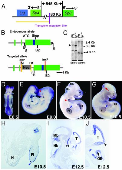
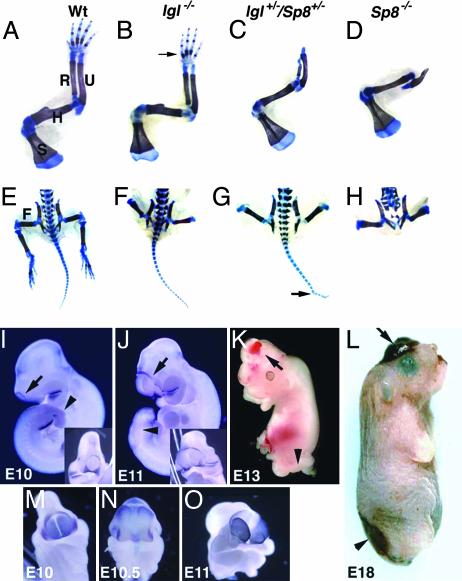
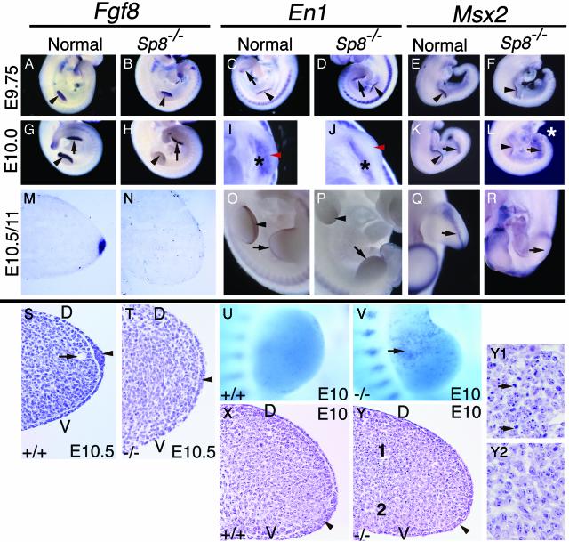
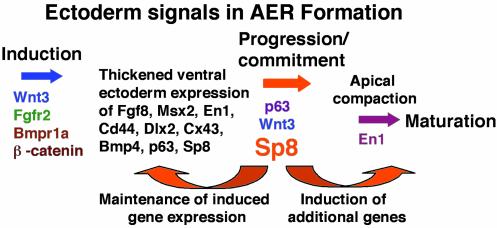
In this report we describe the developmental expression and function of Sp8, a member of the Sp family of zinc finger transcription factors, and provide evidence that the legless transgene insertional mutant is a hypomorphic allele of the Sp8 gene. Sp8 is expressed during embryogenesis in the forming apical ectodermal ridge (AER), restricted regions of the central nervous system, and tail bud. Targeted deletion of the Sp8 gene gives a striking phenotype, with severe truncation of both forelimbs and hindlimbs, absent tail, as well as defects in anterior and posterior neuropore closure leading to exencephaly and spina bifida. Outgrowth of the limb depends on formation of the AER, a signaling center that forms at the limb bud apex. In Sp8 mutants, the AER precursor cells are induced and initially express multiple appropriate marker genes, but expression of these genes is not maintained and progression to a mature AER is blocked. These observations indicate that Sp8 functions downstream of Wnt3, Fgf10, and Bmpr1a in the signaling cascade that mediates AER formation.


Figures

Fig. 1.
Fig. 1.
Structural characterization and expression of the Sp8 gene. (A) Relationship between the Sp8 and Sp4 genes. Yellow line indicates genomic region known to be deleted in the lgl transgene insertional mutant. (B) Structure of the Sp8 gene and the targeting vector. Green boxes (exons), blue box (zinc finger domain). Restriction sites: E, EcoRI; B, BamHI; B2, BglII. (C) Southern blot analysis of wild-type (Wt) and lgl mutant DNA. The blot was hybridized with a 435-bp Sp8 cDNA fragment containing sequences found within exon 2 and the beginning of exon 3. Arrowhead indicates the EcoRI fragment containing the coding region. (DJ) Whole mount and sectioned in situ hybridization using an Sp8 riboprobe. (DF and HJ) Wild-type embryos. (D) Expression is detected in the neuroepithelium at E8.5. (E) On E9.0, expression is heavy around the posterior neuropore/tailbud (red arrowhead) and is first evident in the forelimb (Fl) AER precursor. (F and H) By E10.5, expression is detectable in the telencephalon (T), dorsal and ventral forelimb ectoderm, AER (blue arrowhead), hindlimb (Hl) AER precursor, tail (red arrowhead), medial and lateral nasal processes, isthmus, and ventral spinal cord. H, heart. (G) lgl–/– embryo showing reduction in the level of expression in the forelimb AER, lateral and medial nasal processes, isthmus (red arrow), and the absence of expression in the hindlimb bud AER precursor. (I and J) On E12.5, expression is detected at the midbrain (Mb)/hindbrain (Hb) border, in the olfactory epithelium (OE) and the ventral thalamus (VT). Notably, Sp8 is expressed in the olfactory bulb primordia (J, arrowhead).
Fig. 2.
Fig. 2.
Malformations in Sp8 mutants. (AH) Alizarin red and alcian blue limb skeletal analysis of E18 fetuses. (AD) Forelimb skeletons. (A) S, scapula; H, humerus; R, radius; U, ulna. Arrow in B indicates absence of digit 1. (C and D) The humerus forms normally; however, the ulna is distally truncated. The radius is dysplastic (C) or absent (D) and the autopod is absent. (EH) Caudal portion of fetuses. (E) F, femur. (FH) Structures distal to the femur are absent. (G) Tail vertebral fusions (arrow). (H) In the Sp8–/– fetus, sacral and caudal vertebrae are absent and the lumbar vertebrae are disorganized and abnormal. (IM and O) Sp8 null embryos at the ages indicated. (N) Wild type. (IL) Arrows indicate open anterior neuropore/exencephaly. Arrowheads indicate open posterior neuropore/spina bifida. (I and J) Whole mount in situ with Fgf8 riboprobe arrows indicate ring of cells around open anterior neuropore. (Inset) Frontal view. (MO) Whole mount in situ with Bf1 riboprobe emphasizing progression of telencephalon tissue through open anterior neuropore.
Fig. 3.
Fig. 3.
Disruption in AER morphogenesis in Sp8 null embryos. Embryo ages, genotypes, and probes used in the whole mount and section in situ hybridizations are indicated. Black arrowheads in AY indicate forelimb AER precursor/AER. Black arrows in AR indicate hindlimb AER precursor. Note induction and subsequent loss of Fgf8, Msx2, and En1 expression in the fore and hindlimb buds of Sp8–/– embryos. (I and J) Black asterisks indicate ventral ectoderm expression of En1, red arrowheads indicate normal position of En1 expressing AER precursor cells and absence of expression in Sp8–/– forelimb bud. (L) White asterisk indicates truncated tail bud and open everted neural tissue adjacent to hindlimb buds. (P) Note the absence of En1 expression at distal tips of limb buds and in somites adjacent to the hindlimbs. (Q and R) Hindlimb buds, apical view. (S, T, X, Y, Y1, and Y2). Hematoxylin and eosin-stained 6-μm paraffin sections cut across the anterior/posterior axis of the forelimb. Arrowheads in S, T, X, and Y indicate AER precursor/AER. Arrow in S indicates marginal venous sinus. (U and V) Nile blue sulfate cell death staining, note punctate blue staining in anterior proximal region of Sp8–/– limb indicated by arrow in V. (Y) Note presence of pyknotic nuclei in dorsal proximal region of limb seen at higher magnification in Y1 compared with the proximal ventral mesoderm presented in Y2. D, dorsal; V, ventral.
Fig. 4.
Fig. 4.
Proposed model of genes involved in the process of AER formation. Only the genes in color have been implicated as being required for normal AER formation.

References

    1. Barrow, J. R., Thomas, K. R., Boussadia-Zahui, O., Moore, R., Kemler, R., Capecchi, M. R. & McMahon, A. P. (2003) Genes Dev. 17, 394–409. - PMC - PubMed
    1. Ahn, K., Mishina, Y., Hanks, M. C., Behringer, R. R. & Crenshaw, E. B. I. (2001) Development (Cambridge, U.K.) 128, 4449–4461. - PubMed
    1. Min, H., Danilenko, D. M., Scully, S. A., Bolon, B., Ring, B. D., Tarpley, J. E., DeRose, M. & Simonet, W. S. (1998) Genes Dev. 12, 3156–3161. - PMC - PubMed
    1. Xu, X., Weinstein, M., Li, Cuiling, Naski, M., Cohen, R. I., Ornitz, D. M., Leder, P. & Deng, C. (1998) Development (Cambridge, U.K.) 125, 753–765. - PubMed
    1. Moerlooze, D. L., Spencer-Dene, B., Revest, J.-M., Hajihosseini, M., Rosewell, I. & Dickson, C. (2000) Development (Cambridge, U.K.) 127, 483–492. - PubMed

Publication types