. 2003 Aug 14;1(1):3.
doi: 10.1186/1479-5876-1-3.

Cross-reactivity between HLA-A2-restricted FLU-M1:58-66 and HIV p17 GAG:77-85 epitopes in HIV-infected and uninfected individuals

Affiliations

Cross-reactivity between HLA-A2-restricted FLU-M1:58-66 and HIV p17 GAG:77-85 epitopes in HIV-infected and uninfected individuals

Paula M Acierno et al. J Transl Med. .

Abstract

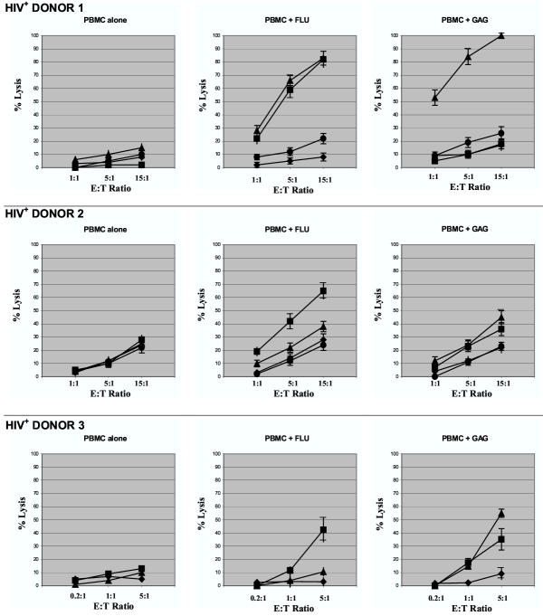
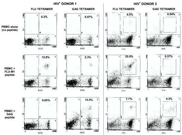
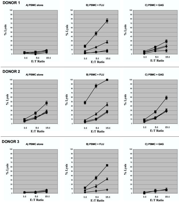
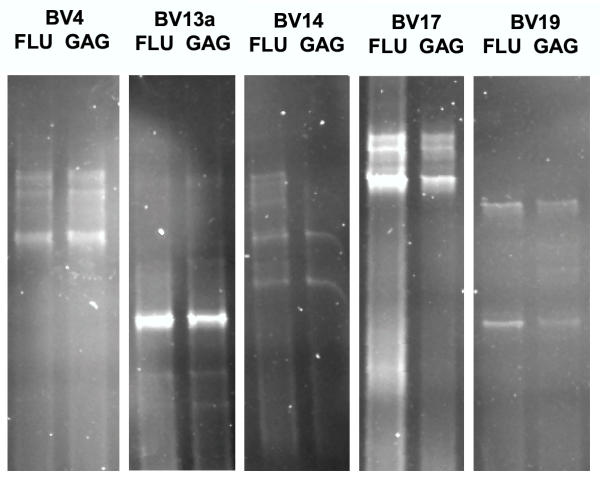
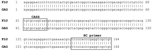
BACKGROUND: The matrix protein of the influenza A virus and the matrix and capsid proteins of the human immunodeficiency virus (HIV) share striking structural similarities which may have evolutionary and biological significance. These similarities led us to hypothesize the existence of cross-reactivity between HLA-A2-restricted FLU-M1:58-66 and HIV-1 p17 GAG:77-85 epitopes. METHODS: The hypothesis that these two epitopes are cross-reactive was tested by determining the presence and extent of FLU/GAG immune cross-reactivity in lymphocytes from HIV-seropositive and seronegative HLA-A2+ donors by cytotoxicity assays and tetramer analyses. Moreover, the molecular basis for FLU/GAG cross-reactivity in HIV-seropositive and seronegative donors was studied by comparing lymphocyte-derived cDNA sequences corresponding to the TCR-beta variable regions, in order to determine whether stimulation of lymphocytes with either peptide results in the expansion of identical T-cell clonotypes. RESULTS: Here, we report evidence of cross-reactivity between FLU-M1:58-66 and HIV-1 p17 GAG:77-85 epitopes following in vitro stimulation of PBMC derived from either HIV-seropositive or seronegative HLA-A2+ donors as determined by cytotoxicity assays, tetramer analyses, and molecular clonotyping. CONCLUSION: These results suggest that immunity to the matrix protein of the influenza virus may drive a specific immune response to an HLA-A2-restricted HIV gag epitope in HIV-infected and uninfected donors vaccinated against influenza.


Figures

Figure 1
Figure 1
Cytotoxic responses by lymphocytes from HIV-seropositive donors. Results are shown of representative cytotoxicity assays (Cr51-release) using stimulated lymphocytes derived from three HLA-A2+, HIV-seropositive donors. Effector cells were mixed with T2 target cells presenting HLA-A2-restricted influenza A-derived FLU-M1:58–66 (■) or HIV-1-derived p17 GAG:77–85 (▲) peptide. T2 cells loaded with GP100:209–217 (●, Donors 1 and 2 only) or no peptide (◆) were used as controls as shown. Effector cells were tested after in vitro stimulation of PBMC with IL-2 and: no peptide (PBMC alone); FLU-M1:58–66; or GAG:77–85 peptide. Data points refer to percent lysis at indicated effector:target ratios.
Figure 2
Figure 2
Tetramer analyses of lymphocytes from HIV-seropositive donors. After in vitro stimulation with IL-2 plus either FLU-M1:58–66 or HIV p17 GAG:77–85 peptide (or no peptide as background), lymphocytes from HLA-A2+, HIV-seropositive Donors 1 and 3 were stained with FITC-labeled anti-CD8 mAb and either HLA-A2/FLU-M1:58–66 tetramer or HLA-A2/GAG:77–85 tetramer (both PE-labeled), as indicated, and viable cells were analyzed by flow cytometry (nonviable cells positive for staining with the dye 7-AAD were excluded). Percentages of CD8+ cells that stain with tetramer appear in the upper-right quadrant of the histograms.
Figure 3
Figure 3
Cytotoxic responses by lymphocytes from HIV-seronegative donors. Results are shown of representative cytotoxicity assays (Cr51-release) using stimulated lymphocytes derived from three HLA-A2+, HIV-seronegative donors. Effector cells were mixed with T2 target cells presenting HLA-A2-restricted influenza A-derived FLU-M1:58–66 (■) or HIV-1-derived p17 GAG:77–85 (▲) peptide. T2 cells loaded with GP100:209–217 (●, Donors 1 and 2 only) or no peptide (◆) were used as controls as shown. Effector cells were tested after in vitro stimulation of PBMC with IL-2 and: no peptide (PBMC alone); FLU-M1:58–66; or GAG:77–85 peptide. Data points refer to percent lysis at indicated effector:target ratios.
Figure 4
Figure 4
Clonotype analysis of FLU- and GAG-stimulated lymphocytes. Some identical FLU-specific clonotypes between FLU- and GAG-stimulated T cells were observed in 5 BV families analyzed by denaturing gradient gel electrophoresis (DGGE). PBMC from a HIV-seronegative Donor 1 were stimulated in vitro with FLU-M1:58–66 or GAG:77–85 peptides, and the resulting FLU-M1:58–66-specific cells purified by binding cells with the cognate TCR to HLA-A2/FLU-M1:58–66 peptide monomers conjugated to magnetic microbeads. RT-PCR was performed on RNA derived from each cell type (FLU-M1:58–66- or GAG:77–85-stimulated) using primers specific for all 24 TCR-β (BV) families. Each band resolved on a DGGE gel represents different clonotypes separated according to their nucleotide composition. The results of five different primers are shown here (BV13a is one of the two variable primers needed to amplify all possible members of the BV13 family).
Figure 5
Figure 5
DNA sequencing of identical TCR-β clonotypes. 100% sequence identity of the TCR-β-variable gene region of FLU- or GAG-stimulated lymphocytes was obtained using a BV17 primer. This sequence includes the CDR3 hypervariable D/J region between the 'CASS' coding sequence and the BC region (both in gray), which greatly influences the HLA/peptide binding specificity of the TCR molecule.

References

    1. Harris A, Sha B, Luo M. Structural similarities between influenza virus matrix protein M1 and human immunodeficiency virus matrix and capsid proteins: an evolutionary link between negative-stranded RNA viruses and retroviruses. J Gen Virol. 1999;80:863–869. - PubMed
    1. Bodmer H, Ogg G, Gotch F, McMichael A. Anti-HLA-A2 antibody-enhancement of peptide association with HLA-A2 as detected by cytotoxic T lymphocytes. Nature. 1989;342:443–446. doi: 10.1038/342443a0. - DOI - PubMed
    1. Tsomides TJ, Aldovini A, Johnson RP, Walker BD, Young RA, Eisen HN. Naturally processed viral peptides recognized by cytotoxic T lymphocytes on cells chronically infected by human immunodeficiency virus type 1. J Exp Med. 1994;180:1283–1293. - PMC - PubMed
    1. Altman JD, Moss PA, Goulder PJ, Barouch DH, McHeyzer-Williams MG, Bell JI, McMichael AJ, Davis MM. Phenotypic analysis of antigen-specific T lymphocytes. Science. 1996;274:94–96. doi: 10.1126/science.274.5284.94. - DOI - PubMed
    1. Salgaller ML, Marincola FM, Cormier JN, Rosenberg SA. Immunization against epitopes in the human melanoma antigen gpl00 following patient immunization with synthetic peptides. Cancer Res. 1996;56:4749–4757. - PubMed