. 2003 Oct 14;100(21):12039-44.
doi: 10.1073/pnas.2034936100. Epub 2003 Oct 3.

Functional map and domain structure of MET, the product of the c-met protooncogene and receptor for hepatocyte growth factor/scatter factor

Affiliations

Functional map and domain structure of MET, the product of the c-met protooncogene and receptor for hepatocyte growth factor/scatter factor

Ermanno Gherardi et al. Proc Natl Acad Sci U S A. .

Abstract

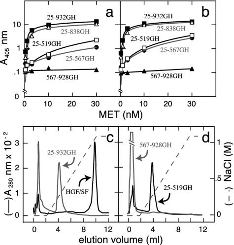
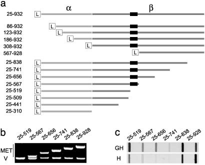
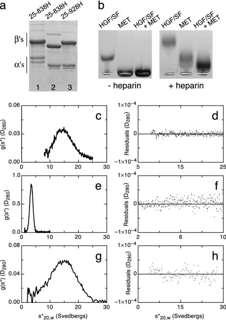
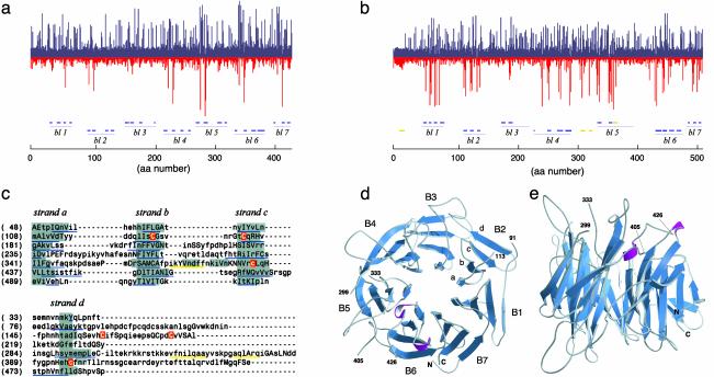
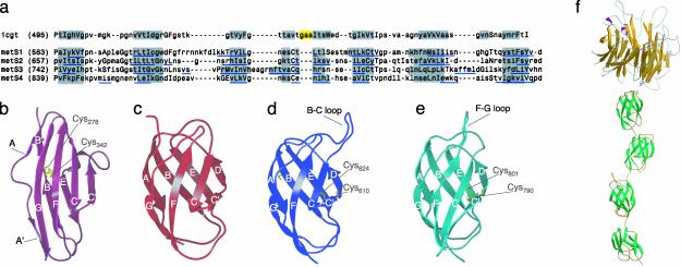
Little is known about the large ectodomain of MET, the product of the c-met protooncogene and receptor for hepatocyte growth factor/scatter factor (HGF/SF). Here, we establish by deletion mutagenesis that the HGF/SF and heparin-binding sites of MET are contained within a large N-terminal domain spanning the alpha-chain (amino acids 25-307) and the first 212 amino acids of the beta-chain (amino acids 308-519). Neither the cystine-rich domain (amino acids 520-561) nor the C-terminal half of MET (amino acids 562-932) bind HGF/SF or heparin directly. The MET ectodomain, which behaves as a rod-shaped monomer with a large Stokes radius in solution, binds HGF/SF in the absence or presence of heparin, and forms a stable HGF/SF-heparin-MET complex with 1:1:1 stoichiometry. We also show that the ligand-binding domain adopts a beta-propeller fold, which is similar to the N-terminal domain of alphaV integrin, and that the C-terminal half contains four Ig domains (amino acids 563-654, 657-738, 742-836, and 839-924) of the unusual structural E set, which could be modeled on bacterial enzymes. Our studies provide 3D models and a functional map of the MET ectodomain. They have broad implications for structure-function of the MET receptor and the related semaphorin and plexin proteins.


Figures

Fig. 2.
Fig. 2.
Binding of MET deletions to HGF/SF (a and b) or heparin (c and d). (a and b) Binding of MET deletions to single-chain (a) or two-chain (b) HGF/SF, as measured in a solid phase assay. (c and d) Binding of three MET constructs (25–519GH, 25–932GH, and 567–928GH) to immobilized heparin. Both full-length MET (25–932GH) and MET 25–519GH showed binding, whereas MET 567–928GH showed none. The strong heparin binding of mature (two-chain) HGF/SF is shown for comparison in c.
Fig. 1.
Fig. 1.
Deletion mapping and expression of MET domains. (a) A schematic view and sequence boundaries of N- and C-terminal deletions of the MET ectodomain. The α- and β-chains are shown in different shades of gray. The L indicates a 21-aa Ig leader used for secretion of MET proteins and the black box corresponds to the cystine-rich sequence (amino acids 520–561) of the MET β chain. (b) The cDNAs corresponding to several C-terminal deletions of the MET ectodomain (top bands, MET) are shown along with a vector band (V). (c) Expression of the same MET deletions in supernatants of stable transfectants of the mouse myeloma line NS0. H and GH indicate monomeric and dimeric MET constructs, respectively.
Fig. 3.
Fig. 3.
Monomeric full-length MET and HGF/SF-MET complexes. (a) SDS/PAGE under reducing conditions of MET 25–838H from NS0 (lane 1) or Lec 8 cells (lane 2) and MET 25–928H from Lec 8 cells. (b) Gel electrophoresis under native conditions of HGF/SF, MET, and HGF/SF-MET complexes, in the absence or presence of heparin (c–h) Velocity sedimentation analysis of HGF/SF (c and d), MET (e and f), and the HGF/SF–heparin–MET complex (g and h). (c, e, and g) Plots of g(s*) against s*20,w. (d, f, and h) Plots of the residuals, from fitting models to the data, against s*20,w. Experiments shown in b, e, and g were carried out with equimolar concentrations of HGF/SF and MET 25–928H derived from Lec 8 cells (4 × 10⁻⁶ M) and a 2.5-fold excess of heparin.
Fig. 4.
Fig. 4.
Predicted structure of the ligand-binding domain of MET. (a and b) HMMs of amino acids 1–438 of the α-chain of the β-propeller domain of integrin αV (1L5G) (a) and amino acids 1–519 of MET. bl, blade. (b) Each column plots the probabilistic distribution of hydrophilic (blue) and hydrophobic (red) amino acids at each position. Column height is proportional to the distance from the generic null distribution. Blue lines below the 1L5G HMM are β-strands in the crystal structure of 1L5G (38). Blue or yellow lines below the MET HMM (and in c below) are β-strands or α-helices predicted with jpred (39). (c) Alignment of the seven-blade β-propeller 3D model of the MET ligand-binding domain. Regions boxed in gray are assigned as β-strands in the model, and residues in small or capital letters are solvent accessible or solvent inaccessible, respectively. The six cysteine residues shown in white on a red background are predicted to form three disulfide bonds between strands b and c of blade 2, in the d-a loop between blades 2 and 3, and between strands c and d of blade 5. (d and e) Ribbon diagrams of the β-propeller model of the ligand-binding domain of MET (amino acids 33–516) viewed from the top and side.
Fig. 5.
Fig. 5.
Sequence and predicted structure of the stalk region of the MET ectodomain. (a) Sequence alignment of domain E of B. stearothermophilus cyclodextrin. Glucanotransferase (1CGT) with four segments of the MET sequence (amino acids 563–656, 657–741, 742–838, and 839–928, which are referred as domains S1, S2, S3d and S4). Regions boxed in gray correspond to β-strands in 1CGT or predicted to form β-strands in the Ig models of MET. Regions boxed in yellow correspond to α-helices in 1CGT. Residues in small or capital letters are solvent accessible or solvent inaccessible, respectively, and segments of the MET sequence predicted as β-strands or α-helices by jpred (39) are underlined in blue. (b and c) Ribbon representations of typical I set (domain 3 of FGF receptor 2, 1e0) (b) and E set (domain E of 1CYG (c) domains. (d and e) Ribbon representations of 3D models of the first (S1) and third (S3) Ig domains of the stalk region of MET. The image shows the long B-C and F-G loops of these domains and the cysteine residues involved in potential intradomain disulfides. (f) Overall view of the MET ectodomain, based on the results of this article. A small cysteine-rich domain located between the β-propeller and Ig domains (amino acids 520–561) is not shown.

References

    1. Blume-Jensen, P. & Hunter, T. (2001) Nature 411, 355–365. - PubMed
    1. Harpaz, Y. & Chothia, C. (1994) J. Mol. Biol. 238, 528–539. - PubMed
    1. Cooper, C. S., Blair, D. G., Oskarsson, M. K., Tainsky, M. A., Eader, L. A. & Vande Woude, G. F. (1984) Cancer Res. 44, 1–10. - PubMed
    1. Park, M., Dean, M., Kaul, K., Braun, M. J., Gonda, M. A. & Vande Woude, G. (1987) Proc. Natl. Acad. Sci. USA 84, 6379–6383. - PMC - PubMed
    1. Bottaro, D. P., Rubin, J. S., Faletto, D. L., Chan, A. M., Kmiecik, T. E., Vande Woude, G. F. & Aaronson, S. A. (1991) Science 251, 802–804. - PubMed

Publication types

MeSH terms

LinkOut - more resources