. 2004 Jan;15(1):71-80.
doi: 10.1091/mbc.e03-08-0586. Epub 2003 Oct 3.

Slx1-Slx4 are subunits of a structure-specific endonuclease that maintains ribosomal DNA in fission yeast

Affiliations

Slx1-Slx4 are subunits of a structure-specific endonuclease that maintains ribosomal DNA in fission yeast

Stéphane Coulon et al. Mol Biol Cell. 2004 Jan.

Abstract

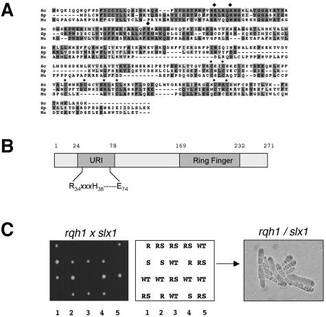
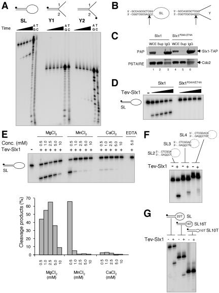
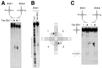
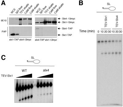
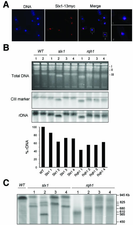
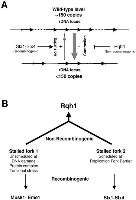
In most eukaryotes, genes encoding ribosomal RNAs (rDNA) are clustered in long tandem head-to-tail repeats. Studies of Saccharomyces cerevisiae have indicated that rDNA copy number is maintained through recombination events associated with site-specific blockage of replication forks (RFs). Here, we describe two Schizosaccharomyces pombe proteins, homologs of S. cerevisiae Slx1 and Slx4, as subunits of a novel type of endonuclease that maintains rDNA copy number. The Slx1-Slx4-dependent endonuclease introduces single-strand cuts in duplex DNA on the 3' side of junctions with single-strand DNA. Deletion of Slx1 or Rqh1 RecQ-like DNA helicase provokes rDNA contraction, whereas simultaneous elimination of Slx1-Slx4 endonuclease and Rqh1 is lethal. Slx1 associates with chromatin at two foci characteristic of the two rDNA repeat loci in S. pombe. We propose a model in which the Slx1-Slx4 complex is involved in the control of the expansion and contraction of the rDNA loci by initiating recombination events at stalled RFs.


Figures

Figure 4.
Figure 4.
Identification of S. pombe Slx4 homolog. (A) Sequence alignments of Slx4 homologs from S. cerevisiae (Sc), S. pombe (Sp; SPAC688.06c), and C. albicans (C.al; Orf6.4697). Dark gray and light gray boxes indicate identical and similar residues, respectively. The peptides identified by MudPIT analysis of Slx1-TAP are underlined. (B) Fivefold serial dilutions of mutant slx1, slx4, or slx1 slx4 cells were incubated on YES agar medium supplemented with indicated amounts of MMS or HU at 30°C. To determine UV survival, cells were ascertained using a hemocytometer, plated on YES agar medium, and exposed to the indicated doses of UV. The agar plates were incubated at 30°C, and percentage of survival was calculated as the number of colonies in irradiated versus nonirradiated control. Data represent the average of two independent trials. (C). Slx4 is essential in rqh1 cells. Left, tetrad analysis of mating between slx4::kanMX6 and rqh1::ura4+ strains. An example of germination products from a slx4 rqh1 spore is presented in right panel. S, slx4; R, rqh1; RS, slx4 rqh1.
Figure 1.
Figure 1.
Identification of S. pombe Slx1. (A) Sequence alignment of Slx1proteins from S. cerevisiae (Sc), S. pombe (Sp; SPAP27G11.15), and human (Hu; MGC5178). Dark gray and light gray boxes indicate identical and similar residues, respectively. Conserved residues of the URI and Ring Finger domains are annotated by black squares and black stars, respectively. (B) Schematic representation of S. pombe Slx1. Positions of the URI domain and Ring finger domain are shown. (C) Genetic interaction between slx1 (S) and rqh1 (R). A slx1::kanMX6 haploid strain was mated with a rqh1::ura4+ haploid strain, and the cells were induced to undergo meiosis and sporulation. Tetrads were dissected onto YES agar. Colonies resulting from five such tetrads were photographed after 3 days growth at 30°C (left). The genotypes of these segregants were determined by replica plating. Double mutant slx1 rqh1 (RS) spores were unable to form colonies. A photomicrograph of cells arising from a germinated slx1 rqh1 spore is shown in the right panel.
Figure 2.
Figure 2.
Structure-specific DNA endonuclease activity associated with Slx1. (A) Three different substrates were incubated for 30 min at 30°C with 3 μl of TEV-Slx1 for 0 s, 30 s, 2.5 min, 10 min, and 50 min. A+G and C+T sequencing ladders were derived from the SL, y1, and y2 oligonucleotides. Reaction products were analyzed by denaturing PAGE. (B) The sites of cleavage were determined from the Maxam-Gilbert sequencing ladders and are indicated by arrows on each DNA structure. (C) Whole cell extract (WCE) derived from strains slx1::kanMx6 slx1wt-TAP:LEU1 and slx1::kanMx6 slx1R34A,E74A-TAP:LEU1 were incubated with IgG-Sepharose beads. TAP-Slx1 and TAP-Slx1R34A,E74A were detected in the WCE, supernatant (Sup), and the pulled down IgG-Sepharose beads (IgG) by Western blotting by using peroxidase-anti-peroxidase antibody (PAP; Sigma-Aldrich, St. Louis, MO). Cdc2 detected with an antibody directed against the Cdc2 PSTAIRE motif was used as a loading control. (D) SL was incubated with 1, 2, 4, 6, and 9 μl of TEV-eluate obtained from slx1/slx1wt-TAP::LEU1 and slx1/slx1R34A,E74A-TAP::LEU1 strains for 30 min at 30°C. Reaction products were analyzed by denaturing PAGE. (E) SL was incubated with 3 μl of TEV-Slx1 in the presence of various amounts of Mg2+, Mn2+, Ca2+, or EDTA and reaction products analyzed by denaturing PAGE. The relative amount of cleavage products was quantified by phosphorimager analysis. (F) The various SL substrates indicated above the gel were incubated with 3 μl of TEV-Slx1 for 30 min at 30°C. (G) Three microliters of TEV-Slx1 was incubated with SL containing ss loops of various sizes as indicated.
Figure 3.
Figure 3.
The Slx1-dependent nuclease cuts but does not resolve HJs. (A) X12-1 and X12-3 substrates were incubated with 3 μl of TEV-Slx1 for 30 min at 30°C. Reaction products were analyzed by denaturing PAGE. (B) The position of the cuts were determined from the Maxam-Gilbert sequencing ladder. (C) Reaction products from reactions carried out in A were analyzed by native PAGE to monitor the conversion of the four-way branched structures into linear duplex products.
Figure 5.
Figure 5.
Slx4 associates with Slx1 to form an active endonuclease. (A) Slx1-TAP and Slx4-TAP fusion proteins were expressed in slx4-myc and slx1-myc strains, respectively. WCE, whole cell extract; IgG flow, flow through from WCE passed over IgG affinity column; TEV eluate, TEV cleavage products from IgG column; CaM flow, flow through from TEV eluate passed over calmodulin affinity column; CaM eluate, eluate from the calmodulin column. These fractions were subjected to immunoblotting revealed by 9E10 anti-myc monoclonal antibody and peroxidase-anti-peroxidase antibody (PAP; Sigma-Aldrich). Note: The loss of the TAP signal in the TEV-eluates and the following fractions is due to the loss of the protein A portion of the TAP-tag after TEV cleavage. (B) The SL substrate was incubated at 30°C for the indicated time with 3 μl of TEV-Slx1 or TEV-Slx4 obtained from slx1-TAP and slx4-TAP strains, respectively. Reaction products were analyzed by denaturing PAGE. (C) The SL substrate was incubated with 1, 2, 4, 6, and 9 μl of TEV-Slx1 obtained from slx1-TAP and slx1-TAP slx4 strains, for 30 min at 30°C, and reaction products were analyzed by denaturing PAGE.
Figure 6.
Figure 6.
Slx1 complex binds to chromatin and is involved in rDNA maintenance. (A) In situ chromatin binding assays performed with a strain overexpressing Slx1-13myc. Slx1-13myc was detected as single or double spots at the periphery of the nucleus. (B) Genomic DNA samples of WT and strains that were freshly deleted for the slx1 or rqh1 genes were subjected to PFGE. The gel was stained with ethidium bromide before Southern blotting. The membrane was hybridized with an rDNA probe and with a probe specific for a region located outside of the rDNA loci on CIII. The signal quantification allowed for determination of the rDNA percentage in each sample taking wild-type templates as references. (C) Genomic DNA samples described in B were digested with SfiI before PFGE and hybridization to an rDNA probe. The SfiI fragments containing the rDNA loci were smaller in the slx1 strains and diffuse in rqh1 strains. The size of S. cerevisiae marker chromosomes is indicated.
Figure 7.
Figure 7.
Schemes rDNA copy number maintenance. (A) The Rqh1-dependent nonrecombinogenic pathway limits the contraction of the rDNA loci due to unscheduled recombination events. The Slx1–Slx4 complex initiates a scheduled recombination process that leads to the expansion of the rDNA loci. (B) Rqh1 would act in a main nonrecombinogenic pathway that rescues two types of stalled forks. Unscheduled stalling of a RF can occur at DNA damage, protein complexes, and torsional stress (fork 1). Another type of stalled fork is generated by a scheduled mechanism that occurs at the RFB (fork 2). In absence of Rqh1, rescue of the stalled forks 1 requires Mus81-Eme1, whereas rescue of fork 2 will necessitate the action of the Slx1–Slx4 complex.

References

    1. Aravind, L., and Koonin, E.V. (2001). Prokaryotic homologs of the eukaryotic DNA-end-binding protein Ku, novel domains in the Ku protein and prediction of a prokaryotic double-strand break repair system. Genome Res. 11, 1365–1374. - PMC - PubMed
    1. Boddy, M.N., Gaillard, P.H., McDonald, W.H., Shanahan, P., Yates, J.R., III, and Russell, P. (2001). Mus81-Eme1 are essential components of a Holliday junction resolvase. Cell 107, 537–548. - PubMed
    1. Boddy, M.N., Lopez-Girona, A., Shanahan, P., Interthal, H., Heyer, W.D., and Russell, P. (2000). Damage tolerance protein Mus81 associates with the FHA1 domain of checkpoint kinase Cds1. Mol. Cell Biol. 20, 8758–8766. - PMC - PubMed
    1. Boddy, M.N., and Russell, P. (2001). DNA replication checkpoint. Curr. Biol. 11, R953–R956. - PubMed
    1. Caspari, T., Murray, J.M., and Carr, A.M. (2002). Cdc2-cyclin B kinase activity links Crb2 and Rqh1-topoisomerase III. Genes Dev. 16, 1195–1208. - PMC - PubMed

Publication types

MeSH terms