. 2004 Jan;15(1):384-96.
doi: 10.1091/mbc.e03-06-0445. Epub 2003 Oct 3.

Scavenger receptor BI (SR-BI) clustered on microvillar extensions suggests that this plasma membrane domain is a way station for cholesterol trafficking between cells and high-density lipoprotein

Affiliations

Scavenger receptor BI (SR-BI) clustered on microvillar extensions suggests that this plasma membrane domain is a way station for cholesterol trafficking between cells and high-density lipoprotein

Yinan Peng et al. Mol Biol Cell. 2004 Jan.

Abstract

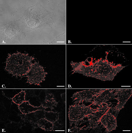
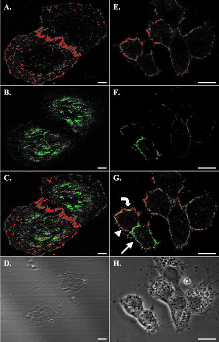
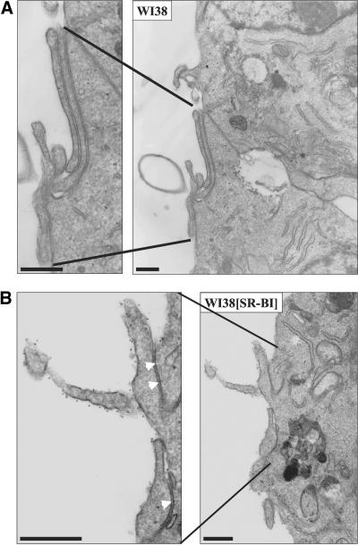
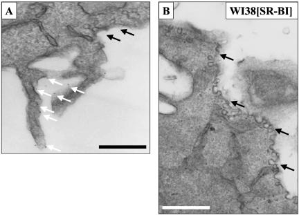
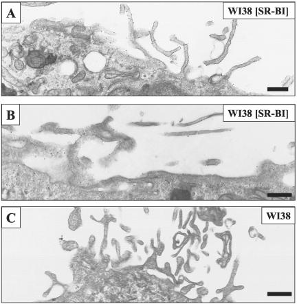
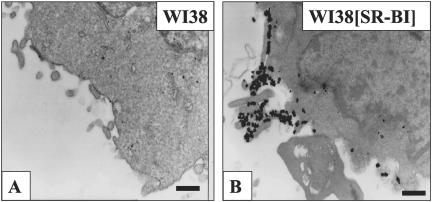
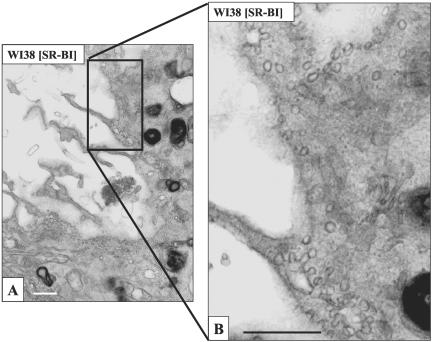
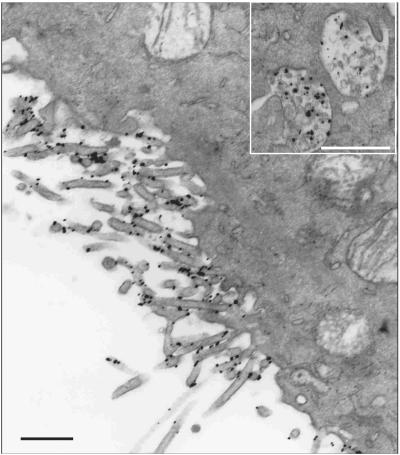
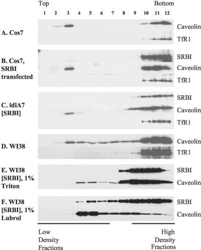
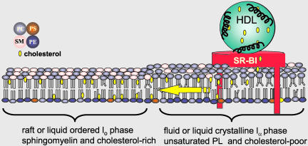
Receptor-mediated trafficking of cholesterol between lipoproteins and cells is a fundamental biological process at the organismal and cellular levels. In contrast to the well-studied pathway of LDL receptor-mediated endocytosis, little is known about the trafficking of high-density lipoprotein (HDL) cholesterol by the HDL receptor, scavenger receptor BI (SR-BI). SR-BI mediates HDL cholesteryl ester uptake in a process in which HDL lipids are selectively transferred to the cell membrane without the uptake and degradation of the HDL particle. We report here the cell surface locale where the trafficking of HDL cholesterol occurs. Fluorescence confocal microscopy showed SR-BI in patches and small extensions of the cell surface that were distinct from sites of caveolin-1 expression. Electron microscopy showed SR-BI in patches or clusters primarily on microvillar extensions of the plasma membrane. The organization of SR-BI in this manner suggests that this microvillar domain is a way station for cholesterol trafficking between HDL and cells. The types of phospholipids in this domain are unknown, but SR-BI is not strongly associated with classical membrane rafts rich in detergent-resistant saturated phospholipids. We speculate that SR-BI is in a more fluid membrane domain that will favor rapid cholesterol flux between the membrane and HDL.


Figures

Figure 1.
Figure 1.
Cells expressing SR-BI showed prominent cell surface HDL binding. Cells were incubated with fluorescently labeled HDL (20 μg/ml) for 30 min on ice and then washed with PBS and fixed with 4% paraformaldehyde. Scale bars, 10 μm. The five cell lines used were WI38 cells (A and B), stably transfected WI38[SR-BI] cells (C), transiently transfected COS-7 cells (D), ACTH treated Y1-BS1 cells (E), and stably transfected ldlA7 cells (F). A and B demonstrate that in the absence of SR-BI expression, no HDL staining was observed. Cells from all SR-BI-transfected cell lines showed prominent cell surface HDL binding in irregularly shaped patches with varying sizes. They also showed heavy staining on cell processes and on extensions connecting the cells.
Figure 2.
Figure 2.
Costaining for SR-BI and caveolin in stably transfected WI38[SR-BI] and ACTH-treated murine adrenal Y1-BS1 cells showed little colocalization of the two proteins. Cells were first incubated with fluorescently labeled HDL as described in Figure 1. Cells were fixed with 4% paraformaldehyde, permeabilized with detergent, incubated with polyclonal antibody against cav-1, and stained with Alexa-488-tagged secondary antibody. Scale bars, 10 μm. (A-D) WI38[SR-BI] cells; (E-H) Y1-BS1 cells. (A and E) SR-BI staining is shown; (B and F) Cav-1 staining is shown. (C and G) The merged images. (D and H) The phase contrast images. Y1-BS1 cells were treated with ACTH for 24 h before staining to maximize SR-BI expression. The images for WI38[SR-BI] cells were taken near the coverslips, whereas the images for the Y1-BS1 cells were taken from the middle of the cells. HDL staining (A and E) showed a patched distribution pattern of SR-BI on the cell membrane, as observed in other cell types. In WI38[SR-BI] cells, cav-1 staining was prominent under the nucleus but was also seen throughout the flattened regions of the cells. In Y1-BS1 cells cav-1 staining was clear on the cell edge in patches (B and F, respectively). In the merged images, some colocalization was observed (C and G, yellow), but the primary impression is the lack of colocalization. There were several prominent features in Y1-BS1 cell staining. Some regions of membrane showed only SR-BI staining (G, arrowhead), or only cav-1 staining (G, arrow), but not both. There were also patches where cav-1 staining appeared to be internal to SR-BI staining (G, curved arrow).
Figure 3.
Figure 3.
Biotinylated HDL specifically labeled SR-BI on microvillar extensions of WI38[SR-BI] cells. Cells were incubated with biotinylated HDL (40 μg/ml) at 4°C for 90 min. Cells were fixed with 2% glutaraldehyde/1% paraformaldehyde and incubated with 10-nm gold-conjugated goat antibiotin secondary antibody, before preparation for electron microscopy. Scale bar, 500 nm. WI38 cells without SR-BI expression did not show biotinylated HDL binding, even when observed under higher magnification (A). In WI38 [SR-BI] cells, biotinylated HDL dotted the surface of microvillar extensions (B), indicating SR-BI was concentrated on these extensions. However, gold was not observed between juxtaposed microvillar extensions, as indicated by the arrowheads in the enlarged portion of B.
Figure 4.
Figure 4.
Biotinylated HDL labeled SR-BI located close to, but not within, morphologically distinct caveolae. Cells were processed as described in Figure 3. Scale bar, 500 nm. In some WI38 [SR-BI] cells labeled with biotinylated HDL, gold (white arrows) was observed on microvillar extensions that were in close proximity to caveolae. However, caveolae (black arrows) did not contain gold label (A). (B) A region of plasma membrane replete with caveolae but lacking gold label.
Figure 5.
Figure 5.
Antibody staining confirmed SR-BI localization on microvillar extensions in WI38[SR-BI] cells. The affinity-purified antibody used in this experiment was raised against the extracellular portion of SR-BI. Cells were fixed and incubated with primary antibody for 1 h and gold-conjugated secondary antibody for 30 min. Cells were then postfixed with 2% glutaraldehyde in PBS and processed for electron microscopy. Scale bar, 500 nm. (A and B) WI38[SR-BI] cells with gold particles mostly decorating microvillar extensions and some on nonmicrovillar regions of membrane. (C) A WI38 cell not expressing SR-BI.
Figure 6.
Figure 6.
Gold HDL binds in patches on WI38 [SR-BI] cell membranes. Cells were incubated with gold HDL (10 μg/ml) at 4°C for 90 min, fixed, and gold-enhanced. Cells were then processed for electron microscopy. WI38 cells were devoid of gold particles (A). WI38 [SR-BI] cells showed extensive gold staining on the cell surface (B). As evident from B, the gold particles did not uniformly stain the cell membrane, but were concentrated on and between microvillar extensions. Scale bars, 500 nm.
Figure 7.
Figure 7.
Gold HDL binds on stacked microvillar extensions and between the cell surface and juxtaposed microvillar extensions in WI38 [SR-BI] cells. Cells were stained with gold HDL as in Figure 6. Scale bars, (A) 200 nm; (B and C) 500 nm. Gold HDL is located on and between stacked or juxtaposed microvillar extensions (arrows) where it is often seen in patches of 100-500-nm dimensions.
Figure 8.
Figure 8.
Gold HDL staining was not seen in caveolae of WI38[SR-BI]. Cells were stained with gold HDL as in Figure 6. (A) A membrane region replete with caveolae and showing some typical rosettes of caveolae; (B) higher magnification view of A. None of the caveolae was stained, even though other regions of the same cell were heavily stained with gold HDL (unpublished data). Scale bar, 500 nm.
Figure 9.
Figure 9.
Gold HDL localized on the cell surface and in multivesicular bodies in Y1-BS1 cells at 37°C. ACTH-treated Y1-BS1 cells were incubated with gold HDL (10 μg/ml) for 60 min at 37°C, fixed, and goldenhanced. Cells were then processed for electron microscopy. Most of the gold label was found on microvillar extensions but some was seen in MVBs within the cells (inset). Scale bars, 500 nm.
Figure 10.
Figure 10.
SR-BI was not shown to be tightly associated with detergent-resistant microdomains. Cells were grown to confluency in 35-mm dishes, lysed with 0.5 ml TNE buffer containing 1% Triton, and incubated on ice for 30 min. Step sucrose gradients were centrifuged at 38,000 rpm (148,305 × g) for 3 h. Twelve fractions were collected from top to bottom as noted in the figure, and distributions of SR-BI, cav-1, and TfR1 were analyzed by immunoblotting. The various cell lines examined are indicated on the figure. (F) The sucrose gradient distribution of SR-BI and cav-1 in WI38[SR-BI] cells solubilized in 1% Lubrol WX.
Figure 11.
Figure 11.
Model of HDL cholesterol trafficking via SR-BI in microvillar membrane domains. The model shows SR-BI localized in a more fluid membrane domain rich in unsaturated phospholipids and poor in sphingolipids and cholesterol (yellow ovals) adjacent to a membrane raft or more liquid-ordered domain rich in sphingolipids and cholesterol. The speculation is that the localization of SR-BI in a more fluid or disordered domain will be energetically favorable for the rapid trafficking of cholesterol between HDL and the membrane and the subsequent redistribution of cholesterol to other membrane domains where cholesterol is sequestered via interactions with saturated acyl tails of sphingolipids. PC, phosphatidylcholine; PS, phosphtidylserine; PE, phosphatidylethanolamine; SM, sphingomyelin.

Similar articles

Cited by

References

    1. Acton, S., Rigotti, A., Landschulz, K.T., Xu, S., Hobbs, H.H., and Krieger, M. (1996). Identification of scavenger receptor SR-BI as a high density lipoprotein receptor. Science 271, 518-520. - PubMed
    1. Arai, T., Wang, N., Bezouevski, M., Welch, C., and Tall, A.R. (1999). Decreased atherosclerosis in heterozygous low density lipoprotein receptor-deficient mice expressing the scavenger receptor BI transgene. J. Biol. Chem. 274, 2366-2371. - PubMed
    1. Babitt, J., Trigatti, B., Rigotti, A., Smart, E., Anderson, R.G.W., Xu, S., and Krieger, M. (1997). Murine SR-BI, a high density lipoprotein receptor that mediates selective lipid uptake, is N-glycosylated and fatty acylated and colocalizes with plasma membrane caveolae. J. Biol. Chem. 272, 13242-13249. - PubMed
    1. Braun, A., Trigatti, B.L., Post, M.J., Sato, K., Simons, M., Edelberg, J.M., Rosenberg, R.D., Schrenzel, M., and Krieger, M. (2002). Loss of SR-BI expression leads to the early onset of occlusive atherosclerotic coronary artery disease, spontaneous myocardial infarctions, severe cardiac dysfunction, and premature death in apolipoprotein E-deficient mice. Circ. Res. 90, 270-276. - PubMed
    1. Brown, D., and London, E. (1998). Structure and origin of ordered lipid domains in biological membranes. J. Membr. Biol. 164, 103-114. - PubMed

Publication types

MeSH terms