. 2004 Jan;15(1):268-80.
doi: 10.1091/mbc.e03-03-0176. Epub 2003 Oct 3.

Targeting of a tropomyosin isoform to short microfilaments associated with the Golgi complex

Affiliations

Targeting of a tropomyosin isoform to short microfilaments associated with the Golgi complex

Justin M Percival et al. Mol Biol Cell. 2004 Jan.

Abstract

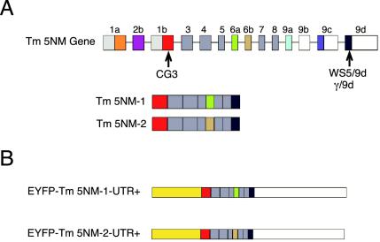
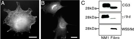
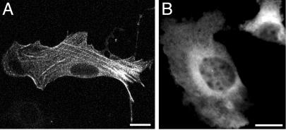
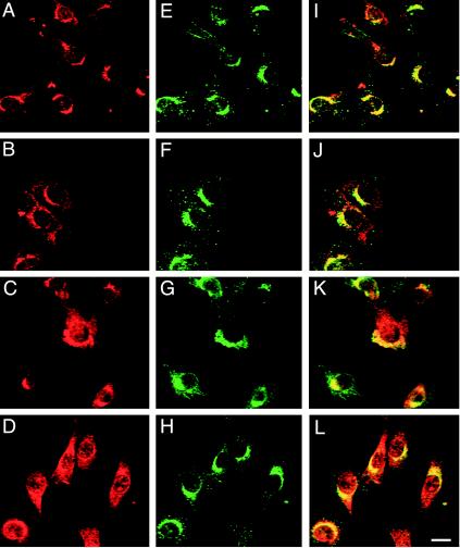
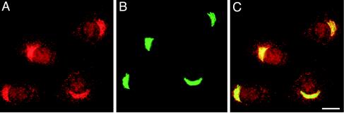
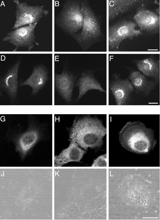
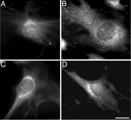
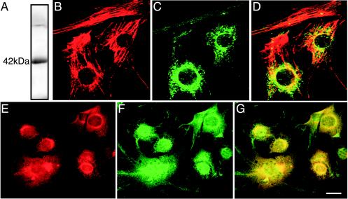
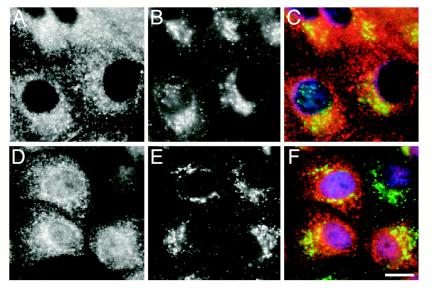
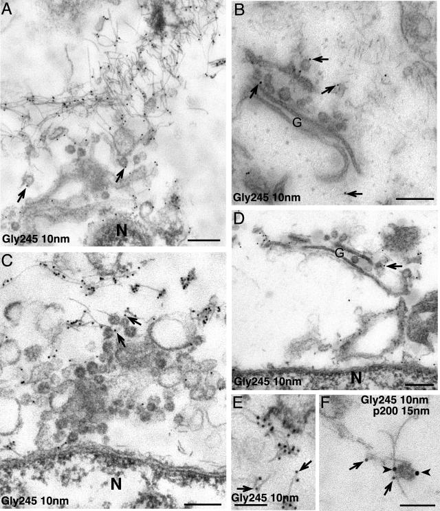
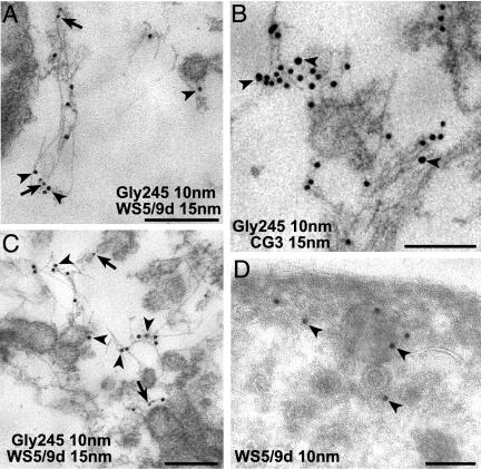
A growing body of evidence suggests that the Golgi complex contains an actin-based filament system. We have previously reported that one or more isoforms from the tropomyosin gene Tm5NM (also known as gamma-Tm), but not from either the alpha- or beta-Tm genes, are associated with Golgi-derived vesicles (Heimann et al., (1999). J. Biol. Chem. 274, 10743-10750). We now show that Tm5NM-2 is sorted specifically to the Golgi complex, whereas Tm5NM-1, which differs by a single alternatively spliced internal exon, is incorporated into stress fibers. Tm5NM-2 is localized to the Golgi complex consistently throughout the G1 phase of the cell cycle and it associates with Golgi membranes in a brefeldin A-sensitive and cytochalasin D-resistant manner. An actin antibody, which preferentially reacts with the ends of microfilaments, newly reveals a population of short actin filaments associated with the Golgi complex and particularly with Golgi-derived vesicles. Tm5NM-2 is also found on these short microfilaments. We conclude that an alternative splice choice can restrict the sorting of a tropomyosin isoform to short actin filaments associated with Golgi-derived vesicles. Our evidence points to a role for these Golgi-associated microfilaments in vesicle budding at the level of the Golgi complex.


Figures

Figure 1.
Figure 1.
Tropomyosin gene organization and EYFP tropomyosin fusion constructs. (A) Organization of the Tm5NM gene (γ-Tm gene). Colored boxes represent exons and white boxes represent 3′ untranslated sequences. Light gray boxes represent 5′ untranslated sequences. The Tm5NM gene encodes Tm5NM-1 and -2, which are identical in amino acid sequence except at an internal alternatively spliced exon. Tm5NM-1 contains exon 6a, whereas Tm5NM-2 contains exon 6b. The recognition sites for the antibodies CG3, WS5/9d, and γ/9d are shown below the appropriate exon. (B) EYFP tropomyosin fusion constructs. Yellow rectangles representing EYFP are fused in frame to the amino termini of Tm5NM-1 and -2. White rectangles represent the 3′ untranslated (UTR) sequence from the 9d exon of the Tm5NM gene.
Figure 2.
Figure 2.
Tropomyosins Tm5NM-1 and -2 are differently targeted in NIH3T3 fibroblasts. NIH3T3 cells were stained with either WS5/9d or γ/9d antibodies. WS5/9d displays perinuclear staining (B), whereas γ/9d primarily stains stress fibers (A). Note that costaining with both antibodies produced very poor images and thus, only individually stained images are shown. (C) Western blots of fibroblast protein and bacterial extract expressing Tm5NM-1 were reacted with CG3, WS5/9d, and γ/9d. Note that unlike γ/9d, WS5/9d does not recognize Tm5NM-1. Scale bars, 15 μm.
Figure 3.
Figure 3.
EYFP-Tm5NM-2 targets to a perinuclear compartment, whereas EYFP-Tm5NM-1 targets to stress fibers. Transient transfection of NIH3T3 fibroblasts with EYFP-Tm5NM-1-UTR+ for 24 h resulted in a predominantly stress fiber distribution (A), whereas cells transiently transfected with EYFP-Tm5NM-2-UTR+ plasmid for 24 h showed characteristic diffuse perinuclear targeting (B). EYFP-Tm5NM-2 was also often found associated with the leading edge. Scale bar, 15 μm.
Figure 4.
Figure 4.
Tm5NM-2 colocalizes with the Golgi complex in synchonized NIH 3T3 fibroblasts. Fibroblasts in G0 and in G1 phases of the cell cycle were double-labeled with anti-FTCD (A-D) and WS5/9d (E-H) antibodies, their subcellular distributions were compared by confocal microscopy, and the merged images are shown (I-L). FTCD (A) and WS5/9d (E) colocalized to a perinuclear compartment in quiescent fibroblasts (I). FTCD and WS5/9d colocalized to the same perinuclear compartment at 3 (B, F, and J), 5 (C, G, and K), and 8 h (D, H, and L) into G1. FTCD protein is far more broadly distributed in the cytoplasm at 8 h (D) than WS5/9d (H), showing that Tm5NM-2 is not on all FTCD-positive structures. The overlapping distribution of Tm5NM-2 with FTCD in fibroblasts suggests that Tm5NM-2 is associated with the Golgi complex. Scale bars, 10 μm.
Figure 5.
Figure 5.
Tropomyosin Tm5NM-2 colocalizes with the Golgi marker GM130. NIH3T3 cells were costained with WS5/9d (A) and anti-GM130 (B) and the merged image is shown in C. The tight colocation confirms the presence of Tm5NM-2 in the Golgi complex. Scale bar, 15 μm.
Figure 6.
Figure 6.
Tm5NM-2 labels the Golgi complex in a Brefeldin A-sensitive manner. Wild-type NIH3T3 cells (A-F) immunostained with WS5/9d or with β-COP antibodies and EYFP-Tm5NM-2 transfected NIH3T3 cells (G-L) were treated with vehicle alone (A, D, and G) or with BFA for 15 min (B, E, and H). Cells were also treated with BFA for 15 min, which was then washed out and followed by a 1-h recovery before fixation (C, F, and I). WS5/9d and β-COP antibody staining of parallel cultures shows perinuclear Golgi staining in both cases (A and D). BFA treatment caused loss of the characteristic perinuclear staining of WS5/9d (B) and β-COP (E). Washout of BFA allowed restoration of the characteristic perinuclear distribution of WS5/9d (C) and β-COP (F). In transfected cells the perinuclear distribution of EYFP-Tm5NM-2 (G) was similarly affected in the presence of BFA (H) and after BFA washout (I). Phase contrast images of the transfected cells in each case highlight the perinuclear concentration of EYFP-Tm5NM-2 (J and L) and its BFA-induced dispersal into the cell periphery (K). Note that lighter exposure of the image shown in H never resembled the patterns shown in G or I. Scale bars, 15 μm.
Figure 7.
Figure 7.
Tm5NM-2 localization is sensitive to nocodazole but not cytochalasin D treatment. Exposure of NIH3T3 cells to 30 μM nocodazole for 1 h resulted in dispersal of Tm5NM-2 throughout the cytoplasm into small puncta (B), whereas the vehicle had no effect (A). In contrast, treatment with 2 μM cytochalasin D for 1 h had no impact on the organization of Tm5NM-2 (D) compared with treatment with vehicle alone (C). Scale bars, 15 μm.
Figure 8.
Figure 8.
Anti-Gly245 antibody shows Golgi-like staining in NIH 3T3 fibroblasts. The anti-Gly245 actin antibody detects a single band at 42 kDa corresponding to both β- and γ-actin in a one-dimensional immunoblot. It sometimes detects a nonspecific slower migrating band (A). Confocal micrographs of the same optical section of NIH 3T3 fibroblasts double-labeled with anti-β-actin (B) and anti-gly245 (C) antibodies and merged image (D). β-actin characteristically detects stress fiber structures (B); however, anti-gly actin antibody preferentially detects puncta localized around the nucleus and in the cytoplasm and only weakly detects stress fibers (C and D). Anti-FTCD (E) and anti-gly245 (F) antibodies show overlapping subcellular distributions (G) in NIH 3T3 fibroblasts, suggesting that the anti-gly245 antibody may be labeling Golgi-associated actin. Scale bar, 10 μm.
Figure 9.
Figure 9.
Short actin filaments and Tm5NM-2 localize to the Golgi complex in MDCK cells. Anti-Gly245 (A) and WS5/9d (D) were double-labeled with the TGN marker p230 on confluent MDCK cells. Both anti-Gly245 and WS5/9d localize to a perinuclear compartment overlapping with p230 (B and E). Merged images C and F show the regions of overlapping distributions. Scale bars, 15 μm.
Figure 10.
Figure 10.
Immunogold labeling of Golgi-associated short microfilaments. Anti-gly245 labeling was performed on perforated MDCK cells where it specifically localizes on short microfilaments that were found clustered around Golgi stacks located in the perinuclear area (A and C). In some cases, anti-gly245 also labels vesicles seen budding from the Golgi complex (B and D). Anti-gly245 labeling is predominantly at the ends of microfilaments where these are easily depicted (E). Double-labeling shows conjoint association of anti-gly245 (arrows) and anti-p200/myosinII (arrowheads) labeling on a Golgi-derived vesicle attached to a short actin filament (F). N, nucleus; G, Golgi. Scale bars, 200 nm.
Figure 11.
Figure 11.
Tropomyosin isoforms localize on Golgi-associated microfilaments and vesicles. In perforated cells, Tm5NM isoforms (Tm5NM-2) labeled with either WS5/9d or CG3 antibodies (arrowheads) are associated with short actin filaments that are colabeled with anti-gly245 (arrows; A-C). These microfilaments are also often found attached to Golgi-derived vesicles (C). On cryosections of fixed cells, WS5/9d labeling is seen directly associated with the surface of coated vesicles in the cytoplasm (D). Scale bars, 200 nm.

Similar articles

Cited by

References

    1. Adami, R., Cintio, O., Trombelta, G., Choquet, D., and Grazi, E. (2002). Effects of chemical modification tropomyosin, and myosin subfragment 1 on the yield strength and critical concentration of F-actin. Biochemistry 41, 5907-5912. - PubMed
    1. Bamburg, J.R. (1999). Proteins of the ADF/cofilin family: essential regulators of actin dynamics. Annu. Rev. Cell Dev. Biol. 15, 185-230. - PubMed
    1. Beck, K.A., Buchanan, J.A., Malhotra, V., and Nelson, W.J. (1994). Golgi spectrin: identification of an erythroid beta-spectrin homologue associated with the Golgi complex. J. Cell Biol. 127, 707-723. - PMC - PubMed
    1. Beck, K.A., Buchanan, J.A., and Nelson, W.J. (1997). Golgi membrane skeleton: identification, localization and oligomerization of a 195 kDa ankyrin associated with the Golgi complex. J. Cell Sci. 110, 1239-1249. - PubMed
    1. Blanchoin, L., Pollard, T.D., and Hitchcock-DeGregori, S.E. (2001). Inhibition of the Arp2/3 complex-nucleated actin polymerization and branch formation by tropomyosin. Curr. Biol. 11, 1300-1304. - PubMed

Publication types

MeSH terms

LinkOut - more resources