. 2003 Sep;203(3):265-76.
doi: 10.1046/j.1469-7580.2003.00214.x.

Ultrastructural correlates of synapse withdrawal at axotomized neuromuscular junctions in mutant and transgenic mice expressing the Wld gene

Affiliations

Ultrastructural correlates of synapse withdrawal at axotomized neuromuscular junctions in mutant and transgenic mice expressing the Wld gene

Thomas H Gillingwater et al. J Anat. 2003 Sep.

Abstract

We carried out an ultrastructural analysis of axotomized synaptic terminals in Wld(s) and Ube4b/Nmnat (Wld) transgenic mice, in which severed distal axons are protected from Wallerian degeneration. Previous studies have suggested that axotomy in juvenile (< 2 months) Wld mice induced a progressive nerve terminal withdrawal from motor endplates. In this study we confirm that axotomy-induced terminal withdrawal occurs in the absence of all major ultrastructural characteristics of Wallerian degeneration. Pre- and post-synaptic membranes showed no signs of disruption or fragmentation, synaptic vesicle densities remained at pre-axotomy levels, the numbers of synaptic vesicles clustered towards presynaptic active zones did not diminish, and mitochondria retained their membranes and cristae. However, motor nerve terminal ultrastructure was measurably different following axotomy in Wld transgenic 4836 line mice, which strongly express Wld protein: axotomized presynaptic terminals were retained, but many were significantly depleted of synaptic vesicles. These findings suggest that the Wld gene interacts with the mechanisms regulating transmitter release and vesicle recycling.


Figures
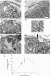
Fig. 1
Fig. 1
Retention of terminal ultrastructure and partial occupancy at axotomized juvenile Wlds NMJs. (A,B) Electron micrographs of wild-type (C57Bl/6) mouse NMJs, 1 day post-axotomy. Fragmented remnants of a motor nerve terminal (black arrow) being phagocytosed in situ by a terminal Schwann cell are shown in panel A (white arrow; scale bar = 1 µm). Panel B shows a vacated motor endplate with retained post-synaptic specializations, loosely capped by a cellular process, presumably from a terminal Schwann cell (scale bar = 1 µm). (C–F) Electron micrographs of 2-month-old Wlds mouse NMJs at 3 days (C,D) and 5 days (E,F) post-axotomy. (C) Nerve terminal with a retained distribution of 50-nm synaptic vesicles (arrow) and mitochondria (M). (D) High-power micrograph taken from C, showing clustering of synaptic vesicles close to active zones (a darkening of the presynaptic membrane opposite the opening of a post-synaptic fold). (E) Enduring nerve terminal bouton at a partially occupied NMJ (as indicated by areas of unoccupied post-synaptic folds; not shown). This example shows retained terminal membranes, synaptic vesicles and mitochondria, as well as accumulation of neurofilaments within its centre (white arrow). Examples of ‘giant’ vesicles (∼125 nm in diameter; black arrow) are also present. (F) Example of a partially occupied endplate, where the remaining bouton on the left neighbours a region of unoccupied post-synaptic specializations (white arrows), capped by the process of a terminal Schwann cell (black arrow). Scale bars = 0.5 µm (C,E,F); 100 nm (D). (G) Levels of partial occupancy at 3, 4 and 7 days post-axotomy at 2-month Wlds mouse neuromuscular junctions (mean ± SD). The dotted curve was generated using a gaussian three-parameter best-fit function in Sigmaplot.
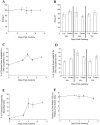
Fig. 2
Fig. 2
Quantification of subcellular organelles at axotomized juvenile (2-month-old) Wlds NMJs. (A) Graph showing the retention of synaptic vesicles at all time points examined following axotomy. (B) Bar chart showing the retention of synaptic vesicles in nerve terminals at both fully and partially occupied endplates, with no loss of vesicles even at the late stages of nerve terminal withdrawal. (C,D) Graph and bar chart showing an increase in the packing densities of synaptic vesicles following axotomy at both fully and partially occupied NMJs. (E) Graph showing the increase in intrabouton neurofilament levels following axotomy, reaching significant levels from controls by 4 days. (F) Graph showing no decline in the numbers of ‘peri-active zone’ synaptic vesicles at any time post-axotomy.
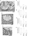
Fig. 3
Fig. 3
Ultrastructural retention and reduced synaptic vesicle densities at 4836 line Wld transgenic NMJs following axotomy. (A,B) Electron micrographs of persistent terminals at 5 days post-axotomy with retained architecture and mitochondria, but sparse synaptic vesicles. (C) Bar chart showing an increase in intraterminal neurofilament levels compared with control preparations, but the increase is less than measured in equivalent Wlds preparations. (D) Bar chart showing a decrease in the numbers of synaptic vesicles (∼50%), compared to controls and 2-month-old Wlds preparations. (E) Bar chart showing that even though there was an increase in neurofilament levels, a decrease in the packing densities of synaptic vesicles was detected compared to controls and 2-month Wlds preparations. (F) Portion of electron micrograph B at higher magnification, showing the paucity of both free and docked synaptic vesicles. (G) Bar chart showing a significant reduction in the numbers of ‘peri-active zone’ vesicles in 4836 Wld preparations (P ≤ 0.0001, unpaired t-test). Scale bars = 0.5 µm.

References

    1. Aberle H, Haghighi AP, Fetter RD, McCabe BD, Magalhaes TR, Goodman CS. Wishful thinking encodes a BMP type II receptor that regulates synaptic growth in Drosophila. Neuron. 2002;33:545–558. - PubMed
    1. Balice-Gordon RJ, Chua CK, Nelson CC, Lichtman JW. Gradual loss of synaptic cartels precedes axon withdrawal at developing neuromuscular junctions. Neuron. 1993;11:801–815. - PubMed
    1. Barker D, Ip MC. Sprouting and degeneration of mammalian motor axons in normal and de-afferentated skeletal muscle. Proc. Roy. Soc. B. 1966;163:538–554. - PubMed
    1. Birks R, Katz B, Miledi R. Physiological and structural changes at the amphibian myoneural junction, in the course of nerve degeneration. J. Physiol. (Lond.) 1960;150:145–168. - PMC - PubMed
    1. Bixby JL. Ultrastructural observations on synapse elimination in neonatal rabbit skeletal muscle. J. Neurocytol. 1981;10:81–100. - PubMed

Publication types

LinkOut - more resources