Comparative Study
. 2003 Nov;140(6):1117-27.
doi: 10.1038/sj.bjp.0705530. Epub 2003 Oct 6.

Histamine induces cytoskeletal changes in human eosinophils via the H(4) receptor

Affiliations
Comparative Study

Histamine induces cytoskeletal changes in human eosinophils via the H(4) receptor

Karen F Buckland et al. Br J Pharmacol. 2003 Nov.

Abstract

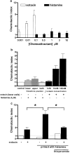
1. Histamine (0.004-2 microm) induced a concentration-dependent shape change of human eosinophils, but not of neutrophils or basophils, detected as an increase in forward scatter (FSC) in the gated autofluorescence/forward scatter (GAFS) assay. 2. The histamine-induced eosinophil shape change was completely abolished by thioperamide (10 microm), an H3/H4 receptor antagonist, but was not inhibited by pyrilamine or cimetidine (10 microm), H1 and H2 receptor antagonists, respectively. The H4 receptor agonists, clobenpropit and clozapine (0.004-2 microm), which are also H3 receptor antagonists, both induced eosinophil shape change, which was inhibited by thioperamide (10 microm). The H3/H4 receptor agonists, imetit, R-alpha-methyl histamine and N-alpha-methyl histamine (0.004-2 microm) also induced eosinophil shape change. 3. Histamine induced actin polymerisation (0.015-10 microm), intracellular calcium mobilisation (10-100 microm) and a significant upregulation of expression of the cell adhesion molecule CD11b (0.004-10 microm) in eosinophils, all of which were inhibited by thioperamide (10-100 microm). In addition, the H4 receptor agonist/H3 receptor antagonist clozapine (20 microm) stimulated a rise in intracellular calcium in eosinophils. 4. Activation of H4 receptors by histamine (1 microm) primed eosinophils for increased chemotactic responses to eotaxin, but histamine (0.1-10 microm) did not directly induce chemotaxis of eosinophils. 5. Pertussis toxin (1 microg ml-1) inhibited shape change and actin polymerisation responses induced by histamine showing that these effects are mediated by coupling to a Galphai/o G-protein. 6. This study demonstrates that human eosinophils express functional H4 receptors and may provide a novel target for allergic disease therapy.


Figures

Figure 1
Figure 1
Histamine induced shape change of human eosinophils. Mixed granulocytes were stimulated with (a) buffer, (b) 1 μM histamine or (c) 1 nM eotaxin and their shape change measured by flow cytometry. Dot plots of the FSC vs SSC of mixed granulocytes show two separate cell populations; eosinophils in black, neutrophils in grey. Autofluorescence was measured in FL-2 fluorescence channel and displayed in histogram plots, on which regions of high (R1=eosinophils) and low (R2=neutrophils) autofluorescence were defined. The mean FSC of eosinophils (R1) increased from 178 (a) to 271 (b) after stimulation with histamine (1 μM) (shift to the right in dot plot) or 267 (c) after stimulation with eotaxin, while the mean FSC of neutrophils (R2) was unchanged. Basophils were detected by flow cytometry as highly positive for CD123 but negative for HLA-DR (dot plots not shown). Histamine induced eosinophil shape change in a concentration-dependent manner, which was selective for (d) eosinophils and induced by (e) all three histamine preparations. Dot plots and histograms are representative of 10 or more experiments. Concentration–response curves are mean±s.e.m.; (d) eosinophils/neutrophils n=10, basophils n=3; (e) n=5.
Figure 2
Figure 2
Effect of histamine and histamine analogues on human eosinophil shape change. Human eosinophils were incubated with receptor antagonists (10 min) or G-protein inhibitors (90 min) prior to stimulation with agonists and eosinophil shape change measured by flow cytometry. Results are expressed as the percent increase in FSC induced by agonist compared to unstimulated cells. (a) Comparison of histamine and eotaxin stimulation of eosinophil shape change; (b) histamine-induced eosinophil shape change is inhibited by thioperamide (10 μM) but not pyrilamine and cimetidine (10 μM). (c) Inhibition of histamine-induced shape change by thioperamide is concentration-dependent. (d) R-α-meH, N-α-meH, imetit, clozapine and clobenpropit, all induce eosinophil shape change, which can be inhibited by (e) thioperamide (10 μM). (f) Histamine-induced shape change is inhibited by pertussis toxin but not cholera toxin (1 μg ml−1); **P<0.01, comparison of entire concentration–response curve in presence of inhibitor vs control. Data shown are mean±s.e.m.: (a) n⩽10; (b), (c) and (f) n=5; (d) and (e) n=4–11.
Figure 3
Figure 3
Histamine stimulates eosinophil shape change in whole-blood eosinophil shape change assay. Whole blood was incubated with or without thioperamide (10 μM) prior to stimulation with agonists and leucocyte shape change analysed by flow cytometry. Results are expressed as the percent increase in FSC induced by agonist compared to unstimulated cells. Data shown are mean±s.e.m., n=5, statistical analysis ***P<0.001 comparison of entire concentration–response curve in presence of thioperamide vs control.
Figure 4
Figure 4
Effect of histamine on actin polymerisation in human eosinophils. Human eosinophils were incubated with receptor antagonists (10 min) or G-protein inhibitors (90 min) prior to stimulation with agonists. Responses were measured by flow cytometry and results are expressed as the percent increase in fluorescence induced by agonist compared to unstimulated cells. (a) Comparison of histamine and eotaxin stimulation of actin polymerisation. (b) Time course of actin polymerisation induced by histamine. (c) Histamine-induced actin polymerisation is inhibited by thioperamide (10 μM) but not pyrilamine and cimetidine (10 μM). (d) Inhibition by pertussis toxin but not cholera toxin (1 μg ml−1) of histamine-induced actin polymerisation; **P<0.01 comparison of entire concentration–response curve in presence of inhibitor vs control. Data shown are mean±s.e.m.: (a) and (c) n=5; (b) n=3–5; (d) n=3.
Figure 5
Figure 5
Histamine induced upregulation of surface CD11b expression and increase in intracellular calcium mobilisation on human eosinophils. (a) Mixed granulocytes were incubated with thioperamide (10 μM) and then stimulated with histamine or eotaxin and CD11b expression analysed by flow cytometry as described in Methods. Results are expressed as percent increase in fluorescence compared to unstimulated cells. Data shown are mean±s.e.m., n=3–7, *P<0.05 comparison of entire concentration–response curve in presence of thioperamide vs control. (b)–(d) Isolated eosinophils were loaded with Fura-2 and then stimulated with agonists. Graphs show actual recorded fluorescence output and are representative of n independent experiments; (b) n=2; (c) n=6; (d) n=4.
Figure 6
Figure 6
Human eosinophil chemotaxis and chemokinesis. (a) Human eosinophils were placed in the upper wells of a micro-Boyden chamber and either eotaxin or histamine was added to lower wells. (b) Human eosinophils were placed in the upper wells of a micro-Boyden chamber and either eotaxin added to the lower wells, or histamine added to the upper, lower or upper and lower wells of the chamber. (c) Human eosinophils were incubated with histamine (1 μM), or histamine and thioperamide (10 μM) and then placed in the upper wells of a micro-Boyden chamber and eotaxin (30 nM) was placed in the lower wells of the chamber. (a)–(c) Cells that had migrated across a polycarbonate filter after 1 h were counted by flow cytometry and expressed as chemotactic index. Data shown are mean±s.e.m. of n experiments each using cells from a different donor and each carried out with three or more replicate wells; (a) n=6; (b) n=3–5; (c) filled bars n=3, open bars n=1. *P<0.05.

Comment in

References

    1. ARRANG J.M., GARBARG M., LANCELOT J.C., LECOMTE J.M., POLLARD H., ROBBA M., SCHUNACK W., SCHWARTZ J.C. Highly potent and selective ligands for histamine H3-receptors. Nature. 1987;327:117–123. - PubMed
    1. ARRANG J.M., GARBARG M., SCHWARTZ J.C. Auto-inhibition of brain histamine release mediated by a novel class (H3) of histamine receptor. Nature. 1983;302:832–837. - PubMed
    1. ASH A.S., SCHILD H.O. Receptors mediating some actions of histamine. Br. J. Pharmacol. 1966;27:427–439. - PMC - PubMed
    1. BISSONNETTE E.Y. Histamine inhibits tumor necrosis factor alpha release by mast cells through H2 and H3 receptors. Am. J. Respir. Cell Mol. Biol. 1996;14:620–626. - PubMed
    1. BOUSQUET J., CHANEZ P., LACOSTE J.Y., BARNEON G., GHAVANIAN N., ENANDER I., VENGE P., AHLSTEDT S., SIMONY-LAFONTAINE J., GODARD P. Eosinophilic inflammation in asthma. N. Engl. J. Med. 1990;323:1033–1039. - PubMed

Publication types

MeSH terms

LinkOut - more resources