. 2003 Oct 6;198(7):1107-17.
doi: 10.1084/jem.20030913.

The strength of persistent antigenic stimulation modulates adaptive tolerance in peripheral CD4+ T cells

Affiliations

The strength of persistent antigenic stimulation modulates adaptive tolerance in peripheral CD4+ T cells

Nevil J Singh et al. J Exp Med. .

Abstract

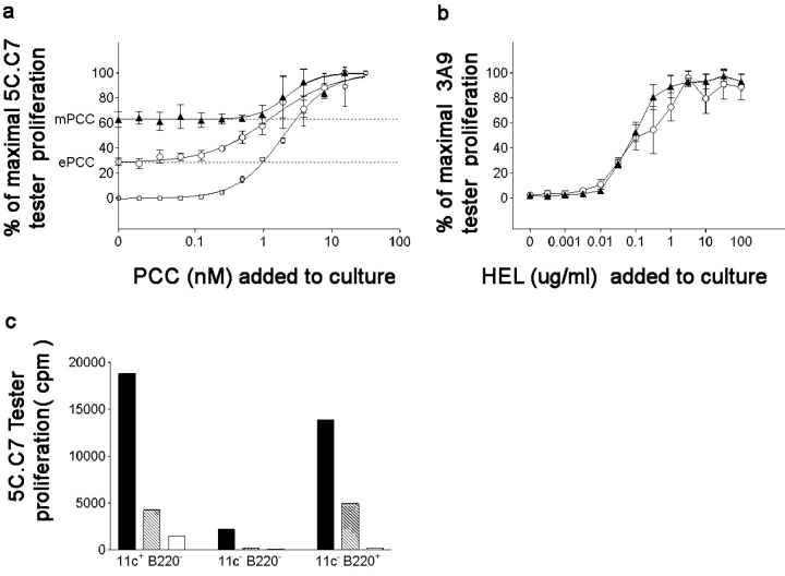
The quantitative adaptation of receptor thresholds allows cells to tailor their responses to changes in ambient ligand concentration in many biological systems. Such a cell-intrinsic calibration of T cell receptor (TCR) sensitivity could be involved in regulating responses to autoantigens, but this has never been demonstrated for peripheral T cells. We examined the ability of monoclonal naive T cells to modulate their responsiveness differentially after exposure to fourfold different levels of persistent antigen stimulation in vivo. T cells expanded and entered a tolerant state with different kinetics in response to the two levels of stimulation, but eventually adjusted to a similar slow rate of turnover. In vivo restimulation revealed a greater impairment in the proliferative ability of T cells resident in a higher antigen presentation environment. We also observed subtle differences in TCR signaling and in vitro cytokine production consistent with differential adaptation. Unexpectedly, the system failed to similarly compensate to the persistent stimulus in vivo at the level of CD69 expression and actin polymerization. This greater responsiveness of T cells residing in a host with a lower level of antigen presentation allows us to demonstrate for the first time an intrinsic tuning process in mature T lymphocytes, albeit one more complex than current theories predict.


Figures

Figure 1.
Figure 1.
In vitro antigen presentation by mPCC splenocytes is greater than that by ePCC splenocytes. (a) Functional presentation of the transgene-derived antigen by mPCC,CD3ɛ−/− (▴) and ePCC,CD3e−/− (○) APCs to T cells in vitro was quantitated by comparing the proliferation of PCC-specific tester T cells to fresh splenocytes from either transgenic or antigen-deficient CD3ɛ−/− (□) mice. Tester T cells used were from a rested, in vitro–preactivated population of 5C.C7 T cells, which are extremely sensitive to relatively subtle variations in antigen presentation. Dotted lines from the y axis denote basal proliferation elicited by each type of APC estimated from a curve fit of the data. (b) Proliferation of 3A9 T cells to HEL presented by splenocytes as in (a) shows that the presentation differences are specific to PCC. (c) Presentation of antigen by the two transgenics is not differentially compartmentalized in splenic APCs. Sorted CD11c+ B220, CD11c B220, or B220+ cells freshly isolated from the mPCC,CD3ɛ−/− (solid bars), ePCC,CD3ɛ−/− (hatched bars), or CD3ɛ−/− mice (open bars) were used to stimulate tester T cells in vitro.
Figure 2.
Figure 2.
Naive T cells expand differentially in the two environments but eventually achieve similar turnover rates. (a) Naive 5C.C7,RAG2−/− T cells transferred into the PCC-hi (▴) or PCC-lo (○) recipients were recovered after various days. The absolute number of recovered T cells on each day was normalized to that on day 1 in each group. Three experiments are averaged on days 1–5 and for subsequent time points showing error bars. (b and c) Different levels of persistent antigen induce T cell cycling with the same lag time, but affect the subsequent rate of proliferation. Dilution of CFSE in the T cells recovered from PCC-hi or PCC-lo hosts was analyzed using the Gett-Hodgkins method to calculate the cell division profile over the first 68 h. Overlay of dotted lines in (c) represents the raw data fitted to an exponential rise to the max function. The best fit parameters (R2 = 0.97–0.99) for the PCC-hi and PCC-lo data are y0 = −11.9 ± 3.3 and −12.2 ± 5.2, a = 19.8 ± 2.5 and 17.5 ± 4.7, and b = 0.966 ± 0.008 and 0.957 ± 0.011, respectively. (d and e) Proliferative expansion is contained earlier in the higher stimulus environment, but is eventually modulated to similar extents in the two hosts. (d) BrdUrd incorporation in the PCC-hi (red lines) or PCC-lo (blue lines) resident T cells was measured after daily injections of BrdUrd between days 7 to 11 (A) or 32 to 36 (B). Green lines represent negative controls. (e) Incremental BrdUrd incorporation over the 5 d of labeling from days 32 to 36.
Figure 3.
Figure 3.
The quantitative impairment of in vivo responsiveness varies with the level of persistent antigen presentation experienced. (a) 5C.C7,RAG2−/− TCR transgenic cells resident for 22–28 d in the PCC-hi host (▴) proliferate poorer than those resident in the PCC-lo host (○) upon retransfer into a fresh PCC-hi environment. Both the adapted T cells are desensitized relative to naive T cells (□) establishing a hierarchy of adaptations in vivo. Data are averaged over three separate experiments. (b) The expansion of adapted T cells was monitored by CFSE dilution over short intervals of time after transfer of 28-d PCC-hi–adapted (P-hi), PCC-lo–adapted (P-lo), or naive (Nv) T cells into fresh PCC-hi mice. Numbers on the left represent the time points sampled in hours. (c) Quantitative analysis of the CFSE profiles in (b) by the Gett-Hodgkins method. (d) Initiation of cell cycling in the transferred T cells quantitated by the disappearance of the zero division CFSE peak reveals two phases of recruitment in the adapted T cells. Both phases, however, decay at greater rates for the PCC-lo–adapted T cells. Overlay of dotted lines on each curve represent the fitting of those parts of the data to a linear regression.
Figure 4.
Figure 4.
Adapted T cells down-regulate cytokine production with different kinetics and to different extents. (a and b) Maximal cytokine production after a 48-h APC plus peptide stimulation of 104 purified T cells recovered from the PCC-hi (▴), PCC-lo (○), or PCC plus LPS-primed (▪) mice at different days after transfer reveals a faster down-regulation of IL-2 (a) and IFN-γ (b) production in the PCC-hi environment. (a) Maximal IL-2 production is expressed as a percentage of that produced by naive T cells. (b) IFN-γ production is expressed as absolute production in picograms per milliliter. Data points in (a) and (b) are averaged over two to five separate experiments for points shown with error bars. (c and d) After stable maximal down-regulation, a subtle difference in the responsiveness of PCC-hi– versus PCC-lo–adapted T cells can be revealed by stimulation with plate-bound anti-CD3 plus soluble CD28. Representative production of IL-2 (c) and IFN-γ (d) by T cells recovered 34 d after transfer. IL-2 production is compared with that by T cells from a naive 5C.C7,RAG2−/− TCR transgenic mice (□).
Figure 5.
Figure 5.
Biochemical alterations in adapted T cells. (a and b) TCR signal transduction assayed at the level of ERK phosphorylation is differentially affected in the PCC-hi–adapted (P-hi, ▴) versus PCC-lo–adapted (P-lo, ○) cells relative to naive T cells (Nv, □). T cells stimulated for various times on anti-CD3–coated plates and analyzed for phosphorylated ERK (p-ERK). Blots were stripped and reprobed for total ERK (t-ERK). (c and d) The percentage of phospho-ERK was quantitated using a PhosphorImager. Adapted samples were recovered 13 (a and c) or 28 d (b and d) after transfer.
Figure 6.
Figure 6.
In vivo T cell reactivation during the adaptive tolerance phase reveals cell-intrinsic differences. (a–c) Surface expression of CD69 on naive 5C.C7,RAG2−/− T cells transferred into the PCC-hi (▴ or red lines) or the PCC-lo (○ or blue lines) recipients reveals an early phase of greater responsiveness in the PCC-hi host, followed by greater responsiveness in the PCC-lo host during the adaptive tolerance phase. (a) Percentage of cells expressing CD69 on various days after transfer and (b) the MFI of CD69 on the positive subset of cells on each day. Green lines represent naive 5C.C7 T cells. (d) T cells were stained for polymerized intracellular actin with Alexa 546–coupled phalloidin, ex vivo.

Similar articles

Cited by

References

    1. Sklar, L.A., and G.M. Omann. 1990. Kinetics and amplification in neutrophil activation and adaptation. Semin. Cell Biol. 1:115–123. - PubMed
    1. Mathison, J.C., G.D. Virca, E. Wolfson, P.S. Tobias, K. Glaser, and R.J. Ulevitch. 1990. Adaptation to bacterial lipopolysaccharide controls lipopolysaccharide-induced tumor necrosis factor production in rabbit macrophages. J. Clin. Invest. 85:1108–1118. - PMC - PubMed
    1. Sentman, C.L., M.Y. Olsson, and K. Karre. 1995. Missing self recognition by natural killer cells in MHC class I transgenic mice. A ‘receptor calibration’ model for how effector cells adapt to self. Semin. Immunol. 7:109–119. - PubMed
    1. Lucas, B., I. Stefanova, K. Yasutomo, N. Dautigny, and R.N. Germain. 1999. Divergent changes in the sensitivity of maturing T cells to structurally related ligands underlies formation of a useful T cell repertoire. Immunity. 10:367–376. - PubMed
    1. Azzam, H.S., J.B. DeJarnette, K. Huang, R. Emmons, C.S. Park, C.L. Sommers, D. El Khoury, E.W. Shores, and P.E. Love. 2001. Fine tuning of TCR signaling by CD5. J. Immunol. 166:5464–5472. - PubMed