. 2003 Oct 13;163(1):35-44.
doi: 10.1083/jcb.200306069. Epub 2003 Oct 6.

Ligand crowding at a nascent signal sequence

Affiliations

Ligand crowding at a nascent signal sequence

Gottfried Eisner et al. J Cell Biol. .

Abstract

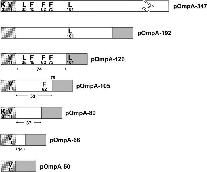
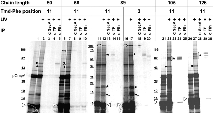
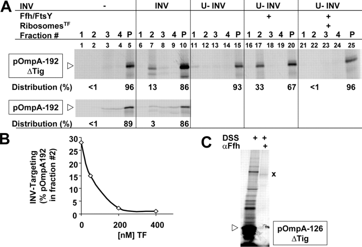
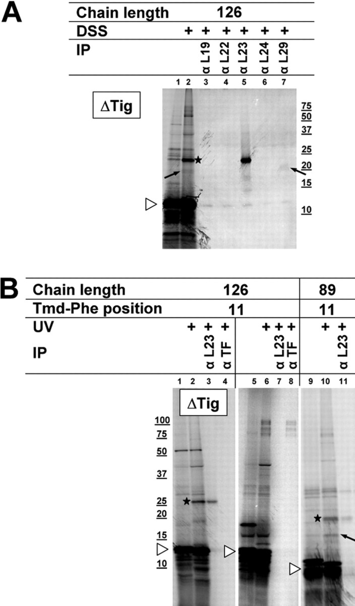
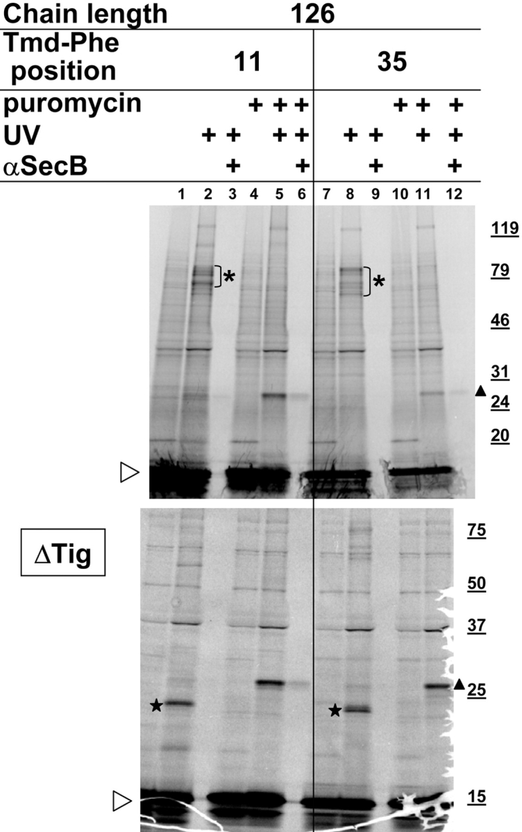
We have systematically analyzed the molecular environment of the signal sequence of a growing secretory protein from Escherichia coli using a stage- and site-specific cross-linking approach. Immediately after emerging from the ribosome, the signal sequence of pOmpA is accessible to Ffh, the protein component of the bacterial signal recognition particle, and to SecA, but it remains attached to the surface of the ribosome via protein L23. These contacts are lost upon further growth of the nascent chain, which brings the signal sequence into sole proximity to the chaperone Trigger factor (TF). In its absence, nascent pOmpA shows extended contacts with L23, and even long chains interact in these conditions proficiently with Ffh. Our results suggest that upon emergence from the ribosome, the signal sequence of an E. coli secretory protein gradually becomes sequestered by TF. Although TF thereby might control the accessibility of pOmpA's signal sequence to Ffh and SecA, it does not influence interaction of pOmpA with SecB.


Figures

Figure 1.
Figure 1.
Compilation of pOmpA constructs used. At the top, the 347–amino acid-long precursor of OmpA is depicted with the hatched box representing the signal sequence. Starting with the first amino acid of the signal sequence, all the positions are indicated at which stop codons were engineered. On top of the numerals, the amino acids of the pOmpA sequence are given that were replaced by the incorporation of Tmd-Phe. Below the precursor of OmpA, nascent chains are listed with the size indicated in number of NH2-terminal residues. The gray boxes represent the COOH-terminal 30 amino acids of each nascent chain predicted to be hidden in the ribosomal exit tunnel. For each nascent chain, the positions of cross-linker tested are indicated. Numbers below the bars correspond to the estimated distances in amino acids between the signal sequence cleavage site and the ribosomal exit site. For pOmpA105, the position of the ribosomal exit site relative to the position of the photoprobe is depicted above the bar. For pOmpA-50, the signal sequence cleavage site is represented by a dotted line.
Figure 2.
Figure 2.
Site-specific incorporation of Tmd-Phe into pOmpA does not interfere with its translocation into membrane vesicles. In addition to wild-type pOmpA (wt), the indicated stop codon mutants located in the mature part and the signal sequence of OmpA were expressed in vitro by the help of suppressor tRNA charged with Tmd-Phe. Radiolabeled proteins were separated by SDS-PAGE and visualized by phosphorimaging. Addition of E. coli INVs and proteinase K (PK) is indicated. Precursor (pOmpA) and mature (OmpA) forms are labeled with open and closed arrowheads, respectively. Note the aberrant electrophoretic mobility of the mutant precursor carrying Tmd-Phe at position 11 within the signal sequence. The precursor of OmpA often resolves on SDS-PAGE into two closely spaced species, the reason for that being unknown. The background processing of the different pOmpAs obtained in the absence of added INVs (lanes 1, 5, 9, and 13) is most likely due to traces of INVs contaminating the ribosome preparation used.
Figure 3.
Figure 3.
Upon growth of the nascent pOmpA chain, the signal sequence changes its molecular environment from SecA and Ffh to TF. Nascent, ribosome-associated chains of pOmpA of 50, 66, 89, 105, and 126 amino acids length were synthesized in vitro. Truncation of the ompA mRNA was achieved by the addition of complementary oligodeoxynucleotides and RNaseH cleavage (Behrmann et al., 1998). Stop codons at the indicated positions were suppressed by tRNAsup charged with Tmd-Phe. The migration on SDS-PAGE of the nascent chains is indicated by white arrowheads. The sizes of marker proteins are given in kD. The expression of some full-size pOmpA resulting from insufficient truncation of mRNA by RNaseH was usually observed. In vitro reactions were supplemented with 270 nM SecA dimer, 234 nM SecB tetramer (468 nM in the case of pOmpA-89), and 80 nM Ffh. UV irradiation (UV) resulted in several adducts, which are marked by various symbols. Identification of cross-links to Ffh (X), SecA (white arrow), and TF (asterisk) by coimmunoprecipitation with specific antibodies is shown. • marks presumed intramolecular cross-links of full-size pOmpA (compare with legend to Fig. 4). Stars and upwards pointing arrows mark cross-links to ribosomal proteins L23 and L29, as demonstrated in Fig. 6.
Figure 4.
Figure 4.
TF interacts with the mature part of nascent pOmpA chains at multiple sites. Nascent, ribosome-associated chains of pOmpA of 126, 192, and 105 amino acids length were synthesized in vitro, and their position on SDS-PAGE is indicated (white arrowheads). UV irradiation (UV) resulted in the appearance of several radiolabeled bands, most of which could be coimmunoprecipitated with anti-TF antibodies (αTF). The asterisk marks the adducts that exhibit apparent molecular masses corresponding to the arithmetic sum between those of TF and each nascent pOmpA chain. The electrophoretic mobility of adducts in the 50-kD range (marked •) changed with the position of Tmd-Phe within the nascent chain, suggesting that they might be derived from intramolecular cross-links of full-size pOmpA.
Figure 5.
Figure 5.
TF interferes with a cotranslational targeting of a bacterial secretory protein. Nascent chains of pOmpA, 192 and 126 amino acids in length, were synthesized in vitro as before (pOmpA-192) or by a cell-free system whose components had been prepared from a TF-null mutant (pOmpA-192, ΔTig). (A) After synthesis in the presence or absence of the components indicated at the top of the panel, pOmpA-192 chains were subjected to flotation gradient centrifugation. The gradient was fractionated into four equal fractions from the top (fraction nos. 1–4), and proteins were precipitated using TCA and analyzed by SDS-PAGE and phosphorimaging. The pellet fraction (P) was directly dissolved in SDS-PAGE loading buffer. The radioactivity of the pOmpA-192 bands of all fractions was quantitated using Imagequant software. Indicated are the numbers for the relevant fraction 2 (membrane fraction) and P (non–membrane-associated material). RibosomesTF, ribosomes prepared from the ΔTig strain and reconstituted with purified TF. (B) The amount of pOmpA-192 synthesized in the ΔTig system and recovered from fraction 2 after flotation centrifugation of an INV-containing sample (compare with A) is plotted against the concentration of purified TF present during synthesis. (C) pOmpA-126 was synthesized in the ΔTig system in the presence of 160 nM Ffh. After synthesis, samples were treated with the cross-linker DSS. αFfh, anti-Ffh antibodies.
Figure 6.
Figure 6.
Transfer of nascent pOmpA from ribosomal protein L23 onto TF. (A) pOmpA-126 was synthesized in the ΔTig system. After cross-linking with DSS, aliquots were immunoprecipitated with antisera raised against the indicated proteins of the large E. coli ribosomal subunit. Adducts to L23 (star) and L29 (upwards pointing arrow) are highlighted. (B) Nascent pOmpA chains of 126 and 89 amino acids in length with the photoprobe incorporated within the signal sequence at position Val-11 were synthesized in the absence or presence of TF as indicated. Cross-links obtained by UV irradiation were identified using antibodies against L23 and TF.
Figure 7.
Figure 7.
Binding of SecB to newly synthesized pOmpA including its signal sequence is not controlled by TF and occurs after release from the ribosome. Chains of pOmpA, 126 amino acids in length with the photoprobe incorporated within the signal sequence at Val-11 or beyond at Leu-35, were synthesized in the presence or absence of TF as indicated. In addition, SecA (270 nM of dimer) and SecB (468 nM of tetramer) were also added. UV-induced cross-linking was performed on nascent chains, resulting in adducts to TF (asterisk) or L23 (star). Both cross-links disappeared upon dissociation of RNCs by puromycin in favor of those to SecB, as identified by immunoprecipitation (triangles).
Figure 8.
Figure 8.
At the ribosome, the interaction partners of a signal sequence change with the length of the nascent chains. Depicted are molecular contacts of the signal sequence of pOmpA at the ribosomal exit site. The large ribosomal subunit is outlined in the top panel. Protein L23 located at the orifice of the ribosomal exit tunnel is indicated. The exiting pOmpA chain is illustrated by a cylinder (signal sequence) followed by a solid line, which represents the part of mature OmpA predicted to have emerged from the ribosome (its size is given in number of amino acids in parentheses). The structure labeled SRP is meant to represent Ffh, the 4.5S RNA is not shown. SecA and TF are indicated. Cross-links are illustrated by stars; for pOmpA-89, cross-links at residues 3 and 11 of the signal sequence are simultaneously depicted. Whereas Ffh and TF have been shown to use L23 as docking site, the presumed ribosomal-binding site of SecA has not been identified. Different from the model showing Ffh and TF simultaneously bound to L23, recent data suggest competitive binding between Ffh and TF to L23 (Ullers et al., 2003). This has also been shown for Ffh and SecA with respect to pOmpA-66 (unpublished data), for which reason binding of the signal sequence to either protein was drawn as two alternative situations (labeled A and B). It should be noted that only single adducts were obtained. Therefore in cases in which the model suggests interaction of the signal sequence with two binding partners (e.g., pOmpA-89), these should be taken as representing two separate populations of adducts. The identified cross-linking partners of the signal sequence are Ffh for pOmpA-50; Ffh and SecA for pOmpA-66; Ffh, SecA, TF, and L23 for pOmpA-89; and TF for pOmpA-105. Upon removal of TF, pOmpA-126 (not depicted) was found cross-linked to L23 and Ffh. The bottom scheme invokes the hypothetical possibility of two TF molecules associating with two different sites of a single nascent chain.

Similar articles

Cited by

References

    1. Beck, E., and E. Bremer. 1980. Nucleotide sequence of the gene ompA coding the outer membrane protein II of Escherichia coli K-12. Nucleic Acids Res. 8:3011–3027. - PMC - PubMed
    1. Beck, K., L.F. Wu, J. Brunner, and M. Muller. 2000. Discrimination between SRP- and SecA/SecB-dependent substrates involves selective recognition of nascent chains by SRP and trigger factor. EMBO J. 19:134–143. - PMC - PubMed
    1. Beha, D., S. Deitermann, M. Muller, and H.G. Koch. 2003. Export of β-lactamase is independent of the signal recognition particle. J. Biol. Chem. 278:22161–22167. - PubMed
    1. Behrmann, M., H.G. Koch, T. Hengelage, B. Wieseler, H.K. Hoffschulte, and M. Muller. 1998. Requirements for the translocation of elongation-arrested, ribosome-associated OmpA across the plasma membrane of Escherichia coli. J. Biol. Chem. 273:13898–13904. - PubMed
    1. Bernabeu, C., and J.A. Lake. 1982. Nascent polypeptide chains emerge from the exit domain of the large ribosomal subunit: immune mapping of the nascent chain. Proc. Natl. Acad. Sci. USA. 79:3111–3115. - PMC - PubMed

Publication types

MeSH terms