. 2003 Oct 14;100(21):12165-70.
doi: 10.1073/pnas.2133253100. Epub 2003 Oct 6.

Nuclear localization of 5-lipoxygenase as a determinant of leukotriene B4 synthetic capacity

Affiliations

Nuclear localization of 5-lipoxygenase as a determinant of leukotriene B4 synthetic capacity

Ming Luo et al. Proc Natl Acad Sci U S A. .

Abstract

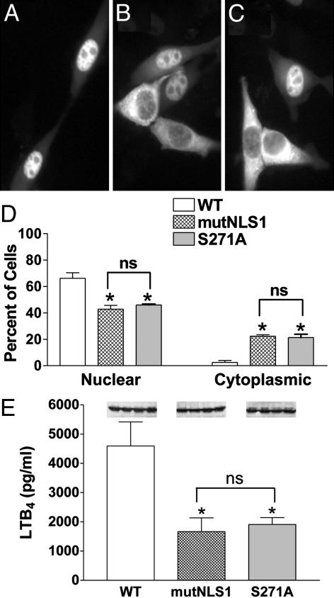
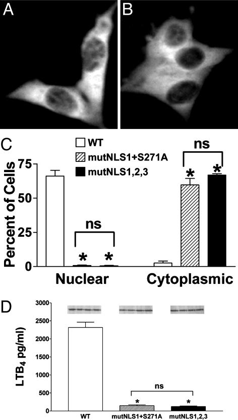
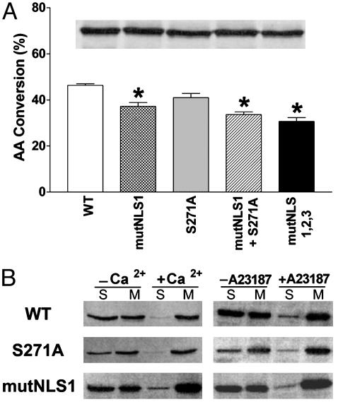
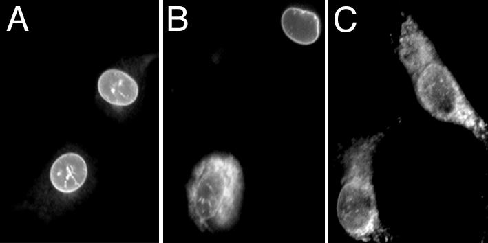
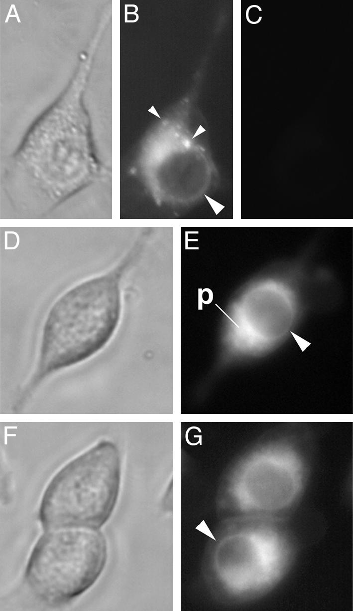
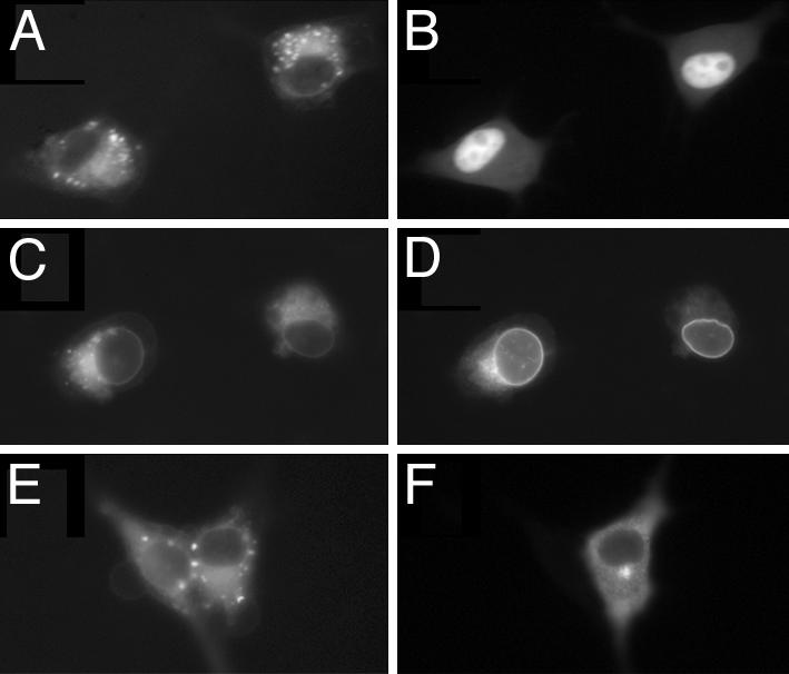
The enzyme 5-lipoxygenase (5-LO) initiates the synthesis of leukotrienes from arachidonic acid. In resting cells, 5-LO can accumulate in either the cytoplasm or the nucleoplasm and, upon cell stimulation, translocates to membranes to initiate leukotriene synthesis. Here, we used mutants of 5-LO with altered subcellular localization to assess the role that nuclear positioning plays in determining leukotriene B4 (LTB4) synthesis. Mutation of either a nuclear localization sequence or a phosphorylation site reduced LTB4 synthesis by 60%, in parallel with reduced nuclear localization of 5-LO. Mutation of both sites together or mutation of all three nuclear localization sequences on 5-LO inhibited LTB4 synthesis by 90% and abolished nuclear localization. Reduced LTB4 generation in mutants could not be attributed to differences in 5-LO amount, enzymatic activity, or membrane association. Instead, 5-LO within the nucleus acts at a different site, the nuclear envelope, than does cytosolic 5-LO, which acts at cytoplasmic and perinuclear membranes. The significance of this difference was suggested by evidence that exogenously derived arachidonic acid colocalized with activated nuclear 5-LO. These results unequivocally demonstrate that the positioning of 5-LO within the nucleus of resting cells is a powerful determinant of the capacity to generate LTB4 upon subsequent activation.


Figures

Fig. 1.
Fig. 1.
Different mutations that reduce nuclear localization of 5-LO also reduce LTB4 synthesis. 3T3 cells were transfected with WT, mutNLS1, or S271A 5-LO/GFP and, after 16 h, evaluated for 5-LO localization and LTB4 synthesis. Representative fields show WT 5-LO/GFP (A), mutNLS1 5-LO/GFP (B), or S271A 5-LO/GFP (C) from one experiment and are representative of 10 experiments. (D) Quantitative analysis of transfected populations of cells expressing WT, mutNLS1, or S271A 5-LO/GFP, with localization determined as described in Experimental Procedures. Data are means ± SE of n = 4 independent transfections per construct (*, P < 0.05 vs. WT; ns, not statistically different). (E) LTB4 synthesis. Transfected cells were stimulated with 10 μM A23187 and 10 μMAA at 37°C for 15 min, and LTB4 in conditioned media was measured by ELISA. Data are means ± SE (n = 4) (*, P < 0.05 vs. WT; ns, not statistically different). (Inset) Protein expression of the examined constructs assessed by immunoblotting. Results are from one experiment and are representative of three.
Fig. 2.
Fig. 2.
Different combinations of mutations that eliminate nuclear localization of 5-LO also strongly reduce LTB4 synthesis. 3T3 cells were transfected with WT, mutNLS1+S271A, or mutNLS1,2,3 5-LO/GFP and, after 16 h, evaluated for 5-LO localization and LTB4 synthesis. Representative fields show mutNLS1+S271A 5-LO/GFP (A) or mutNLS1,2,3 5-LO/GFP (B) from one experiment and are representative of eight experiments. (C) Quantitative analysis of transfected populations of cells expressing WT, mutNLS1+S271A, or mutNLS1,2,3 5-LO/GFP, with localization determined as described in Experimental Procedures. Data are means ± SE of n = 4 independent transfections per construct (*, P < 0.05 vs. WT; ns, not statistically different). (D) LTB4 synthesis. Transfected cells were stimulated with 10 μM A23187 and 10 μMAA at 37°C for 15 min, and LTB4 in conditioned media was measured by ELISA. Data are means ± SE (n = 4) (*, P < 0.05 vs. WT; ns, not statistically different). (Inset) Protein expression of the examined constructs assessed by immunoblotting. Results are from one experiment and are representative of three.
Fig. 3.
Fig. 3.
Effect of mutations of 5-LO/GFP on enzymatic activity and membrane association. (A) Cell-free enzymatic activity. Cell lysates from 3T3 cells expressing the different 5-LO/GFP constructs were tested for their capacity to convert AA to 5-hydroperoxyeicosatetraenoic acid (5-HPETE)/5-hydroxyeicosatetraenoic acid (5-HETE), using comparable amounts of 5-LO protein (indicated by immunoblot evaluation, Inset). Data are means ± SE of n = 3 independent transfections per construct (*, P < 0.05 vs. WT). (B)Ca2+-dependent membrane translocation of expressed 5-LO/GFP proteins both in vitro and in intact cells. 3T3 cells expressing WT, S271A, or mutNLS1 5-LO/GFP were fractionated with or without Ca2+ or stimulated with or without 10 μM A23187 (20 min, 37°C) before fractionation. Cells were fractionated into soluble (S) and microsomal (M) fractions and were examined by immunoblotting using antibodies against GFP based on equal loading (10 μg per well) of total protein.
Fig. 4.
Fig. 4.
Nuclear and cytosolic 5-LO translocate to different subcellular sites after activation. Transfected cells were stimulated with 10 μM A23187 (15 min, 37°C) and fluorescently imaged. Representative fields of cells expressing activated WT 5-LO/GFP (A), S271A 5-LO/GFP (B), and mutNLS1+S271A 5-LO/GFP (C) are shown.
Fig. 5.
Fig. 5.
Exogenous AA accumulates at the perinuclear and nuclear membranes. AA/PDAM (10 μM) was added to 3T3 cells, and cells were imaged by phase contrast (A, D, and F) and fluorescent (B, E, and G) microscopy after 15 min. Time-matched fluorescent imaging of cells before addition of AA/PDAM (C) indicated minimal autofluorescence. Large arrowheads (B, E, and G) indicate nuclear envelope; small arrowheads (B) indicate punctate fluorescence suggestive of lipid bodies. p, perinuclear membranes. Sets of images are from independent experiments.
Fig. 6.
Fig. 6.
Exogenous AA colocalizes strongly with activated WT 5-LO/GFP. Cells were transfected with WT or (mutNLS1+S271A) 5-LO/GFP; AA/PDAM was added before (A and B) or after (C–F) cell stimulation with A23187. (A) AA localization in cells expressing WT 5-LO/GFP before activation. (B) 5-LO/GFP localization in cells shown in A.(C) AA localization in cells expressing WT 5-LO/GFP and activated before AA addition. (D) 5-LO/GFP localization in cells shown in C. (E) AA localization in cells expressing (mutNLS1+S271A) 5-LO/GFP and activated before AA addition. (F) 5-LO/GFP localization in cells shown in E.

References

    1. Funk, C. D. (2001) Science 294, 1871–1875. - PubMed
    1. Bailie, M., Standiford, T., Laichalk, L., Coffey, M., Strieter, R. & Peters-Golden, M. (1996) J. Immunol. 157, 5221–5224. - PubMed
    1. Chen, N., Restivo, A. & Reiss, C. S. (2001) J. Neuroimmunol. 120, 94–102. - PubMed
    1. Chen, X., Sheller, J., Johnson, E. & Funk, C. (1994) Nature 372, 179–182. - PubMed
    1. Goulet, J., Snouwaert, J., Latour, A., Coffman, T. & Koller, B. (1994) Proc. Natl. Acad. Sci. USA 91, 12852–12856. - PMC - PubMed

Publication types

MeSH terms

LinkOut - more resources