. 2003 Oct 14;100(21):12384-9.
doi: 10.1073/pnas.2135336100. Epub 2003 Oct 6.

Y box-binding protein 1 induces resistance to oncogenic transformation by the phosphatidylinositol 3-kinase pathway

Affiliations

Y box-binding protein 1 induces resistance to oncogenic transformation by the phosphatidylinositol 3-kinase pathway

Andreas G Bader et al. Proc Natl Acad Sci U S A. .

Abstract

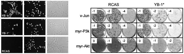
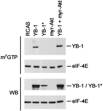
Y box-binding protein 1 (YB-1) is a multifunctional protein that can act as a regulator of transcription and of translation. In chicken embryo fibroblasts transformed by the oncoproteins P3k (phosphatidylinositol 3-kinase) or Akt, YB-1 is transcriptionally down-regulated. Expression of YB-1 from a retroviral vector induces a strong cellular resistance to transformation by P3k or Akt but does not affect sensitivity to transformation by other oncoproteins, such as Src, Jun, or Qin. The YB-1-expressing cells assume a tightly adherent, flat phenotype, with YB-1 localized in the cytoplasm, and show a greatly reduced saturation density. Both cap-dependent and cap-independent translation is inhibited in these cells, but the activity of Akt remains unaffected, suggesting that YB-1 functions downstream of Akt. A YB-1 protein with a loss-of-function mutation in the RNA-binding motif no longer binds to the mRNA cap structure, is localized in the cell nucleus, does not induce the flat cellular phenotype, and fails to interfere with P3k- or Akt-induced oncogenic transformation. This mutant also does not inhibit cap-dependent or cap-independent translation. These results suggest that YB-1 acts like a rapamycin mimic, inhibiting translational events that are required in phosphatidylinositol 3-kinase-driven oncogenic transformation.


Figures

Fig. 1.
Fig. 1.
Transcriptional expression of YB-1 in CEF transformed by various oncoproteins. Real-time PCR measuring YB-1 mRNA levels was performed as described in Materials and Methods. YB-1 mRNA expression in RCAS-transfected cells was designated 1.0.
Fig. 2.
Fig. 2.
Morphology and growth of YB-1-expressing cells. (A) A micrograph of YB-1-expressing CEF is shown (phase-contrast optics, ×6.3 objective lens). (B) Proliferation of YB-1-expressing cells. CEF were stably transfected with RCAS-YB-1 (▪) or with RCAS (▴) and cultured for 10 days. Then, equal numbers of cells were seeded onto 35-mm wells. The next day (day 0), and on days as indicated, cells were counted. The results represent the mean of three independent experiments; error bars mark the standard deviation.
Fig. 3.
Fig. 3.
YB-1 mediated interference with oncogenic transformation. CEF were transfected with RCAS(B)-YB-1 and RCAS(B) and cultured for 10 days. Then, equal numbers of cells were seeded onto 35-mm wells and infected with oncogenic viruses carrying subgroup A envelope proteins as indicated. The log10 of the virus dilution is shown in the corner of each well. Cells were fed with agar medium every 2 days and stained with 2% crystal violet after 13 (v-Src) and 17 (v-Jun, myr-P3k, and myr-Akt) days, respectively.
Fig. 4.
Fig. 4.
Phosphorylation of Akt targets in YB-1-expressing cells. Protein extracts were taken from actively dividing CEF transfected with RCAS expressing YB-1 or myr-Akt inserts, and from CEF transfected with RCAS-YB-1 plus RCAS-myr-Akt. Lysates were probed with phosphospecific antibodies directed against proteins as shown. Overexpression of myr-Akt and YB-1 proteins was confirmed by using hemagglutinin- and FLAG-specific antibodies, respectively. Tubulin served as loading control.
Fig. 5.
Fig. 5.
YB-1* with a mutated RNA-binding motif. (Left) CEF expressing either FLAG-tagged YB-1 or YB-1* proteins were fixed in 3.7% formaldehyde and treated with anti-FLAG and FITC-conjugated secondary antibody as shown on the left. 4′,6-Diamidino-2-phenylindole (DAPI)-stained nuclei and phase-contrast images are shown in the center and right, respectively. RCAS-transfected cells were used as a negative control. (Right) YB-1* in a transformation interference assay. Equal numbers of CEF transfected with RCAS-YB-1* and vector only were seeded onto 35-mm wells and challenged with transforming viruses encoding v-Jun, myr-Akt, and myr-P3k. Numbers in the corner of each well indicate the log10 of viral dilutions.
Fig. 6.
Fig. 6.
Cap-binding activity of YB-1. Lysates of CEF expressing the indicated proteins and CEF transfected with RCAS only were analyzed in a m7GTP pull-down assay. Bound FLAG-YB-1 and 4E proteins were detected by Western blotting. A Western blot (WB) analysis using 40 μg of total protein was carried out to confirm the expression of nonbound proteins. eIF, eukaryotic initiation factor.
Fig. 7.
Fig. 7.
YB-1 inhibits cap-dependent and cap-independent translation. CEF, expressing proteins encoded by RCAS vectors as indicated, were transiently transfected with pHCV-IRES containing the bicistronic reporter CAT/luc. Luciferase activity reflects internal ribosome entry site-dependent translation (formula image
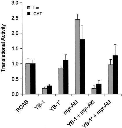
); CAT activity represents cap-dependent translation (▪). Experiments were carried out three times; standard deviations are shown as error bars. The translational activity of cells transfected with the empty vector was designated 1.0.

References

    1. Bellacosa, A., Testa, J. R., Staal, S. P. & Tsichlis, P. N. (1991) Science 254, 274-277. - PubMed
    1. Chang, H. W., Aoki, M., Fruman, D., Auger, K. R., Bellacosa, A., Tsichlis, P. N., Cantley, L. C., Roberts, T. M. & Vogt, P. K. (1997) Science 276, 1848-1850. - PubMed
    1. Alessi, D. R. & Downes, C. P. (1998) Biochim. Biophys. Acta 1436, 151-164. - PubMed
    1. Wymann, M. P. & Pirola, L. (1998) Biochim. Biophys. Acta 1436, 127-150. - PubMed
    1. Scheid, M. P. & Woodgett, J. R. (2001) Nat. Rev. Mol. Cell Biol. 2, 760-768. - PubMed

Publication types

MeSH terms