. 2003 Oct 15;31(20):e125.
doi: 10.1093/nar/gng125.

Sequence-specific fluorescent labeling of double-stranded DNA observed at the single molecule level

Affiliations

Sequence-specific fluorescent labeling of double-stranded DNA observed at the single molecule level

Bénédicte Géron-Landre et al. Nucleic Acids Res. .

Abstract

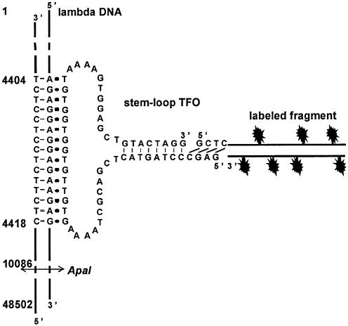
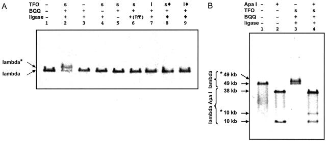
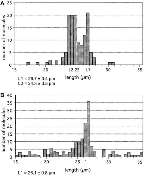
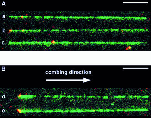
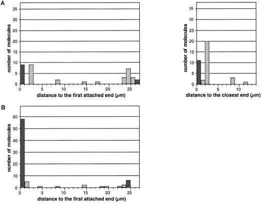
Fluorescent labeling of a short sequence of double-stranded DNA (dsDNA) was achieved by ligating a labeled dsDNA fragment to a stem-loop triplex forming oligonucleotide (TFO). After the TFO has wound around the target sequence by ligand-induced triple helix formation, its extremities hybridize to each other, leaving a dangling single-stranded sequence, which is then ligated to a fluorescent dsDNA fragment using T4 DNA ligase. A non-repeated 15 bp sequence present on lambda DNA was labeled and visualized by fluorescence microscopy after DNA combing. The label was found to be attached at a specific position located at 4.2 +/- 0.5 kb from one end of the molecule, in agreement with the location of the target sequence for triple helix formation (4.4 kb from one end). In addition, an alternative combing process was noticed in which a DNA molecule becomes attached to the combing slide from the label rather than from one of its ends. The method described herein provides a new tool for the detection of very short sequences of dsDNA and offers various perspectives in the micromanipulation of single DNA molecules.


Figures

Figure 1
Figure 1
Description of the labeling strategy used in this study. The sequence of the 59mer stem–loop TFO is shown. The 59mer oligonucleotide can form a triple helix by binding to a 15 bp oligopurineoligopyrimidine target sequence, located between 4404 and 4418 bp from the extremity of the phage. A fluorescently labeled fragment made by PCR is digested by a class IIS restriction enzyme in order to produce a sticky end that can be ligated to the 5′-end of the TFO. The restriction site for ApaI is indicated.
Figure 2
Figure 2
Attachment of the labeled fragment to its specific target site as revealed by pulsed-field gel electrophoresis. (A) Lambda DNA (previously treated in order to prevent concatemerization) was incubated with the stem–loop (s) or the linear (l) TFO and the DNA fragment in the presence of BQQ and the enzymatic ligation reaction was performed. The TFO was omitted in lanes 1 and 3. BQQ was omitted in lanes 1 and 4. The ligase was omitted in lanes 1 and 5. Sample shown in lane 6 was not heated before ligation. In lanes 8 and 9, the stem–loop (s, filled lozenge) or linear (l, filled lozenge) TFO had been ligated (filled lozenge) to the labeled fragment before performing the incubation with lambda DNA. (B) Lambda DNA loaded on lanes 1 and 3 was identical to that of lanes 1 and 2 in (A), respectively. These unlabeled and labeled phage molecules were digested with ApaI (lanes 2 and 4), which generates a 38 kb and a 10 kb fragment. Bands *49 kb and *10 kb correspond to the shifted (labeled) 49 kb lambda DNA and 10 kb fragment, respectively.
Figure 3
Figure 3
Analysis of the length of combed lambda DNA molecules. Lambda DNA molecules either labeled (A) or untreated (B) were combed on polystyrene coated slides. Images were captured and analysed. 135 (A) and 159 (B) molecules were measured.
Figure 4
Figure 4
Visualisation of labels on combed lambda molecules. DNA was stained with YOYO-1 (green). The labeled DNA fragments contained Alexa Fluor 546 (red); they therefore appear as yellow spots. The bars represent 5 µm. A montage of different patterns of combing and labeling is produced: (A) aligned longer (26–27.5 µm) molecules. The combed molecules were aligned so that the red internal labels were located on the left side, but they were equally distributed on both sides with respect to the combing direction (see text). (B) Aligned shorter molecules (23.5–25.5 µm). In this case, combed molecules were oriented with respect to the combing direction, i.e. the left side is the side where the DNA molecule sticks first to the glass surface, before being stretched in the direction indicated by the arrow.
Figure 5
Figure 5
Histograms presenting the position of labels on lambda DNA molecules. Distances were measured between the label and the end of the molecule that becomes attached first to the slide during the combing process. Data are shown for the first set of molecules (26.0–27.5 µm) (A, left) and for the second set (23.5–25.5 µm) (B). The labels located exactly at the extremity of the molecules are scored in black, whereas those located at an internal position are scored in grey. For the first set of molecules, the distance between the label and the closest end of the molecule is also presented (A, right).

References

    1. Tiner W.J. Sr, Potaman,V.N., Sinden,R.R. and Lyubchenko,Y.L. (2001) The structure of intramolecular triplex DNA: atomic force microscopy study. J. Mol. Biol., 314, 353–357. - PubMed
    1. Strick T., Allemand,J., Croquette,V. and Bensimon,D. (2000) Twisting and stretching single DNA molecules. Prog. Biophys. Mol. Biol., 74, 115–140. - PubMed
    1. Wang M.D., Schnitzer,M.J., Yin,H., Landick,R., Gelles,J. and Block,S.M. (1998) Force and velocity measured for single molecules of RNA polymerase. Science, 282, 902–907. - PubMed
    1. Harada Y., Funatsu,T., Murakami,K., Nonoyama,Y., Ishihama,A. and Yanagida,T. (1999) Single-molecule imaging of RNA polymerase–DNA interactions in real time. Biophys. J., 76, 709–715. - PMC - PubMed
    1. Strick T.R., Croquette,V. and Bensimon,D. (2000) Single-molecule analysis of DNA uncoiling by a type II topoisomerase. Nature, 404, 901–904. - PubMed

Publication types

Substances