. 2003 Oct;69(10):5941-9.
doi: 10.1128/AEM.69.10.5941-5949.2003.

Utilization of acyl-homoserine lactone quorum signals for growth by a soil pseudomonad and Pseudomonas aeruginosa PAO1

Affiliations

Utilization of acyl-homoserine lactone quorum signals for growth by a soil pseudomonad and Pseudomonas aeruginosa PAO1

Jean J Huang et al. Appl Environ Microbiol. 2003 Oct.

Abstract

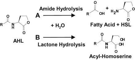
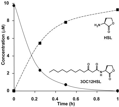
Acyl-homoserine lactones (AHLs) are employed by several Proteobacteria as quorum-sensing signals. Past studies have established that these compounds are subject to biochemical decay and can be used as growth nutrients. Here we describe the isolation of a soil bacterium, Pseudomonas strain PAI-A, that degrades 3-oxododecanoyl-homoserine lactone (3OC12HSL) and other long-acyl, but not short-acyl, AHLs as sole energy sources for growth. The small-subunit rRNA gene from strain PAI-A was 98.4% identical to that of Pseudomonas aeruginosa, but the soil isolate did not produce obvious pigments or AHLs or grow under denitrifying conditions or at 42 degrees C. The quorum-sensing bacterium P. aeruginosa, which produces both 3OC12HSL and C4HSL, was examined for the ability to utilize AHLs for growth. It did so with a specificity similar to that of strain PAI-A, i.e., degrading long-acyl but not short-acyl AHLs. In contrast to the growth observed with strain PAI-A, P. aeruginosa strain PAO1 growth on AHLs commenced only after extremely long lag phases. Liquid-chromatography-atmospheric pressure chemical ionization-mass spectrometry analyses indicate that strain PAO1 degrades long-acyl AHLs via an AHL acylase and a homoserine-generating HSL lactonase. A P. aeruginosa gene, pvdQ (PA2385), has previously been identified as being a homologue of the AHL acylase described as occurring in a Ralstonia species. Escherichia coli expressing pvdQ catalyzed the rapid inactivation of long-acyl AHLs and the release of HSL. P. aeruginosa engineered to constitutively express pvdQ did not accumulate its 3OC12HSL quorum signal when grown in rich media. However, pvdQ knockout mutants of P. aeruginosa were still able to grow by utilizing 3OC12HSL. To our knowledge, this is the first report of the degradation of AHLs by pseudomonads or other gamma-Proteobacteria, of AHL acylase activity in a quorum-sensing bacterium, of HSL lactonase activity in any bacterium, and of AHL degradation with specificity only towards AHLs with long side chains.


Figures

FIG. 1.
FIG. 1.
Two mechanisms by which AHLs can be inactivated. A, cleavage of the amide bond by bacterial AHL acylase yields HSL and the corresponding fatty acid (21, 23). The AHL amide bond is chemically stable under conditions of nonextreme temperature and pH. B, cleavage of the lactone ring by bacterial AHL lactonase yields the corresponding acyl-homoserine (7, 46). The lactone ring is also subject to chemical hydrolysis; the chemical half-life of the ring is ca. 10[7−pH] days, i.e., it is less stable with increased alkalinity. The acyl side chain diversity of known, naturally occurring AHLs has been reviewed (11).
FIG. 2.
FIG. 2.
rRNA-based phylogeny of strain PAI-A. Construction of the phylogram used 1,120 unambiguously aligned nucleotide positions in a 10,000-step Tree-Puzzle 5.0 maximum-likelihood analysis (36, 39). The bar represents evolutionary distance as 0.01 changes per nucleotide position, determined by measuring the lengths of the horizontal lines connecting the species. The numbers provide support for the robustness of the adjacent nodes. The arrow points to the short node from which the five strains within the shaded box radiate. See Materials and Methods for GenBank accession numbers.
FIG. 3.
FIG. 3.
LC/APCI-MS analysis of a cell-free fluid sampled from a P. aeruginosa culture utilizing C10HSL as a sole energy source in MES 5.5 medium. The details for resolving AHLs from their degradation products and other media components are described in Materials and Methods. (A) Chromatogram showing the separation of homoserine and/or HSL, MES buffer, and decanoyl-HSL (left axis). The hatch marks correspond to changes in methanol/water solvent ratios during the course of the run (right axis). (B) The mass spectrum of the first peak resolves homoserine from homoserine lactone; note that the peak tail can overlap with, but can be resolved from, the component in the second peak. (C) Mass spectrum of the second peak, morpholinoethane sulfonic acid (MES buffer). (D) Mass spectrum of the third peak, decanoyl-HSL. This method can be applied to separate and determine the concentrations of a number of other AHLs and any acyl-homoserine degradation products (not shown).
FIG. 4.
FIG. 4.
Growth of P. aeruginosa PAO1 in ammonia-replete MES 5.5 media containing 1 mM 3OC12HSL as the sole energy source. Substrate consumption and product accumulation were determined via LC/APCI-MS. Note that since the 3OC12HSL substrate was poorly soluble at the initial concentrations employed, virtually no AHL was observed in the culture fluid at the time of inoculation. As growth progressed, a transient spike of AHL in solution was observed. HSL accumulated throughout the growth phase but was degraded upon entry into stationary phase yielding a transient intermediate, homoserine. 3OC12-homoserine concentrations remained static throughout the course of the experiment and never exceeded 0.1% of the initial AHL concentration (not plotted). Culture pH was examined and found to be well controlled throughout.
FIG. 5.
FIG. 5.
E. coli cells expressing recombinant pvdQ degrade 3OC12HSL and generate stoichiometric amounts of HSL. Substrate disappearance and product accumulation were determined by using the LC/APCI-MS method (for an example, see Fig. 3.). Induced cells containing plasmid pPvdQ-PROTet were washed and suspended in MOPS (pH 7.2) buffered medium to a final OD600 of 1.2. AHL degradation and HSL accumulation were not observed over the duration of the experiment in either heat-killed suspensions of the same cells, or in no-cell controls (not shown).
FIG. 6.
FIG. 6.
Growth and accumulation of endogenous 3OC12HSL by P. aeruginosa PAO1 wild type (▴, ▵), and a recombinant derivative constitutively expressing pvdQ (▪, □). Because of the organic complexities of LB, sampled cell-free culture fluids were extracted with ethyl acetate before LC/APCI-MS analysis; the limit of detection for 3OC12HSL was 75 nM and was plotted in place of zero. Cultures were grown at 30°C in LB. Under similar culture conditions, a pvdQ knockout mutant grew and accumulated 3OC12HSL in parallel with the wild type (not shown).

Similar articles

Cited by

References

    1. Anzai, Y., H. Kim, J. Y. Park, H. Wakabayashi, and H. Oyaizu. 2000. Phylogenetic affiliation of the pseudomonads based on 16S rRNA sequence. Int. J. Syst. Evol. Microbiol. 50:1563-1589. - PubMed
    1. Anzai, Y., Y. Kudo, and H. Oyaizu. 1997. The phylogeny of the genera Chryseomonas, Flavimonas, and Pseudomonas supports synonymy of these three genera. Int. J. Syst. Bacteriol. 47:249-251. - PubMed
    1. Bennasar, A., R. Rossello-Mora, J. Lalucat, and E. R. Moore. 1996. 16S rRNA gene sequence analysis relative to genomovars of Pseudomonas stutzeri and proposal of Pseudomonas balearica sp. nov. Int. J. Syst. Bacteriol. 46:200-205. - PubMed
    1. Cronin, C. N., and W. S. McIntire. 1999. pUCP-Nco and pUCP-Nde: Escherichia-Pseudomonas shuttle vectors for recombinant protein expression in Pseudomonas. Anal. Biochem. 272:112-115. - PubMed
    1. Domenech Fernandez-Garayzabal, J. F., P. Lawson, J. A. Garcia, M. T. Cutuli, M. Blanco, A. Gibello, M. A. Moreno, M. D. Collins, and L. Dominguez. 1997. Winter disease outbreak in sea-bream (Sparus aurata) associated with Pseudomonas anguilliseptica infection. Aquaculture 156:317-326.

Publication types

MeSH terms

LinkOut - more resources