. 2003 Oct 15;22(20):5390-402.
doi: 10.1093/emboj/cdg521.

Interaction of fascin and protein kinase Calpha: a novel intersection in cell adhesion and motility

Affiliations

Interaction of fascin and protein kinase Calpha: a novel intersection in cell adhesion and motility

N Anilkumar et al. EMBO J. .

Abstract

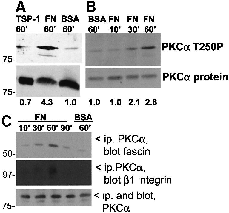
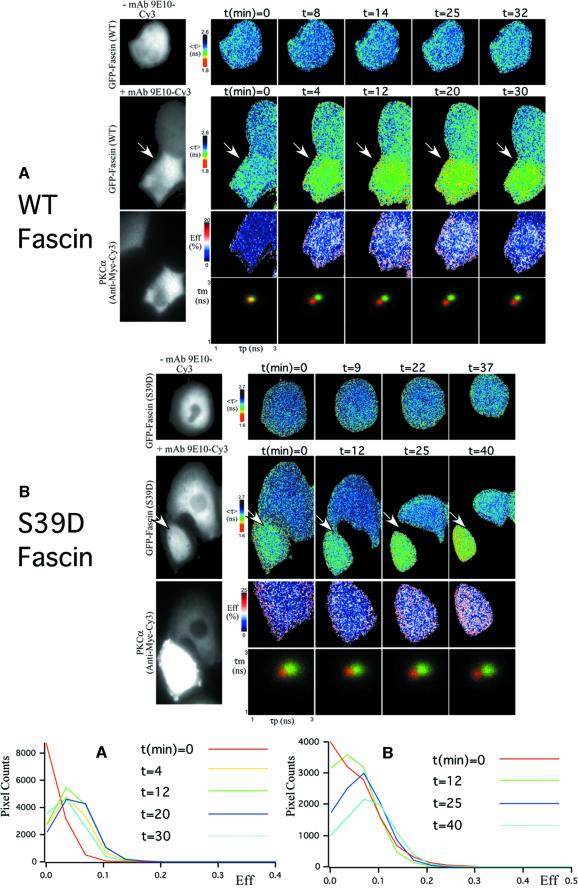
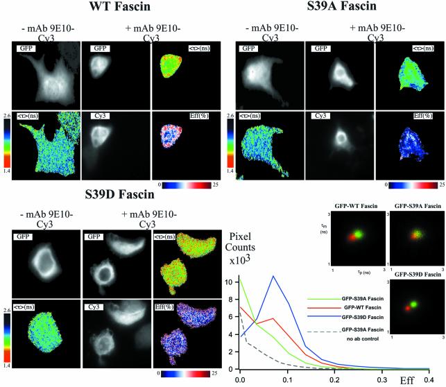
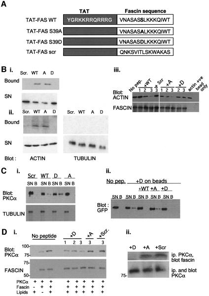
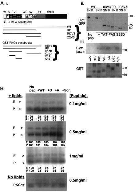
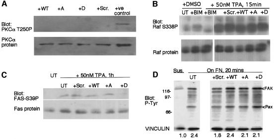
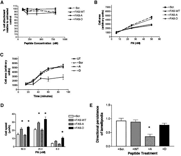
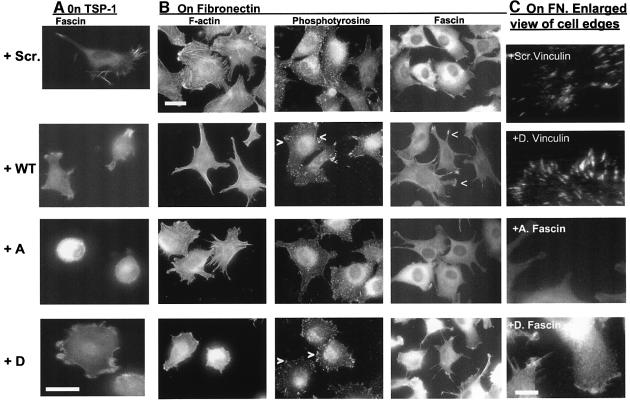
Coordination of protrusive and contractile cell-matrix contacts is important for cell adhesion and migration, but the mechanisms involved are not well understood. We report an unexpected direct association between fascin, an actin-bundling component of filopodia, microspikes and lamellipodial ribs, and protein kinase Calpha (PKCalpha), a regulator of focal adhesions. The association is detectable by protein-protein binding in vitro, by coimmunoprecipitation from cell extracts, and in live cells as fluorescence resonance energy transfer detected by fluorescence imaging lifetime microscopy. The interaction is physiologically regulated by the extracellular matrix context of cells, depends on activation of PKCalpha and is mediated by the C1B domain of PKCalpha. Strikingly, a fascin mutant, fascin S39D, associates constitutively with PKCalpha. Through use of a newly developed set of membrane-permeable peptides that separately inhibit either fascin/PKCalpha or fascin/actin binding, we have uncovered that specific blockade of the fascin/PKCalpha interaction increases cell migration on fibronectin in conjunction with increased fascin protrusions and remodeling of focal adhesions. These results identify the fascin-PKCalpha interaction as an important novel intersection in the regulation and networking of cell-matrix contacts.


Figures

None
Fig. 1. Activation of PKCα by ECM adhesion. (A) C2C12 cells were plated for 1 h on surfaces coated with BSA, 50 nM FN or 50 nM TSP-1. Western blots of whole-cell lysates were probed with antibodies to activated PKCα or PKCα total protein. The mean relative signal intensity, normalized against protein load by densitometric scanning, is shown below. (B) Time-course of PKCα activation in FN-adherent cells, determined by the same methods as in (A). (C) Time-course of association of fascin and β1 integrin with PKCα. Detergent-soluble fractions of C2C12 cells were immunoprecipitated for PKCα and associated fascin or β1 integrin detected by immunoblot. All data are representative of three independent experiments.
None
Fig. 2. Association of fascin and PKCα detected in live cells by FLIM/FRET techniques. (A) Real-time FLIM between EGFP–WT fascin and Myc-tagged PKCα. In the +mAb 9E10-Cy3 field of GFP–fascin-expressing cells, only one of the two was injected with a visible amount of Cy3-conjugated mAb 9E10 (arrowed). Changes in donor fluorescence lifetimes, <τ> (the average of τp and τm), were monitored with time in both control and antibody-injected cells following treatment with 400 nM TPA. The FRET efficiency (Eff) pseudocolour cell map (Eff = 1 – τdad, where τda is the lifetime map of the donor in the presence of acceptor and τd is the average lifetime <τ> of the donor in the corresponding control, uninjected cell at each time point) is shown for each individual sampling point. The cumulative lifetimes (of all the pixels) of GFP–fascin alone (green) and that measured in the presence of the acceptor fluorophore (red) are plotted on the τp versus τm 2D histograms for each time point. At t = 0, the donor lifetimes in antibody-injected cells were the same as those of control cells, giving rise to the yellow overlap area. After TPA treatment, there was a time-dependent shortening of GFP fluorescence lifetime according to both τp and τm in the antibody-injected cell. The bottom left-hand panel shows the pixel counts versus Eff (%) profiles (Eff histograms) for the full time-course. (B) A similar experiment to that in (A), performed using MCF-7 cells microinjected with GFP-tagged S39D fascin and Myc-tagged PKCα constructs. In the +mAb 9E10-Cy3 field of GFP–fascin-expressing cells, only one of the two was injected with a sufficient amount of Cy3-conjugated mAb 9E10 (arrowed). Eff histogram analysis (lower right-hand panel) was performed using lifetimes obtained from this cell.
None
Fig. 3. Influence of phosphoregulation of fascin on its association with PKCα. MCF-7 cells transiently coexpressing either GFP–WT fascin, GFP–fascin S39A or GFP–fascin S39D, and myc-tagged PKCα, for 36 h were left untreated or stimulated with 400 nM TPA for 15 min. Cells were fixed in 4% paraformaldehyde and either left as controls (–anti-myc-Cy3) or stained with Cy3-conjugated anti-myc antibody (+anti-myc-Cy3). The pixel counts versus Eff (%) profiles (bottom right-hand panel) summarize all the FRET efficiency data for each construct (measured at each pixel of the cell from at least five donor and acceptor-positive cells), as well as for unstained, cotranfected cells as a negative control.
None
Fig. 4. Biochemical properties of TAT-FAS peptides. (A) Design and amino acid sequences of TAT-FAS peptides. (B) Binding specificity of FAS-TAT peptides for actin. Pull-downs were performed with the different biotinylated TAT-FAS peptides bound to streptavidin–agarose as indicated. Candidate partner proteins were detected by immunoblot of SDS–PAGE gels. For each sample in (i) and (ii), the whole of the bound fraction and one-fifth of the supernatant (SN) was loaded per lane. (i) Binding to purified F-actin; (ii) binding specificity for actin in cell lysates; and (iii) competition of actin binding to purified fascin-His by free peptides. For each peptide: lane 1, 100 nM; lane 2, 300 nM; lane 3, 600 nM free peptide. Beads, Ni-NTA without fascin; actin +ve, cell extract. (C) Binding specificities of TAT-FAS peptides for PKCα. (i) Pull-down assays with TAT-FAS peptides bound to streptavidin–agarose as indicated were carried out on lysates of MCF-7 cells expressing GFP–PKCα-myc and immunoblotted for PKCα. Samples were reprobed for α-tubulin to demonstrate binding specificity. (ii) Cross-competition for PKCα binding to TAT–fascin S39D peptide–beads by free peptides. Cell extracts were preincubated with free TAT–fascin peptides as indicated for 1 h at 4°C before addition of TAT-FAS S39D–beads. For each sample, the whole of the bound fraction (B) and one-fifth of the supernatant (SN) was loaded per lane. (D) (i) Direct binding of fascin to active PKCα. Ni-NTA beads were left unloaded or loaded with 400 ng of purified recombinant fascin, washed, and incubated with 100 ng recombinant PKCα in the absence or presence of activating lipids and free TAT-FAS peptides as indicated. For each peptide: lane 1, 100 nM; lane 2, 300 nM; lane 3, 600 nM free peptide. (ii) Competition of fascin coimmunoprecipitation with PKCα by TAT-FAS S39D. C2C12 cells were preloaded with 300 nM TAT-FAS peptides as indicated, plated on 50 nM FN for 1 h, then immunoprecipitated for PKCα. Associated fascin was detected by immunoblot.
None
Fig. 5. Mapping the fascin-binding site on PKCα. (A) (i) Schematic diagram of the GFP-tagged or GST-tagged PKCα constructs. (ii) Pull-down of GFP–PKCα proteins, expressed in MCF-7 cells, by TAT-FAS–S39D peptide–beads. Western blots of bound proteins were probed with antibody to GFP. Only forms of PKCα containing the V1-PS-C1-V2 region (aa 1–289) bound to the peptide. (iii) Pull-down of fascin from C2C12 cells by GST–PKCα affinity matrices. The indicated purified GST–PKCα fusion proteins were bound to glutathione–agarose and incubated with C2C12 extracts. Western blots of bound proteins were probed with antibodies to fascin or to GST. Only proteins containing the C1B domain bound to fascin. (B) TAT-FAS peptides do not alter kinase activity of PKCα. In vitro kinase reactions were run for 30 min at 30°C with ezrin as exogenous substrate, in the absence or presence of activating lipids or TAT-FAS peptides as indicated. The reactions were resolved on 10% polyacrylamide gels and incorporation of [32P]ATP detected by autoradiography. Band intensities for ezrin (E) and PKCα (P) were quantified by scanning densitometry and were taken as 100% in the presence of lipids and the absence of peptides for each band. Band intensities in the presence of the peptides were expressed as a percentage of the control value and are shown under each lane. All results are representative of at least three independent experiments.
None
Fig. 6. Effects of TAT-FAS peptides on cell signaling pathways. (A) Thirty micrograms of extracts from adherent C2C12 cells, preloaded for 2 h with 200 nM peptides as indicated, were probed by immunoblot for the content of active (T250 phosphorylated) PKCα. Five hundred nanograms of purified, autophosphorylated PKCα was used as a positive control for T250P antibody reactivity. (B) C2C12 cells were left untreated (UT), treated with 50 nM bisindolylmaleimide (BIM), or stimulated with 50 nM TPA (TPA) for 15 min as indicated. Cells pretreated for 1 h with 200 nM TAT-FAS peptides as indicated were also stimulated with TPA. Thirty micrograms of cell extracts were probed by immunoblot for Raf S338P and reprobed for Raf-1 protein. Peptide loading without TPA did not affect Raf-1 phosphorylation (not shown). (C) Thirty micrograms of extracts from adherent C2C12 cells, untreated, or stimulated with 50 nM TPA for 1 h, with or without 200 nM TAT-FAS peptides as indicated, were probed by immunoblot for S39-phosphorylated fascin and reprobed for fascin protein. Cells treated with peptides in the absence of TPA resembled untreated controls (not shown). (D) C2C12 cells were placed in suspension for 2 h (Sus), left untreated (UT) or preloaded for 1 h with 200 nM peptide as indicated. Untreated and peptide-treated cells were plated on FN for 20 min in the continued presence of the peptides. Thirty micrograms of cell extracts were probed by immunoblot for phosphotyrosine or for vinculin as a loading control. Focal adhesion kinase (FAK) and paxillin (pax) were identified in separate experiments by immunoprecipitation with anti-phosphotyrosine followed by immunoblot with antibodies specific for FAK or paxillin. Relative signal intensities for FAK-ptyr, normalized against protein load, are listed below. All data are representative of three independent experiments.
None
Fig. 7. Effects of TAT-FAS peptides on FN-induced cell adhesion and migration. (A) Introduction of TAT-FAS peptides into C2C12 cells does not affect initial attachment to FN. Cells were preloaded with peptides at the indicated concentrations for 1 h and cell attachment quantitated after 1 h at 37°C on surfaces coated with 50 nM FN in the continued presence of peptides. Each point is the mean of three independent experiments; bars represent the SEM. (B) Effects of TAT-FAS peptides on cell area. C2C12 cells were preloaded with 200 nM peptides for 1 h and plated for 1 h at 37°C on surfaces coated with the indicated concentrations of FN in the continued presence of peptides, then fixed and imaged by phase-contrast microscopy. Cell perimeters were outlined morphometrically and areas calculated. Each point represents the mean of triplicate experiments (60–80 cells in total); bars indicate the SEM. (C) Effect of TAT-FAS peptides on cell spreading on FN. C2C12 cells were preloaded with 200 nM peptides for 1 h, plated onto 50 nM FN at 37°C for the indicated times in the continued presence of peptides, then fixed and stained for F-actin. Cell perimeters were outlined morphometrically and areas calculated. Each point represents the mean of triplicate experiments (80–100 cells in total); bars indicate the SEM. (D) Effects of TAT-FAS peptides on FN-induced cell migration. C2C12 cells were preloaded with 200 nM peptides for 1 h, plated for 1 h at 37°C on surfaces coated with the indicated concentrations of FN and imaged for 2 h by time-lapse phase-contrast microscopy in the continuing presence of peptides. Migration speeds were calculated by measuring the change in position of cell nuclei over time, with exclusion of mitotic cells. Each bar represents the mean of triplicate experiments (60–80 cells in total); bars indicate the SEM. Differences significant to at least P = 0.05 are indicated by an asterisk. (E) Effects of TAT-FAS peptides on lamellipodial persistence. Directional persistence of cell migration was calculated as the ratio of the number of changes in direction of lamellipodial extensions, divided by the total distance moved in the 2-h filming period. Each bar represents the mean of triplicate experiments (60–80 cells in total); bars indicate the SEM. Cells treated with TAT-FAS S39A had significantly decreased lamellipodial persistence (P <0.001).
None
Fig. 8. Effects of TAT-FAS peptides on F-actin organization and matrix contact structures. C2C12 cells were preloaded for 1 h with 200 nM peptides as indicated, plated onto 50 nM TSP-1 (A), or 50 nM FN (B and C), for 1 h in the continued presence of peptides and fixed and stained for (A) fascin, or (B) F-actin, fascin or phosphotyrosine. Bars, 10 µm. (C) Higher magnification views of cell edges. Bar, 5 µm. Images are representative of six independent experiments.

References

    1. Adams J.C. (1995) Formation of stable microspikes containing actin and the 55kDa actin-bundling protein, fascin, is a consequence of cell adhesion to thrombospondin-1: implications for the antiadhesive activities of thrombospondin-1. J. Cell Sci., 108, 1977–1990. - PubMed
    1. Adams J.C. (1997) Characterisation of cell–matrix adhesion requirements for the formation of fascin microspikes. Mol. Biol. Cell, 8, 2345–2363. - PMC - PubMed
    1. Adams J.C. (2001) Cell–matrix contact structures. Cell. Mol. Life Sci., 58, 371–392. - PMC - PubMed
    1. Adams J.C. and Lawler,J. (1994) Cell-type specific adhesive interactions of skeletal myoblasts with thrombospondin-1. Mol. Biol. Cell, 5, 423–437. - PMC - PubMed
    1. Adams J.C. and Schwartz,M.A. (2000) Stimulation of fascin spikes by thrombospondin-1 is mediated by the GTPases Rac and Cdc42. J. Cell Biol., 150, 807–822. - PMC - PubMed

Publication types

MeSH terms