. 2003 Oct 15;22(20):5412-21.
doi: 10.1093/emboj/cdg526.

Phosphatidylinositol-4,5-bisphosphate, PIP2, controls KCNQ1/KCNE1 voltage-gated potassium channels: a functional homology between voltage-gated and inward rectifier K+ channels

Affiliations

Phosphatidylinositol-4,5-bisphosphate, PIP2, controls KCNQ1/KCNE1 voltage-gated potassium channels: a functional homology between voltage-gated and inward rectifier K+ channels

G Loussouarn et al. EMBO J. .

Abstract

Phosphatidylinositol-4,5-bisphosphate (PIP(2)) is a major signaling molecule implicated in the regulation of various ion transporters and channels. Here we show that PIP(2) and intracellular MgATP control the activity of the KCNQ1/KCNE1 potassium channel complex. In excised patch-clamp recordings, the KCNQ1/KCNE1 current decreased spontaneously with time. This rundown was markedly slowed by cytosolic application of PIP(2) and fully prevented by application of PIP(2) plus MgATP. PIP(2)-dependent rundown was accompanied by acceleration in the current deactivation kinetics, whereas the MgATP-dependent rundown was not. Cytosolic application of PIP(2) slowed deactivation kinetics and also shifted the voltage dependency of the channel activation toward negative potentials. Complex changes in the current characteristics induced by membrane PIP(2) was fully restituted by a model originally elaborated for ATP-regulated two transmembrane-domain potassium channels. The model is consistent with stabilization by PIP(2) of KCNQ1/KCNE1 channels in the open state. Our data suggest a striking functional homology between a six transmembrane-domain voltage-gated channel and a two transmembrane-domain ATP-gated channel.


Figures

None
Fig. 1. KCNQ1/KCNE1 channel rundown is accelerated by divalent cations and slowed by PIP2. (A) Representative inside-out recording of KCNQ1/KCNE1 currents at various time after excision in a control solution (145 mM K-gluconate, 10 mM K-HEPES, 1 mM K-EGTA, pH 7.2). The membrane potential was stepped from a holding potential of –80 to +40 mV (1000 ms) and then back to –40 mV (500 ms), every 5 s. Zero current is indicated by a solid line. (B) Average time-dependent currents measured at the end of the depolarizing step relative to their maximum value and plotted against various times after patch excision. Patches were excised in control solution (open squares, n = 9), control solution plus 0.6 mM free Mg2+ (open circles, n = 6), control solution plus 1 mM free Mg2+ and Ca2+ (open triangles, n = 5) or control solution plus 5 µg/ml PIP2 (filled squares, n = 5). (C) Mean ± SEM values of τrundown determined from a monoexponential fit of rundowns in the different conditions presented in (B). *P < 0.001 compared with control. (D) Individual time-dependent currents measured at the end of the depolarizing step relative to their maximum value measured after patch excision. Three individual traces are presented, one with PIP2 application just before excision, and two with PIP2 application at 3 or 12 min after excision (arrows).
None
Fig. 2. PIP2-dependent, but not PIP2-independent, rundown is associated with an acceleration of the deactivation. (A and B) Representative normalized inside-out recordings of KCNQ1/KCNE1 currents at various time after excision in the absence (A) or presence (B) of PIP2 (5 µg/ml). Activating pulse currents measured at different time after excision were normalized to 1 at the end of the depolarizing step, in order to compare activation kinetics (left part of the curve). Deactivating tail currents at different time after excision were normalized to 1 at the beginning of the repolarizing step, so as to compare deactivation kinetics (right part of the curve). (C) Mean ± SEM values of τact determined from a fit of the activating current based on the Hodgkin and Huxley model for a voltage-dependent potassium channel activation (cf. Materials and methods). Black bars (0 µg/ml PIP2) and white bars (5 µg/ml PIP2) represent the average τact as a function of the remaining current during rundown (100, 75, 50 and 25% stand for the ranges 75–100%, 50–75%, 25–50% and 0–25%, respectively (n = 5–16). (D) Mean ± SEM values of τdeact measured from a monoexponential fit of the deactivating tail current. Black bars (0 µg/ml PIP2) and white bars (5 µg/ml PIP2) represent the average τdeact as a function of the remaining current during rundown (n = 4–16). *P < 0.01.
None
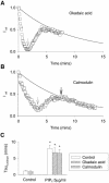
Fig. 3. PIP2-independent rundown is okadaic acid and calmodulin insensitive. (A) Representative PIP2-dependent and -independent rundowns of the KCNQ1/KCNE1 current in the presence of 300 nM okadaic acid. The KCNQ1/KCNE1 current is calculated as in Figure 1B and D. The arrow indicates PIP2 application to the cytosolic side of the patch. The solid line represents the average rundown in presence of PIP2 in control cells. (B) Representative PIP2-dependent and -independent rundown of the KCNQ1/KCNE1 current. The first arrow indicates PIP2 application to the cytosolic side of the patch, the second arrow indicates 500 nM CaM cytosolic application. The solid line represents the average rundown in presence of PIP2 in control cells. (C) Mean ± SEM values of τrundown from a monoexponential fit of rundowns in the different conditions presented in (A) and (B) (n = 5–9). *P < 0.001 compared to control.
None
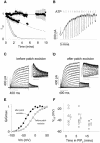
Fig. 4. The effects of PIP2 on the voltage-dependency of KCNQ1/KCNE1 currents. (A) Average time-dependent currents measured at the end of the depolarizing step relative to their maximum value measured after patch excision. Patches were excised in control solution (open circles, n = 9), control solution plus 1.4 mM MgATP (0.6 mM free Mg2+; filled circles, n = 3), control solution plus 1.4 mM MgATP plus 5 µg/ml PIP2 (filled squares, n = 9) and control solution plus 5 µg/ml PIP2 (dashed line, n = 5). Gaps correspond to acquisition interruptions for other protocols recordings. (B) Representative effects of PIP2 on Kir6.2/SUR1 channels expressed in a COS-7 cell excised patch. PIP2 was applied as indicated by the arrow. The solid line indicates zero current level. Two hundred micromolar cytosolic ATP repetitive applications are indicated by open boxes. (C and D) Superimposed KCNQ1/KCNE1 currents recorded when membrane potential was stepped, in 10-mV increments, from a holding potential of –80 mV to various potentials between –100 and +60 mV, and then stepped back to –40 mV. Voltage protocols were performed (C) before and (D) 10 min after patch excision in a MgATP + PIP2-containing solution. Insets, detail of the tail current at –40 mV. Horizontal bar 200 ms, vertical bar 100 pA. (E) Activation curves calculated from the tail current amplitude presented in (C) and (D) before (open circles) and after (filled circles) patch excision in a MgATP + PIP2-containing solution. (F) Potential for half-maximal activation (V0.5) calculated from activation curves such as presented in (E) before and at 3 and >5 min after patch excision. *P < 0.05 compared with value at t = 0.
None
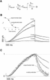
Fig. 5. A model based on the stabilization of the open state by PIP2 recapitulates the characteristics of the KCNQ1/KCNE1 currents. (A) The kinetic scheme presented here is based on previous models of Kir6.2/SUR1 channel regulation by PIP2. This simple model is based on the assumption that PIP2 does not affect the voltage sensor (kS4, k′S4), but only a closed state to an open state transition when the four voltage sensors are in the permissive state. Hence kS4, k′S4 and k′ are PIP2-independent and only kPIP2 varies during the simulated rundown. kS4= 3.56/s during activation and is negligible during deactivation; k′S4 = 7.47/s during deactivation and is negligible during activation; and k′ = 87.3/s. (B) Current traces from Figure 1A, to which the leak current was subtracted, were superimposed with the simulated current (solid lines). kPIP2 was fixed to 592.74, 176.43, 25.84 and 4.53/s to best fit the decrease in current amplitude during rundown, as shown in the inset. Inset: simulated (circles) and observed current (solid line) amplitudes as a function of time after patch excision. (C) Traces in (B) were normalized to compare the observed and simulated kinetics of activation and deactivation.
None
Fig. 6. Metabolic poisoning of cardiomyocytes leads to rundown of IKs. (A) The membrane potential was stepped from a holding potential of –40 to +40 mV (3 s) and then back to –40 mV (1 s), every 4.5 s. Zero current is indicated by a solid line. Cells were perfused permanently with glibenclamide (3 µM), E4031 (0.3 µM) and CoCl2 (1.8 mM) to inhibit IKATP, IKr and Ica,L, respectively. Addition of dinitrophenol (DNP; 10 µM) is followed by a decrease in IKs. Note the variation of the steady state current at –40 mV, suggesting that another channel may be affected by DNP as well, such as the inward rectifier IK1. (B) Normalization of the tail current of IKs shows no changes in deactivation kinetics.
None
Fig. 7. Alignment of the pore and M2/S6 domains of KCNQ1 with Kir6.2 showing the similar clustering of positive charges at the bottom of the putative pore-lining domains S6 (for KCNQ1) and M2 (for Kir6.2). The boxed histidine is aligned with the KCNQ2 histidine that decreases PIP2 affinity when neutralized (see text).

Similar articles

Cited by

References

    1. Barhanin J., Lesage,F., Guillemare,E., Fink,M., Lazdunski,M. and Romey,G. (1996) K(V)LQT1 and lsK (minK) proteins associate to form the I(Ks) cardiac potassium current. Nature, 384, 78–80. - PubMed
    1. Baró I. and Escande,D. (1989) A long lasting Ca2+-activated outward current in guinea-pig atrial myocytes. Pflugers Arch., 415, 63–71. - PubMed
    1. Baukrowitz T and Fakler,B. (2000) K(ATP) channels: linker between phospholipid metabolism and excitability. Biochem. Pharmacol., 60, 735–740. - PubMed
    1. Baukrowitz T., Schulte,U., Oliver,D., Herlitze,S., Krauter,T., Tucker,S.J. Ruppersberg,J.P. and Fakler,B. (1998) PIP2 and PIP as determinants for ATP inhibition of KATP channels. Science, 282, 1141–1144. - PubMed
    1. Becq F. (1996) Ionic channel rundown in excised membrane patches. Biochim. Biophys Acta, 1286, 53–63. - PubMed

Publication types

MeSH terms