. 2003 Oct 15;22(20):5446-58.
doi: 10.1093/emboj/cdg529.

CHIP activates HSF1 and confers protection against apoptosis and cellular stress

Affiliations

CHIP activates HSF1 and confers protection against apoptosis and cellular stress

Qian Dai et al. EMBO J. .

Abstract

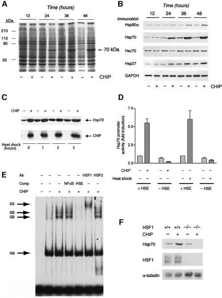
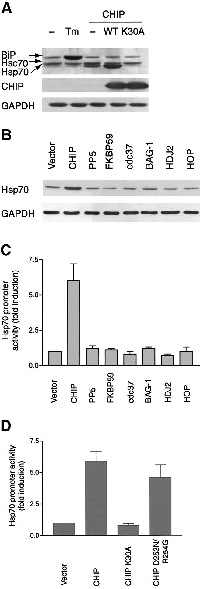
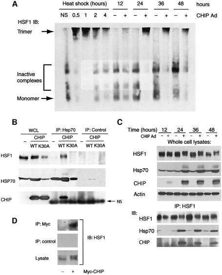
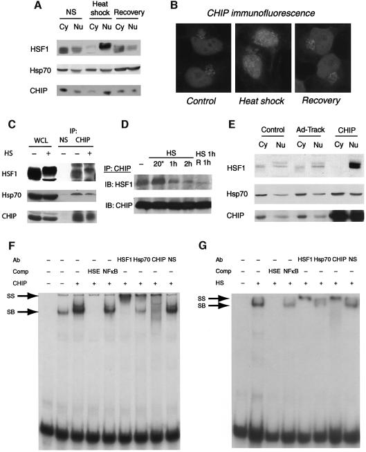
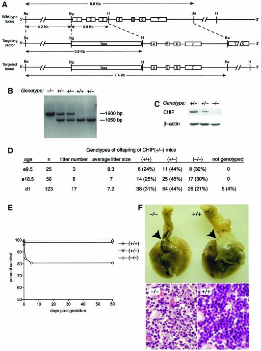
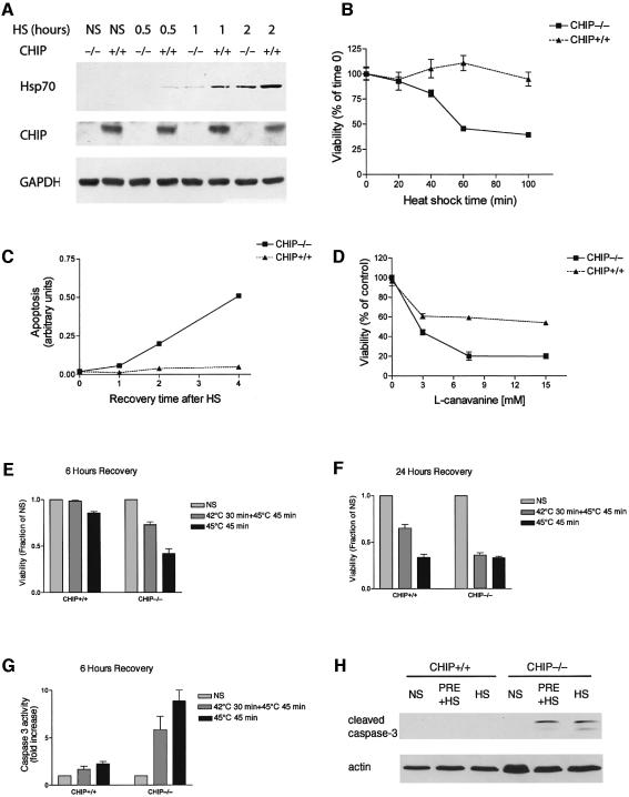
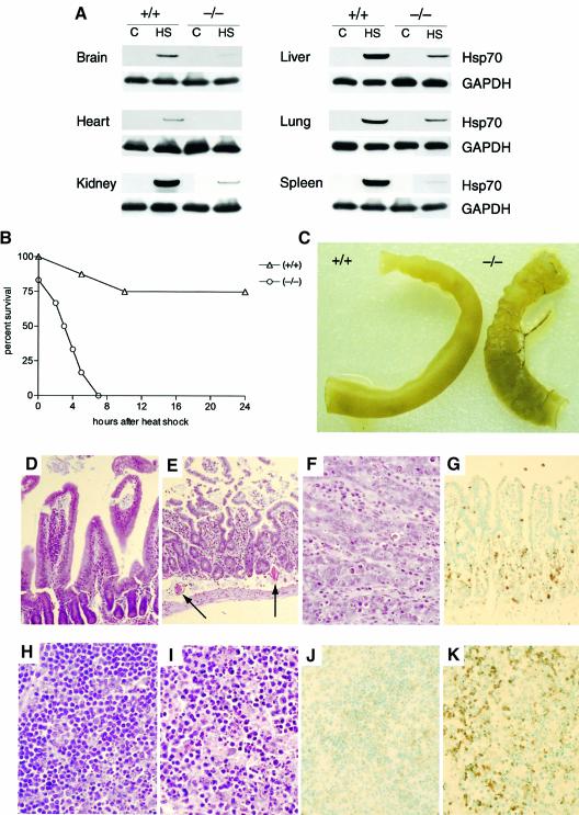
Induction of molecular chaperones is the characteristic protective response to environmental stress, and is regulated by a transcriptional program that depends on heat shock factor 1 (HSF1), which is normally under negative regulatory control by molecular chaperones Hsp70 and Hsp90. In metazoan species, the chaperone system also provides protection against apoptosis. We demonstrate that the dual function co-chaperone/ubiquitin ligase CHIP (C-terminus of Hsp70-interacting protein) regulates activation of the stress-chaperone response through induced trimerization and transcriptional activation of HSF1, and is required for protection against stress-induced apoptosis in murine fibroblasts. The consequences of this function are demonstrated by the phenotype of mice lacking CHIP, which develop normally but are temperature-sensitive and develop apoptosis in multiple organs after environmental challenge. CHIP exerts a central and unique role in tuning the response to stress at multiple levels by regulation of protein quality control and transcriptional activation of stress response signaling.


Figures

None
Fig. 1. Activation of HSF1 by CHIP. (A) Coomassie Blue gel of lysates from COS7 cells infected with an adenovirus expressing CHIP demonstrates increased expression of a 70-kDa protein compared with cells infected with a control adenovirus. (B) Western blot analysis indicates that expression of the chaperones Hsp27, Hsp90α and especially Hsp70 is increased by CHIP. (C) COS7 cells infected for 24 h with an adenovirus expressing CHIP or a control adenovirus were subjected to heat shock at 42°C for the indicated times. Hsp70 induction by CHIP was comparable to, and not additive with, that induced by heat shock. (D) Hsp70 promoter:reporter constructs containing (+HSE) or lacking (–HSE) the HSF1 response element were transiently co-transfected with CHIP expression vectors and reporter activity was determined 48 h after transfection, with or without heat shock (42°C). CHIP transactivates the Hsp70 promoter in an HSE-dependent fashion, as does heat shock. (E) HSF1 DNA binding activity to the HSE was assessed by gel shift assay. Expression of CHIP enhances binding of two specific bands (SB), which are competed away by unlabeled HSE but not by an NFκB consensus sequence. The binding activities can be supershifted (SS) with an HSF1, but not an HSF2, antibody. NS, nonspecific. (F) Upregulation of Hsp70 in response to CHIP 48 h after transfection was determined in wild-type or HSF1-null murine fibroblasts by western blot analysis.
None
Fig. 2. Activation of HSF1 is specific to CHIP and dependent on chaperone interactions. (A) Western blot analysis using an antibody that recognizes BiP, Hsc70 and Hsp70 demonstrates that only wild-type CHIP, but not CHIP K30A, increases Hsp70 expression after adenoviral infection, whereas BiP levels are increased by tunicamycin (Tm) but not by CHIP. (B and C) Only overexpression of CHIP, but not other co-chaperones [protein phosphatase 5 (PP5), FKBP59, cdc37, BAG-1, HDJ2 or HOP], increases Hsp70 protein expression (B) and promoter activity (C) after transient transfection. (D) Hsp70 promoter activity is increased by wild-type CHIP and by a mutant lacking ubiquitin ligase activity (CHIP D253N/R254G), but not by a mutant that does not interact with Hsp70/Hsp90 (CHIP K30A).
None
Fig. 3. CHIP-dependent HSF1:chaperone complexes. (A) HSF1 expression was examined by non-denaturing gel electrophoresis in COS7 cells after heat shock (42°C) or after infection with CHIP-expressing adenovirus (or a control adenovirus). Depletion of inactive complexes [which consist of dimers and Hsp70-bound monomers (Liu and Thiele, 1999; Guo et al., 2001)] and quantitative accumulation of the activated trimeric form of HSF1 occurs in CHIP-expressing cells. (B) COS7 cell lysates were prepared 24 h after infection with wild-type (WT) or CHIP or the K30A mutant (or a control adenovirus), and whole cell lysates (WCL) or immunoprecipitates (IP) were probed by western blotting for the presence of HSF1, Hsp70 and CHIP. (C) COS7 cells were infected with CHIP adenovirus as in (B) for the indicated times and whole cell lysates or HSF1 immunoprecipitates were probed for HSF1, Hsp70 or CHIP. Hsp70 is stably associated with activated HSF1 in CHIP-expressing cells and CHIP could be precipitated with Hsp70 in these HSF1 immunoprecipitates. (D) HSF1 co-immunoprecipitates with CHIP following transient transfection of Myc-tagged CHIP in COS7 cells.
None
Fig. 4. Nuclear transport and DNA binding of HSF1 induced by CHIP. (A) The subcellular localization of HSF1 and CHIP before and after heat shock (42°C for 30 min) or heat shock plus recovery for 1 h was determined by western blot analysis of nuclear (Nu) and cytoplasmic (Cy) fractions. (B) Immunocytochemical analysis of endogenous CHIP expression after heat shock (42°C for 30 min). The CHIP antibody stains red. (C) Association of endogenous CHIP and HSF1 in HeLa cells before and after heat shock (HS). WCL, whole cell lysate; NS, non-specific antibody. (D) Kinetics of association of HSF1 with CHIP after heat shock (HS) or heat shock plus recovery (R) for the indicated times. (E) The subcellular redistribution of HSF1 and Hsp70 was determined by western blot analysis of nuclear and cytoplasmic fractions of HeLa cells that were untreated (Control) or infected with a GFP-expressing adenovirus (Ad-Track) or with a virus expressing CHIP for 24 h. (F) Gel shift analysis of DNA-binding-competent HSF1 complexes with or without CHIP overexpression (24 h after infection) using HSE as a probe. Specific binding (SB) of complexes was determined by competition (Comp) with HSE or an NFκB binding site. DNA-bound HSF1 complexes were probed with antibodies to HSF1, Hsp70, CHIP or preimmune serum (NS). (G) Gel shift analysis of HSF1 complexes before or after heat shock (HS) for 30 min.
None
Fig. 5. Viability of CHIP (–/–) mice generated by homologous recombination. (ACHIP gene, targeting vector and disrupted allele The PGK–Neo cassette was inserted in place of a 0.8 kb BglII (BA)–HpaI (H) fragment, resulting in the deletion of the first three exons of CHIP. (B) Recombinant clones were initially identified by Southern blot analysis with 3′ external probes and F1 mice by Southern blot analysis with external probes or by PCR analysis for wild-type and mutant alleles. (C) Deletion of CHIP expression was confirmed by western blot analysis. (D) Tabular results of intercrosses of CHIP (+/–) parents. (E) Survival curves of live-born mice from CHIP (+/–) intercrosses indicate that a fraction of CHIP (–/–) mice die in the immediate peripartum period. (F) The thymus gland (arrows) overlying the heart is atrophied in organ preparations of deceased neonatal CHIP (–/–) mice, but not in wild-type mice (upper panels) or in surviving CHIP (–/–) mice (not shown). Histologic examination reveals hypocellularity and shrunken, pyknotic nuclei in atrophic thymuses from CHIP (–/–) mice, characteristic of thymocyte apoptosis (lower panels).
None
Fig. 6. Impaired stress response and increased apoptosis in CHIP-deficient fibroblasts. (A) Western blot analysis of CHIP (+/+) or CHIP (–/–) fibroblasts after heat shock (HS) at 42°C for the indicated times. Induction of Hsp70 by heat shock is attenuated in cells lacking CHIP. (B and C) The viability of CHIP (–/–) fibroblasts is markedly decreased in response to heat stress (45°C) for the indicated times (B). The decrease in viability is associated with increased accumulation of oligonucleosomes during recovery from heat shock (45°C for 45 min) for the indicated times as a marker of apoptosis (C). (DCHIP (–/–) cells are also sensitive to challenge with l-canavanine, an amino acid analog that causes accumulation of misfolded proteins. (E and F) Viability in CHIP (–/–) and CHIP (+/+) cells was determined after 45 min of heat shock (45°C) with or without preconditioning at 42°C for 30 min and 6 h (E) or 24 h (F) of recovery at 37°C, as indicated. CHIP (–/–) fibroblasts suffer decrements in viability in the presence or absence of preconditioning. NS, without heat stress. (G and H) Caspase 3 activity (G) and cleaved caspase 3 levels (H) were measured using a colorimetric assay after thermal challenge (HS) with or without thermal preconditioning (PRE), as indicated.
None
Fig. 7. Impaired stress response and stress-induced apoptosis in CHIP (–/–) mice. (A) Induction of Hsp70 in organs from CHIP (+/+) or (–/–) mice before (C) or 24 h after transient heat shock (HS) to 42°C. (B) Survival curves of CHIP (–/–) and CHIP (+/+) mice (n = 16) subjected to sustained core hyperthermia of 42°C for 15 min. No CHIP (–/–) mice recovered from hyperthermia. (C) Pathologic analysis of CHIP (–/–) mice after hyperthermia demonstrated friability and hyperemia of the small intestine. (DG). Histologic analysis of small intestinal mucosal of CHIP (+/+) (D) and CHIP (–/–) (E–G) mice after hyperthermic challenge. Normal villous structure is preserved in CHIP (+/+) mice (D), whereas atrophy of the terminal villi and thrombosis (arrows) are noted in CHIP (–/–) mice (E). Dense, shrunken nuclei are observed in villous tissues of CHIP (–/–) mice (F) that stain TUNEL-positive (G), indicating stress-induced apoptosis. (HK) Histologic analysis of splenic tissues from CHIP (+/+) (H and J) and CHIP (–/–) (I and K) mice after hyperthermic challenge. Hypocellularity and pyknosis of splenocytes from CHIP (–/–) mice (I) stain strongly TUNEL-positive (K), whereas the architecture and viability of splenocytes from CHIP (+/+) mice is preserved after hyperthermia (H and J).

References

    1. Aravind L. and Koonin,E.V. (2000) The U box is a modified RING finger—a common domain in ubiquitination. Curr. Biol., 10, R132–R134. - PubMed
    1. Ballinger C.A., Connell,P., Wu,Y., Hu,Z., Thompson,L.J., Yin,L.-Y. and Patterson,C. (1999) Identification of CHIP, a novel tetratricopeptide repeat-containing protein that interacts with heat shock proteins and negatively regulates chaperone functions. Mol. Cell. Biol., 19, 4535–4545. - PMC - PubMed
    1. Beere H.M. and Green,D.R. (2001) Stress management: heat shock protein-70 and the regulation of apoptosis. Trends Cell Biol., 11, 6–10. - PubMed
    1. Bercovich B., Stancovski,I., Mayer,A., Blumenfeld,N., Laszlo,A., Schwartz,A.L. and Ciechanover,A. (1997) Ubiquitin-dependent degradation of certain protein substrates in vitro requires the molecular chaperone Hsc70. J. Biol. Chem., 272, 9002–9010. - PubMed
    1. Bharadwaj S., Ali,A. and Ovsenek,N. (1999) Multiple components of the HSP90 chaperone complex function in regulation of heat shock factor 1 in vivo. Mol. Cell. Biol., 19, 8033–8041. - PMC - PubMed

Publication types

MeSH terms