. 2003 Oct 15;22(20):5471-9.
doi: 10.1093/emboj/cdg512.

Abl tyrosine kinases are required for infection by Shigella flexneri

Affiliations

Abl tyrosine kinases are required for infection by Shigella flexneri

Elizabeth A Burton et al. EMBO J. .

Erratum in

  • EMBO J. 2003 Nov 3;22(21):5962

Abstract

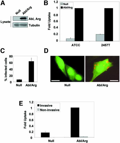
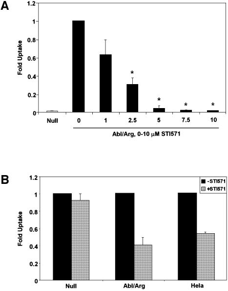
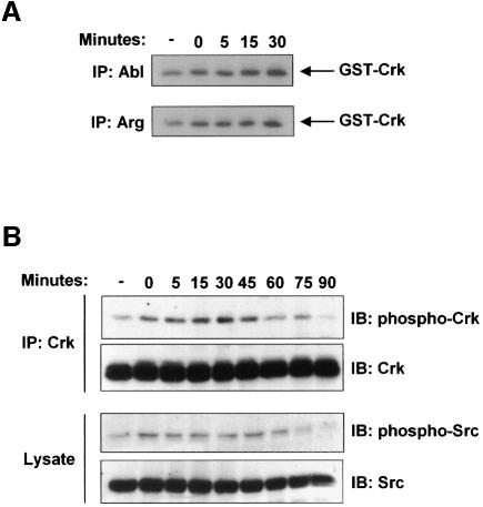
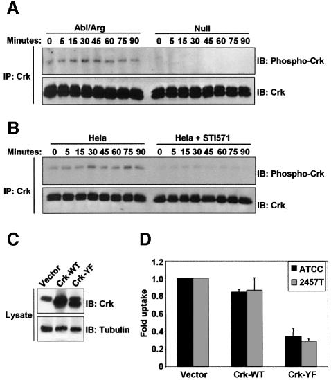
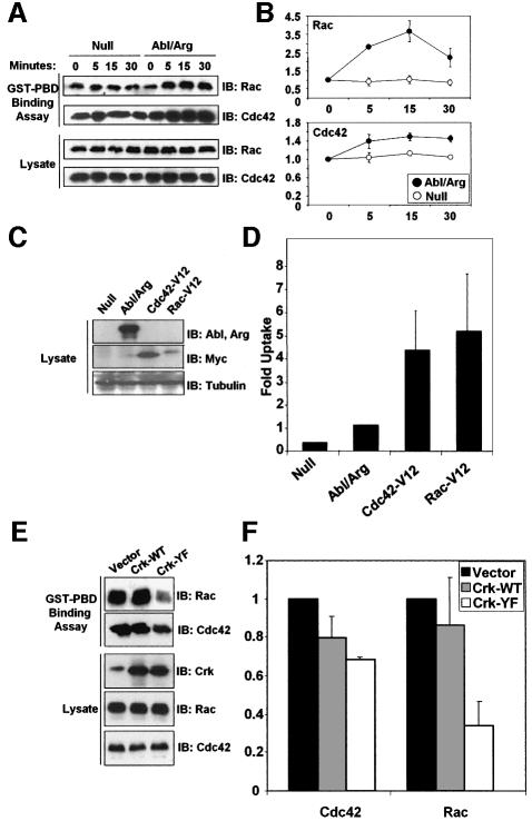
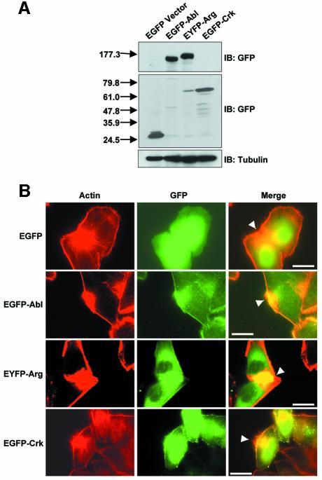
Infection by the opportunistic bacterial pathogen Shigella flexneri stimulates tyrosine phosphorylation of host cell proteins, but the kinases involved and their effects on the regulation of cell signaling pathways during bacterial entry remain largely undefined. Here, we demonstrate a requirement for the Abl family of tyrosine kinases during Shigella internalization. Family members Abl and Arg are catalytically activated upon Shigella infection, accumulate at the site of bacterial entry, and are required for efficient bacterial uptake, as internalization is blocked upon targeted deletion of these kinases or treatment with a specific pharmacological inhibitor. We identify the adapter protein Crk as a target for Abl kinases during Shigella uptake, and show that a phosphorylation-deficient Crk mutant significantly inhibits bacterial uptake. Moreover, we define a novel signaling pathway activated during Shigella entry that links Abl kinase phosphorylation of Crk to activation of the Rho family GTPases Rac and Cdc42. Together, these findings reveal a new role for the Abl kinases, and suggest a novel approach to treatment of Shigella infections through inhibition of host cell signaling pathways.


Figures

None
Fig. 1. Abl and Arg are required for Shigella internalization. (A) MEFs from mice lacking both Abl and Arg were reconstituted with either vector alone (Null) or with Abl and Arg expression constructs (Abl/Arg). The expression of Abl and Arg was confirmed by western blotting with anti-Abl 8E9, which recognizes the catalytic domain of both Abl and Arg. Anti-β-tubulin immunoblotting was used to demonstrate equal protein loading. (B) Null (gray bars) or Abl/Arg cells (black bars) were infected with S.flexneri strains ATCC® serotype 2a (ATCC) or 2457T, and bacterial uptake was measured by the gentamicin protection assay. Results shown correspond to three independent experiments, each performed in triplicate. (C) Null or Abl/Arg cells plated on coverslips were infected with S.flexneri 2457T, and incubated with gentamicin to eliminate extracellular bacteria. The percentage of infected cells was quantitated by immunofluorescence microscopy. (D) Null or Abl/Arg cells (both GFP-positive, due to stable expression of MIGR1 plasmids) plated on coverslips were infected with Shigella 2457T and incubated with gentamicin to eliminate extracellular bacteria. Cells were immunostained with anti-GFP (green) and anti-Shigella (red) antibodies, and visualized by immunofluorescence microscopy. Calibration bars = 50 µm. (E) Null or Abl/Arg cells were infected with either invasive (black bars) or plasmid-cured, non-invasive (gray bars) variants of S.flexneri 2457T, and bacterial uptake was measured by the gentamicin protection assay.
None
Fig. 2. Abl and Arg kinase activities are required for Shigella uptake. (A) MEFs lacking (Null, gray bar) or expressing Abl and Arg (Abl/Arg, black bars) were infected with S.flexneri ATCC® serotype 2a in the presence of 0–10 µM STI571, and bacterial uptake was measured by the gentamicin protection assay. The asterisks represent concentrations of STI571 at which the decrease in uptake is statistically significant (P < 0.05). (B) MEFs either lacking (Null) or re-expressing Abl and Arg (Abl/Arg) and HeLa cells were infected with S.flexneri 2457T in the absence (black bars) or presence (gray bars) of 10 µM STI571, and bacterial uptake was measured by the gentamicin protection assay. Results shown correspond to three independent experiments, each performed in triplicate, and are normalized with respect to the 0 µM STI571 treatment.
None
Fig. 3. Abl and Arg are catalytically activated during Shigella infection. (A) Abl and Arg were immunoprecipitated from lysates of NIH-3T3 cells that were either uninfected (-) or infected with S.flexneri 2457T for 0–30 min. The immunoprecipitates were used in an in vitro kinase assay, using GST–Crk as a substrate. (B) NIH-3T3 cells were infected with S.flexneri 2457T for 0–90 min. Anti-Crk immunoprecipitates were examined by immunoblotting with anti-phospho-Crk-Y221 or anti-Crk (upper panels). Total lysates were examined by immunoblotting with anti-phospho-Src-Y418 or anti-Src (lower panels).
None
Fig. 4. Phosphorylation of Crk by the Abl kinases is required for Shigella internalization. (A) Cells lacking (Null) or re-expressing Abl and Arg (Abl/Arg) were infected with S.flexneri 2457T for noted times. Anti-Crk immunoprecipitates were examined by immunoblotting with anti-phospho-Crk-Y221 (upper panel) and anti-Crk (lower panel). (B) HeLa cells were serum-starved for 3 h in the presence or absence of STI571, and infected for the indicated times with S.flexneri 2457T. Anti-Crk immunoprecipitates were examined by immunoblotting with anti-phospho-Crk-Y221 (upper panel) and anti-Crk (lower panel). (C) MIGR1 vector, Crk-WT, and Crk-Y222F were introduced into NIH-3T3 cells. Crk expression was analyzed by immunoblotting with anti-Crk (upper panel). Anti-β-tubulin immunoblotting was used to assess equal protein loading (lower panel). (D) NIH-3T3 cells expressing MIGR1 vector, Crk-WT, or Crk-Y222F were infected with S.flexneri strains ATCC® serotype 2a (black bars) or 2457T (gray bars), and bacterial uptake was measured by the gentamicin protection assay. Results shown correspond to three independent experiments, each performed in triplicate.
None
Fig. 5. The Abl kinases act upstream of activation of Cdc42 and Rac during Shigella infection. (A) Cells lacking (Null) or re-expressing Abl and Arg (Abl/Arg) were infected with S.flexneri 2457T for noted times. Lysates were incubated with GST–PBD to precipitate GTP-bound Cdc42 and Rac, and the bound proteins were analyzed by immunoblotting with anti-Rac and anti-Cdc42 (upper panels). Cellular lysates were examined by immunoblotting with anti-Rac and anti-Cdc42 (lower panels). (B) The GST–PBD binding assays to assess Rac (upper panel) or Cdc42 (lower panel) activation in the Null and Abl/Arg cells were quantitated by densitometry. Results correspond to three independent experiments. (C) Activated forms of Cdc42 and Rac (Cdc42-V12 and Rac-V12) were introduced into cells lacking Abl and Arg. Expression of Abl and Arg was analyzed by western blotting with anti-Abl 8E9 (upper panel). Expression of myc-tagged Cdc42-V12 and Rac-V12 was analyzed by immunoblotting with anti-myc (middle panel). Anti-β-tubulin immunoblotting was used to assess equal protein loading (lower panel). (D) Null, Abl/Arg and Null cells expressing Cdc42-V12, and Rac-V12 were infected with S.flexneri, and bacterial uptake was analyzed by the gentamicin protection assay. Results represent three independent experiments, each performed in triplicate. (E) NIH-3T3 cells expressing MIGR1 vector, Crk-WT or Crk-Y222F were infected with S.flexneri 2457T for 30 min, and analyzed for Cdc42 and Rac activities using the GST–PBD binding assay, as in (A). (F) The GST–PBD binding assays to assess Cdc42 or Rac activities in cells expressing Crk-WT or Crk-Y222F were quantitated by densitometry. Results correspond to three independent experiments.
None
Fig. 6. Abl, Arg, and Crk localize to the site of bacterial entry. (A) HeLa cells were transfected with EGFP vector, EGFP–Abl, EYFP–Arg or EGFP–Crk, as noted. Cells were lysed and analyzed for fusion protein expression by immunoblotting with anti-GFP (upper panels). Anti-β-tubulin immunoblotting was used to assess equal protein loading (lower panel). (B) HeLa cells expressing EGFP vector, EGFP–Abl, EYFP–Arg, or EGFP–Crk were infected with S.flexneri for 30 min, and analyzed by immunofluorescence microscopy. Sites of bacterial entry were identified by staining the bacteria with DAPI (data not shown) and staining the actin foci with rhodamine–phalloidin (red, left panels). Abl, Arg and Crk localization was performed by staining with anti-GFP (green, middle panels). These images were merged to show co-localization of Abl, Arg and Crk with the focus of actin at the site of bacterial entry (yellow, right panels). The sites of actin foci formation and co-localization with Abl, Arg and Crk are noted by the arrowheads. Calibration bar (shown in the right panel only) = 50 µm.

References

    1. Abassi Y.A. and Vuori,K. (2002) Tyrosine 221 in Crk regulates adhesion-dependent membrane localization of Crk and Rac and activation of Rac signaling. EMBO J., 21, 4571–4582. - PMC - PubMed
    1. Adam T., Arpin,M., Prevost,M.C., Gounon,P. and Sansonetti,P.J. (1995) Cytoskeletal rearrangements and the functional role of T-plastin during entry of Shigella flexneri into HeLa cells. J. Cell Biol., 129, 367–381. - PMC - PubMed
    1. Adam T., Giry,M., Boquet,P. and Sansonetti,P. (1996) Rho-dependent membrane folding causes Shigella entry into epithelial cells. EMBO J., 15, 3315–3321. - PMC - PubMed
    1. Bagrodia S., Taylor,S.J., Jordon,K.A., Van Aelst,L. and Cerione,R.A. (1998) A novel regulator of p21-activated kinases. J. Biol. Chem., 273, 23633–23636. - PubMed
    1. Bateman J., Shu,H. and Van Vactor,D. (2000) The guanine nucleotide exchange factor trio mediates axonal development in the Drosophila embryo. Neuron, 26, 93–106. - PubMed

Publication types