. 2003 Oct 15;22(20):5530-9.
doi: 10.1093/emboj/cdg534.

Genetic analysis of NF-kappaB/Rel transcription factors defines functional specificities

Affiliations

Genetic analysis of NF-kappaB/Rel transcription factors defines functional specificities

Alexander Hoffmann et al. EMBO J. .

Abstract

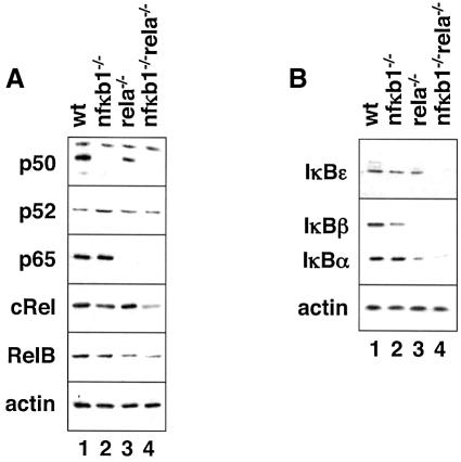
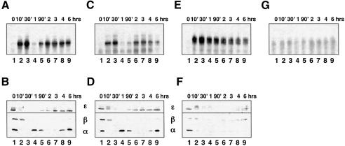
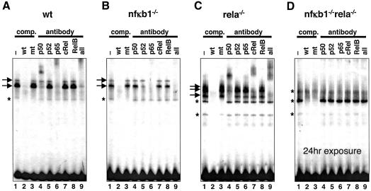
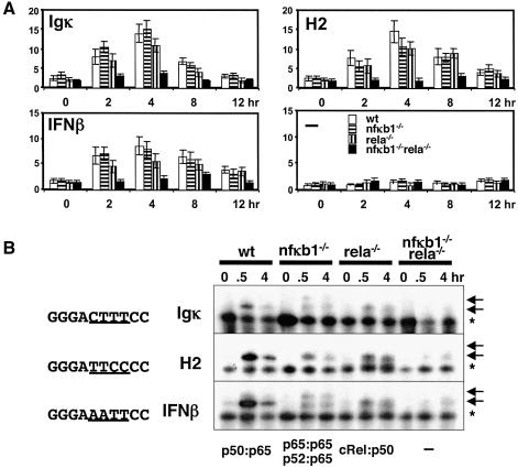
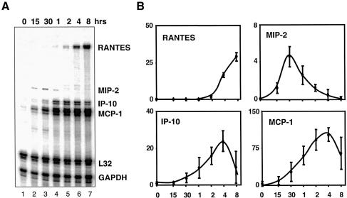
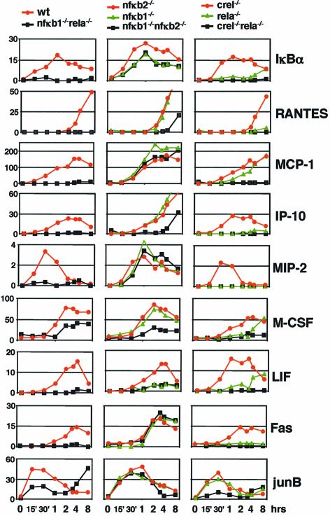
The NF-kappaB transcription factors consist of dimeric proteins of the Rel homology family. They activate many promoters containing highly divergent kappaB-site sequences. We have generated cell lines lacking individual and multiple NF-kappaB proteins and used them to establish interactions between components of the NF-kappaB-IkappaB signaling system. Functional compensation within the family of dimers was evident in knockout cell lines. Analysis of transiently transfected genes gave an impression of promiscuity that was not borne out by analysis of endogenous genes. Using TNFalpha as an inducer, a panel of endogenous genes showed a wide range of subunit specificities as well as highly variable kinetics of induction. Comparing the function and subunit specificity of genes with the sequence of the kappaB DNA-binding site we found little correlation, indicating that NF-kappaB family member specificity for endogenous promoters is not solely encoded by the kappaB site sequence itself.


Figures

None
Fig. 1. RHD proteins in NF-κB knockout cells. Western blots of cells derived from wild-type, nfκb1–/–, rela–/– and nfκb1–/–/rela–/– mouse embryos with antibodies directed against p50, p52, p65, cRel, RelB and actin (A), and IκBε, -β, -α and actin (B) as indicated. Specific bands are shown, but additional bands were detectable with some antibodies.
None
Fig. 2. TNFα induction of κB-binding activity in NF-κB knockout cells. EMSA was used to monitor nuclear NF-κB in wild-type (A), nfκb1–/– (C), rela–/– (E) and nfκb1–/–rela–/– (G) cells in the indicated (in min and h) time course following the onset of TNFα stimulation. The cytoplasmic portion from wild-type (B), nfκb1–/– (D), relA–/– (F) was probed in western blots to monitor concurrent degradation and synthesis of IκB proteins, IκBα and IκBβ (bottom panels) and IκBε (top panels).
None
Fig. 3. Molecular composition of κB-binding activity in NF-κB knockout cells. Immediate early protein–DNA complexes induced by TNFα in wild-type (A), nfκb1–/– (B), rela–/– (C) and nfκb1–/–rela–/– (D) cells and detected by EMSA in Figure 2 are indicated by arrows in lanes 1, while constitutive complexes are indicated by asterisks. They are specific for κB-sites as shown by competition with double-stranded wild-type and mutant oligonucleotides (lanes 2 and 3) and are probed with antibodies directed against the indicated RHD proteins (lanes 4–9). This results in ‘supershift’ or ablation of the characteristic protein–DNA complex. We conclude that the κB-binding activity in wild-type cells consists of p50:p65 heterodimer and p65 homodimer, in nfκb1–/– cells p52:p65 heterodimer and p65 homodimer, in rela–/– cells p50 homodimer, cRel:p50 heterodimer, and most likely, but not unambiguously cRel homodimer. While experiments shown in panels A, B and C were exposed to film for 6 h, panel D shows a 24 h exposure.
None
Fig. 4. Compensation on different κB-site sequences in extra-chromosomal plasmids. Reporter plasmids driven by the c-fos core promoter alone or fused to two κB-sites derived from the Igκ the MHC H2 and IFNβ promoter, as indicated in (A), were assayed in response to TNFα stimulation for the indicated time in wild-type (empty bars), nfκb1–/– (horizontally striped bars), rela–/– (vertically striped bars) and nfκb1–/–rela–/– (black bars). EMSA (B) was used to monitor κB-binding activity in TNFα-stimulated NF-κB knockout cells with indicated κB-site containing double-stranded oligonucleotide probes. Induced, specific bands are indicated by arrows, a prominent constitutive protein–DNA complex is indicated by an asterisk.
None
Fig. 5. Quantitative monitoring of gene expression by multiplex RPA. Multiple double-stranded RNA products indicative of mRNA transcripts derived from indicated NF-κB-dependent and housekeeping genes following TNFα stimulation of wild-type fibroblasts resolved by electrophoresis (A). Three independent experiments with independently derived wild-type 3T3 cell lines were quantitated by PhosphoImager and mRNA abundance was graphed for the indicated chemokine genes in arbitrary units relative to housekeeping genes L32 and GAPDH (B).
None
Fig. 6. Transcriptional induction by TNFα of NF-κB target genes in NF-κB knockout cells. Results from a representative RPA experiment are graphed for the indicated genes in three panels. The left panel shows data from wild-type (red line) and nfκb1–/–rela–/– cells (black line), the center panel from nfκb2–/– (red), nfκb1–/– (green) and nfκb1–/–nfκb2–/– (black), and the right panel from crel–/– (red), rela–/– (green), and rela–/–crel–/– (black) cells.

References

    1. Aaronson S.A. and Todaro,G.J. (1968) Development of 3T3-like lines from Balb-c mouse embryo cultures: transformation susceptibility to SV40. J. Cell Physiol., 72, 141–148. - PubMed
    1. Beg A.A., Sha,W.C., Bronson,R.T., Ghosh,S. and Baltimore,D. (1995) Embryonic lethality and liver degeneration in mice lacking the RelA component of NF-κB. Nature, 376, 167–170. - PubMed
    1. Berkowitz B., Huang,D.B., Chen-Park,F.E., Sigler,P.B. and Ghosh,G. (2002) The x-ray crystal structure of the NF-κB p50.p65 heterodimer bound to the interferon β–κB site. J. Biol. Chem., 277, 24694–24700. - PubMed
    1. Bren G.D., Solan,N.J., Miyoshi,H., Pennington,K.N., Pobst,L.J. and Paya,C.V. (2001) Transcription of the RelB gene is regulated by NF-κB. Oncogene, 20, 7722–7733. - PubMed
    1. Caamano J.H., Rizzo,C.A., Durham,S.K., Barton,D.S., Raventos-Suarez,C., Snapper,C.M. and Bravo,R. (1998) Nuclear factor (NF)-κB2 (p100/p52) is required for normal splenic microarchitecture and B cell-mediated immune responses. J. Exp. Med., 187, 185–196. - PMC - PubMed

Publication types