. 2003 Oct 15;22(20):5551-60.
doi: 10.1093/emboj/cdg516.

Monocytic cells hyperacetylate chromatin protein HMGB1 to redirect it towards secretion

Affiliations

Monocytic cells hyperacetylate chromatin protein HMGB1 to redirect it towards secretion

Tiziana Bonaldi et al. EMBO J. .

Abstract

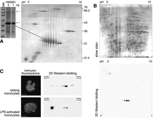
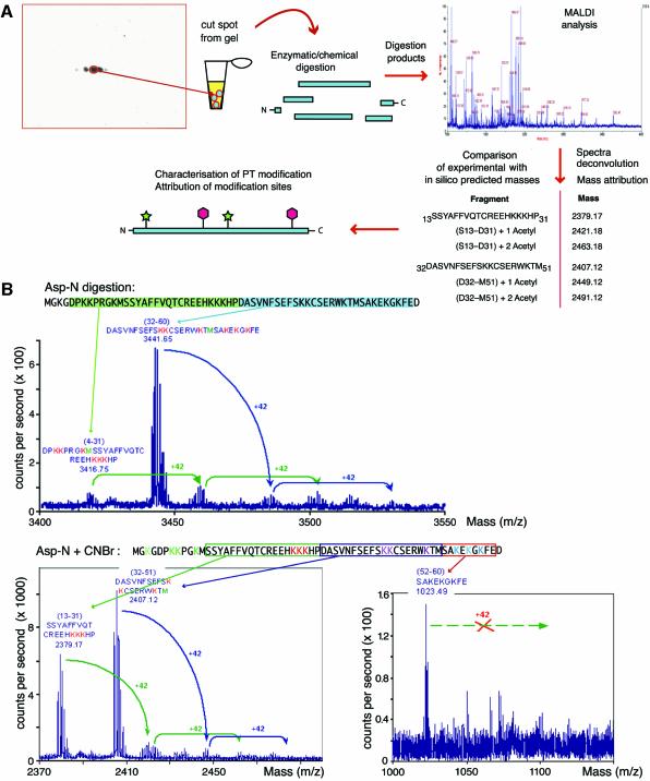
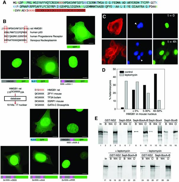
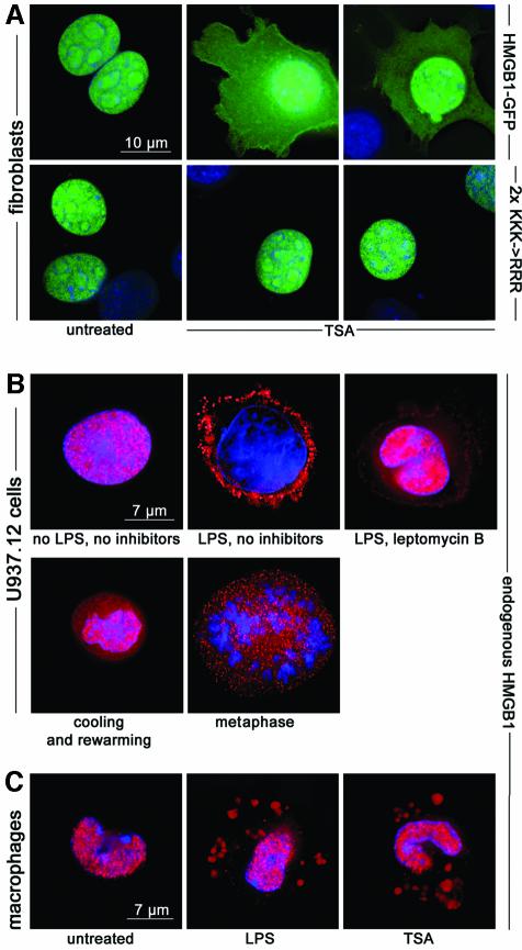
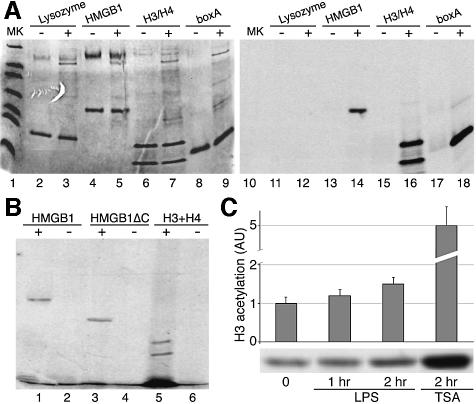
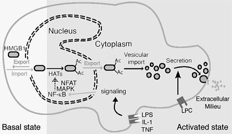
High Mobility Group 1 protein (HMGB1) is a chromatin component that, when leaked out by necrotic cells, triggers inflammation. HMGB1 can also be secreted by activated monocytes and macrophages, and functions as a late mediator of inflammation. Secretion of a nuclear protein requires a tightly controlled relocation program. We show here that in all cells HMGB1 shuttles actively between the nucleus and cytoplasm. Monocytes and macrophages acetylate HMGB1 extensively upon activation with lipopolysaccharide; moreover, forced hyperacetylation of HMGB1 in resting macrophages causes its relocalization to the cytosol. Cytosolic HMGB1 is then concentrated by default into secretory lysosomes, and secreted when monocytic cells receive an appropriate second signal.


Figures

None
Fig. 1. HMGB1 is multiply acetylated in thymus and activated monocytes. (A) HMGB1 purified from calf thymus gives rise on a monodimensional gel (SDS–PAGE, Coomassie stain, right panel) to two bands (arrows). The minor band contains ADP-ribosylated HMGB1. Fifty micrograms of the same sample of HMGB1 was subjected to 2D gel electrophoresis (silver stain, left panel); 2D Protein Marker from Bio-Rad was loaded together with HMGB1, generating a matrix of spots at the molecular weights listed on the right. (B) Total mouse thymus contains many isoforms of HMGB1. About 300 µg of protein from a thymus total extract (from a 17-day-old mouse embryo) were loaded on twin 2D gels; the gel shown at the top was silver stained, while the one on the bottom was blotted and assayed with anti-HMGB1 antibody. (C) LPS-activated human monocytes hyperacetylate HMGB1 and accumulate it in cytoplasmic vesicles. Monocytes purified from peripheral blood were cultured overnight, with or without LPS. Aliquots of activated and control monocytes were then fixed and immunostained with anti-HMGB1 antibody (red). HMGB1 is nuclear in unstimulated monocytes, as opposed to nuclear plus vesicular in LPS-activated monocytes. Bar represents 7 µm. Aliquots of untreated and LPS-activated monocytes were freeze-thawed, and about 400 µg of total protein extract was loaded onto 2D gels, blotted and immunodetected with anti-HMGB1. Note the major additional HMGB1 spot in activated monocytes.
None
Fig. 2. Strategy for 2D/MALDI-MS analysis of multiply modified HMGB1. (A) Spots were excised from 2D gels, proteolysed and analysed by MALDI-TOF. A mass was attributed to each peptide in the mixture; the masses corresponding to peptides predicted in silico were selected as ‘anchors’, and we searched the complex spectra for further peaks corresponding to anchor masses plus multiples of 42 (the molecular weight of an acetyl group). The analysis of two peptides is shown; the procedure was iterated for all peptides. We also mined the spectra for evidence of phosphorylation, methylation and glycosylation, without finding any. (B) An example of the multi-step digestion strategy developed to assign acetylation sites in HMGB1. Spots from 2D gels were digested in gel with protease Asp-N. One aliquot was analysed by MALDI-TOF: the arrows identify the peaks corresponding to unmodified fragment. We then looked for peaks with the molecular weight of unmodified fragments plus multiples of 42. The maximum number of acetyl moieties on each fragment was thus determined. Another aliquot of Asp-N digested HMGB1 was further digested with CNBr, and the products were analysed similarly. We proceeded with further cleavages until we could obtain identifiable fragments where all lysines were acetylated, or none was.
None
Fig. 3. HMGB1 has two segments with NLS activity and two NESs. (A) Final attribution of acetylation sites on the HMGB1 sequence. Lysines marked in red (8 out of 43) are frequently modified; lysines in blue are never modified (20/43); lysines in green (9/43) are modified with a low frequency; lysines in violet (6/43) are uncharacterized, because the peptides that contained them were not found in spectra. HMG boxes are highlighted in blue and the acidic tail in red. (B) Identification of peptides with NLS activity. Amino acids 27–43 of HMGB1 match perfectly with bipartite NLSs. This presumptive bipartite NLS was fused to GFP and expressed in mouse fibroblasts: while the NLS1–GFP fusion is predominantly nuclear, GFP alone is broadly diffuse. Mutation of lysines (K) 27, 28 and 29 of HMGB1–GFP into alanines (KKK→AAA-1) does not alter the nuclear localization. Sequence segments of HMGB1 were then launched into the PredictNLS database: the 178–184 segment matched with 19 proteins, 17 of them nuclear. This constitutes a good indication for a potential NLS, which we named NLS2. Its fusion to GFP (NLS2–GFP) showed a predominantly nuclear distribution. Mutation of lysines 181, 182 and 183 of HMGB1–GFP into alanines (KKK→AAA-2) does not alter the nuclear localization of the fluorescent protein. Mutation of all six lysines in the two clusters into alanines (2×KKK→AAA) or glutamines as a mimic of acetyl-lysine (2×KKK→QQQ) caused a partial cytosolic localization. Mutation of the six lysines to arginines (2×KKK→RRR) did not alter the nuclear localization. Bar represents 10 µm. (C) HMGB1 diffuses rapidly from the nucleus to the cytoplasm. Heterokaryons were formed by fusing HMGB1-expressing HeLa cells (human) and Hmgb1–/– mouse embryonic fibroblasts. Human cytokeratin and HMGB1 were stained red and green, respectively; nuclei were stained blue with Hoechst 33342. Cells with human cytokeratin staining and two nuclei, one of which with bright Hoechst-positive heterochromatic spots characteristic of mouse cells, were considered heterokaryons. Initially, only the human nucleus in the heterokaryons contained HMGB1 (t = 0). After incubation at 37°C for 4 h (t = 4 h), HMGB1 re-equilibrated from human to mouse nuclei. (D) HMGB1 exits the nucleus both by passive and active transport. Leptomycin B substantially reduces, but does not abolish, HMGB1 transfer between human and mouse nuclei in heterokaryons. For each treatment (leptomycin and control) 150 heterokaryons were evaluated; the 50% level is equivalent to complete equilibration. (E) Both HMG boxes interact directly with CRM1 exportin. Labelled CRM1 protein was mixed with beads bearing GST–NS2 immobilized onto glutathione–Sepharose or BSA, tail-less HMGB1 (BoxA+B) or individual boxes covalently linked to Sepharose. Aliquots representing the input material (In), the fourth wash (W4), the material remaining bound to beads after five washes (bound, B), and the unbound material (output, O) were electrophoresed and autoradiographed. CRM1 binds to all beads save the negative control BSA. Inclusion of 0.4 µM leptomicin B in the binding buffer prevents binding of CRM1 to both GST–NS2 and the NESs contained in the HMG boxes (lanes 18–23).
None
Fig. 4. HMGB1 moves to the cytoplasm following TSA treatment. (A) Exposure of mouse fibroblasts to 10 ng/ml TSA for 1 h causes a significant relocation of HMGB1–GFP to the cytoplasm; no vesicles are recognizable. The mutation of six lysines to arginines (2×KKK→RRR) prevents the cytoplasmic accumulation of HMGB1–GFP, even after TSA treatment. (B) U937.12 cells were cultured for 3 h with or without LPS or leptomycin B. Cells were then fixed and immunostained with anti-HMGB1 antibody (red). HMGB1 is exclusively nuclear in resting U937.12 cells, whereas it is predominantly vesicular in LPS-activated cells. Leptomycin blocks nuclear export, and consequently vesicular accumulation. A significant fraction of HMGB1 is contained in the cytoplasm and in vesicles in resting U937.12 cells that have been incubated 3 h at 4°C to promote passive diffusion of hypoacetylated HMGB1 to the cytoplasm and rewarmed to 37°C for 5 min to resume active transport (‘cooling and rewarming’). Likewise, a significant fraction of HMGB1 is contained in the cytoplasm and in vesicles when U937.12 cells enter M phase and free hypoacetylated HMGB1 into the cytoplasm after nuclear membrane breakdown (‘metaphase’). (C) Mouse peritoneal macrophages contain HMGB1 in the nucleus, but after stimulation with LPS, a fraction of HMGB1 is transferred to cytoplasmic vesicles. Incubation with TSA, but in the absence of LPS, also causes the transfer of HMGB1 to vesicles.
None
Fig. 5. HMGB1 is acetylated by histone acetyltransferases. (A) The indicated purified proteins were incubated with gluthatione–Sepharose-bound GST–PCAF (+), or control gluthatione–Sepharose (–), and [14C]acetyl-CoA. After incubation, the reaction products in the supernatant were separated from the beads and electrophoresed. The gel was Coomassie stained (left), dried and autoradiographed (right). (B) Native PCAF was immunoprecipitated from mouse 3T3 fibroblasts and incubated with recombinant histones H3 and H4, recombinant full-length HMGB1 and its tail-less derivative HMGB1ΔC. [14C]acetyl-CoA was added to the reaction mix in lanes 2, 4 and 6. (C) U937 cells were incubated with LPS for the indicated times, and the cell extracts assayed by western blotting with anti-acetyl H3 antibody. For comparison, the extent of H3 acetylation after incubation with TSA is also shown. The histogram shows the average and standard error of three independent experiments (the difference after LPS treatment is significant, P < 0.05).
None
Fig. 6. The control of HMGB1 secretion in professional inflammatory cells. In all cells, including resting inflammatory cells, HMGB1 shuttles between nucleus and cytoplasm; nuclear import is active, and the protein migrates back to the cytoplasm via passive diffusion and CRM1-mediated active export. When HMGB1 is underacetylated, the rate of nuclear import exceeds that of rediffusion plus export, and the protein appears predominantly or solely nuclear. Upon activation of inflammatory cells through binding of IL-1β, TNF-α, LPS or HMGB1 itself to their own receptors, the NF-κB and MAP kinase (MAPK) pathways are activated. Phosphorylated MAPKs migrate to the nucleus, where directly or via adaptor proteins they activate histone acetylases or inhibit deacetylases. This in turn promotes acetylation of HMGB1. Exported acetyl-HMGB1 cannot return to the nucleus. Myeloid cells are equipped with secretory lysosomes, a variety of lysosomes that can be secreted upon appropriate stimulation and that can accumulate IL-1β or HMGB1, presumably through specific transporters embedded in the lysosomal membrane. Upon binding of LPC (an inflammatory lipid) to its own receptor, the secretory lysosomes carrying HMGB1 fuse with the plasma membrane and secrete their cargo.

References

    1. Agresti A. and Bianchi,M.E. (2003) HMGB proteins and gene expression. Curr. Opin. Genet. Dev., 13, 170–178. - PubMed
    1. Andersson U. et al. (2000) High mobility group 1 protein (HMG-1) stimulates proinflammatory cytokine synthesis in human monocytes. J. Exp. Med., 192, 565–570. - PMC - PubMed
    1. Andrei C., Dazzi,C., Lotti,L., Torrisi,M.R., Chimini,G. and Rubartelli,A. (1999) The secretory route of the leaderless protein interleukin 1β involves exocytosis of endolysosome-related vesicles. Mol. Biol. Cell, 10, 1463–1475. - PMC - PubMed
    1. Askjaer P. et al. (1999) RanGTP-regulated interactions of CRM1 with nucleoporins and a shuttling DEAD-box helicase. Mol. Cell. Biol., 19, 6276–6285. - PMC - PubMed
    1. Blott E.J. and Griffiths,G.M. (2002) Secretory lysosomes. Nat. Rev. Mol. Cell Biol., 3, 122–131. - PubMed

Publication types

MeSH terms