. 2003 Oct 15;22(20):5633-42.
doi: 10.1093/emboj/cdg535.

Distinct regulators for Plk1 activation in starfish meiotic and early embryonic cycles

Affiliations

Distinct regulators for Plk1 activation in starfish meiotic and early embryonic cycles

Takayuki Okano-Uchida et al. EMBO J. .

Abstract

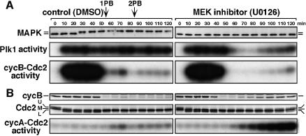
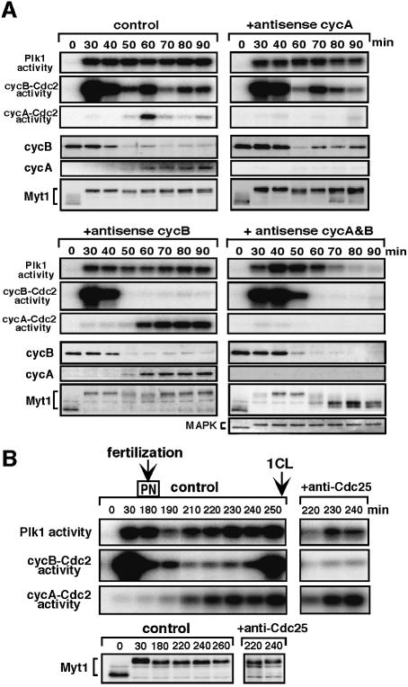
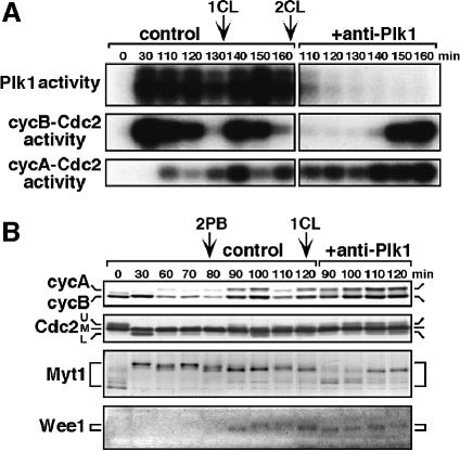
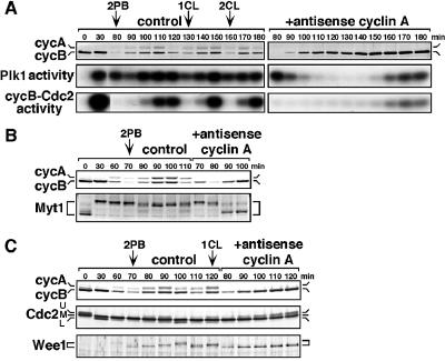
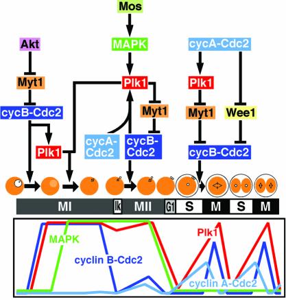
The Polo-like kinase, Plk, has multiple roles in regulating mitosis. In particular, Plk1 has been postulated to function as a trigger kinase that phosphorylates and activates Cdc25C prior to the activation of cyclin B-Cdc2 and thereby initiates its activation. However, the upstream regulation of Plk1 activation remains unclear. Here we have studied the interplay between Plk1 and Cdc2 through meiotic and early embryonic cycles in starfish. Distinct kinases, cyclin B-Cdc2, MAPK along with cyclin B- and/or cyclin A-Cdc2 and cyclin A-Cdc2, were unique upstream regulators for Plk1 activation at meiosis I, meiosis II and embryonic M-phase, respectively, indicating that Plk1 is not the trigger kinase at meiotic reinitiation. When Plk1 was required for cyclin B-Cdc2 activation, the action of Plk1 was mediated primarily through suppression of Myt1 rather than through activation of Cdc25. We propose that Plk1 can be activated by either cyclin A- or cyclin B-Cdc2, and its primary target is Myt1.


Figures

None
Fig. 1. Specificity of antibodies against starfish Plk1 and Wee1. Lysates from immature oocytes (lanes 1, 3 and 4), oocytes at metaphase of meiosis I (lanes 2 and 5), unfertilized mature eggs (lane 6) and eggs at metaphase of the first cleavage cycle (lane 7) were separated on a 10% SDS–PAGE gel and blotted onto nitrocellulose. Each blot was probed with affinity-purified anti-Plk1 antibody (lanes 1–3) and anti-Wee1 antibody (lanes 4–7). Molecular weight markers are indicated in kDa.
None
Fig. 2. Dynamics of Plk1 protein levels and kinase activity during starfish meiotic and cleavage cycles. Extracts were prepared from unfertilized (A) or fertilized (B) oocytes and eggs at 10 min intervals after 1-MeAde addition, and immunoblotted with anti-Plk1 (upper) and anti-MAPK (B, lowest) antibodies. Immunoprecipitates with the anti-Plk1 antibody were assayed for phosphorylation of α-casein (middle). Extracts were also assayed for phosphorylation of histone H1 (lower). Arrows indicate the time of GVBD, the first (1PB) and second (2PB) polar body emission, and the first (1CL) and second (2CL) cleavage. The arrowhead, the time of insemination.
None
Fig. 3. Suppression of H1 kinase activation prevents Plk1 activation at reinitiation of meiosis I. (A) Extracts were prepared at 3 min intervals after 1-MeAde addition, and Plk1 (upper) and histone H1 kinase (lower) activities were assayed as in Figure 2. (B) Immature oocytes were uninjected (control) or injected with either neutralizing anti-Cdc25 antibody (+anti-Cdc25) or p13suc1 protein (+p13suc1), and then treated with 1-MeAde. Extracts were prepared from groups of 10 oocytes at 10 min intervals, and histone H1 kinase (upper) and Plk1 (middle) activities were determined. Phosphorylation states of Cdc25 were analyzed by immunoblots with the anti-Cdc25 antibody (lower).
None
Fig. 4. The anti-Plk1 antibody prevents Plk1 activation and causes abortive spindle formation, but does not suppress cyclin B–Cdc2 activation at reinitiation of meiosis I. (A) Immature oocytes were injected with anti-Plk1 antibody (lanes 2–4, 300, 200 and 100 pg, respectively), control IgG (lane 5, 300 pg) or uninjected (lane 6), and then treated with 1-MeAde. Extracts were prepared from groups of 10 oocytes (after the occurrence of GVBD, lanes 2–6; immature, lane 1). Plk1 activity was detected on an autoradiogram (upper) or quantified in the excised bands by liquid scintillation counting (lower). (B) Extracts were prepared at 5 min intervals after 1-MeAde addition from groups of five oocytes that were uninjected (control, open circle) or injected with anti-Plk1 antibody (+anti-Plk1, closed circle), and histone H1 kinase activity was determined. (C) Time course of GVBD in oocytes that were uninjected (open squares) or injected with anti-Plk1 antibody (closed squares) was monitored during preparation of the extracts in (B). (D) Oocytes that were injected with control IgG (left) or anti-Plk1 antibody (right) were fixed at 20 min after GVBD. Green, immunofluorescence staining with anti-β-tubulin antibody; blue, DAPI staining of DNA.
None
Fig. 5. Lack of Plk1 activity does not affect cyclin B–Cdc2 inactivation at exit from meiosis I but prevents its reactivation at meiosis II through dephosphorylation of Myt1. Immature oocytes were uninjected (control) or injected with anti-Plk1 antibody (+anti-Plk1), and then treated with 1-MeAde. (A) At the indicated times, extracts were prepared from groups of 10 oocytes and immunoprecipitates with anti-Plk1, anti-cyclin B and anti-cyclin A antibodies were assayed for their associated kinase activities. (B) At the indicated times, groups of 30 oocytes were recovered and immunoblotted with anti-cyclin B and anti-Cdc2 antibodies. Cdc2-U and L represent cyclin B-associated inactive and active forms, respectively. (C) Groups of 40 oocytes that were uninjected (–) or injected (+) with anti-Plk1 antibody were sampled at the indicated times and immunoblotted with anti-Cdc25, anti-Myt1 and anti-MAPK antibodies.
None
Fig. 6. Inhibition of MAPK activation causes Plk1 inactivation after exit from meiosis I. Immature oocytes were first preincubated for 10 min in seawater containing DMSO (left) or 50 µM MEK inhibitor, U0126 (right), and then treated with 1-MeAde. (A) Extracts were prepared at 10 min intervals and immunoblotted with anti-MAPK antibody (upper), or immunoprecipitated with anti-Plk1 (middle) and anti-cyclin B (lower) antibodies for kinase assays. (B) The same extracts as in A were immunoblotted with anti-cyclin B (upper) and anti-Cdc2 (middle) antibodies, or assayed for cyclin A–Cdc2 kinase activity (lower).
None
Fig. 7. Plk1 activity depends on either cyclin A–Cdc2 or cyclin B–Cdc2 after meiosis I but not on cyclin B–Cdc2 at entry into M-phase of the first cleavage cycle. (A) Immature oocytes were uninjected (control) or injected with cyclin A and/or cyclin B antisense oligonucleotides (+antisense), and then treated with 1-MeAde. At the indicated times, extracts were prepared from groups of 10 oocytes and immunoprecipitated with anti-Plk1, anti-cyclin B and anti-cyclin A antibodies for assay of their associated kinase activities, or immunoblotted with anti-cyclin B, anti-cyclin A, anti-Myt1 and anti-MAPK antibodies. (B) Mature eggs at the female pronucleus stage were injected with control IgG (control) or neutralizing anti-Cdc25 antibody (+anti-Cdc25), and then inseminated. At the indicated times, extracts were prepared from groups of 10 eggs and immunoprecipitated with anti-Plk1, anti-cyclin B and anti-cyclin A antibodies for assay of their associated kinase activities, or immunoblotted with anti-Myt1 antibody.
None
Fig. 8. Prevention of Plk1 reactivation after meiosis II reverses hyperphosphorylation of Myt1 and suppresses cyclin B–Cdc2 activation at entry into M-phase of the first cleavage cycle. Maturing oocytes were fertilized at meiosis I, and then uninjected (control) or injected with anti-Plk1 antibody (+anti-Plk1) at the beginning of the second polar body emission (80 min). (A) Extracts were prepared at the indicated times from groups of five oocytes and eggs, and assayed for Plk1, cyclin B–Cdc2 and cyclin A–Cdc2 kinase activities. (B) Groups of 17 oocytes and eggs were sampled at the indicated times and analyzed by immunoblotting with anti-cyclin A, anti-cyclin B, anti-Cdc2, anti-Myt1 and anti-Wee1 antibodies.
None
Fig. 9. Inhibition of cyclin A synthesis prevents Plk1 activation, Wee1 phosphorylation and cyclin B–Cdc2 activation at entry into M-phase of the first cleavage cycle. Immature oocytes were uninjected (control) or injected with cyclin A antisense oligonucleotide (+antisense cyclin A), treated with 1-MeAde, and then fertilized at the end of meiosis I. (A) At indicated times, extracts were prepared from groups of eight oocytes and eggs, and immunoblotted with anti-cyclin A and anti-cyclin B antibodies (upper), or assayed for Plk1 (middle) and cyclin B–Cdc2 (lower) kinase activities. (B) Groups of 25 oocytes and eggs were sampled at the indicated times and analyzed by immunoblotting with anti-cyclin A, anti-cyclin B, and anti-Myt1 antibodies. (C) Groups of 16 oocytes and eggs were sampled at the indicated times and analyzed by immunoblotting with anti-cyclin A, anti-cyclin B, anti-Cdc2 and anti-Wee1 antibodies.
None
Fig. 10. Regulatory pathways for cyclin B–Cdc2 activation during starfish meiotic and early cleavage cycles. At reinitiation of meiosis I (MI), Akt inhibits Myt1 to cause the activation of cyclin B–Cdc2, which induces Plk1 activation. Plk1 is required for spindle formation but not for cyclin B–Cdc2 activation. At entry into meiosis II (Ik and MII), MAPK, along with cyclin A–Cdc2 and/or cyclin B–Cdc2, maintains Plk1 activity, which promotes further activation of cyclin B–Cdc2 through suppression of Myt1. MAPK may also contribute to maintenance of Cdc25 and suppression of Myt1. At entry into M-phase of the first cleavage cycle (M), cyclin A–Cdc2 promotes both Plk1 activation to cause Myt1 suppression and Wee1 phosphorylation, both of which result in cyclin B–Cdc2 activation. Thus, interplay among Plk1, cyclin B–Cdc2, cyclin A–Cdc2 and MAPK is unique at each of three types of cell cycle transition.

Similar articles

Cited by

References

    1. Abrieu A., Brassac,T., Galas,S., Fisher,D., Labbe,J.C. and Doree,M. (1998) The Polo-like kinase Plx1 is a component of the MPF amplification loop at the G2/M-phase transition of the cell cycle in Xenopus eggs. J. Cell Sci., 111, 1751–1757. - PubMed
    1. Brazil D.P. and Hemmings,B.A. (2001) Ten years of protein kinase B signalling: a hard Akt to follow. Trends Biochem. Sci., 26, 657–664. - PubMed
    1. Carmena M., Riparbelli,M.G., Minestrini,G., Tavares,A.M., Adams,R., Callaini,G. and Glover,D.M. (1998) Drosophila polo kinase is required for cytokinesis. J. Cell Biol., 143, 659–671. - PMC - PubMed
    1. Coleman T.R. and Dunphy,W.G. (1994) Cdc2 regulatory factors. Curr. Opin. Cell Biol., 6, 877–882. - PubMed
    1. Devault A., Fesquet,D., Cavadore,J.C., Garrigues,A.M., Labbe,J.C., Lorca,T., Picard,A., Philippe,M. and Doree,M. (1992) Cyclin A potentiates maturation-promoting factor activation in the early Xenopus embryo via inhibition of the tyrosine kinase that phosphorylates cdc2. J. Cell Biol., 118, 1109–1120. - PMC - PubMed

Publication types