

doi: 10.1523/JNEUROSCI.23-27-09133.2003.
sup-9, sup-10, and unc-93 may encode components of a two-pore K+ channel that coordinates muscle contraction in Caenorhabditis elegans
Affiliations
- PMID: 14534247
- PMCID: PMC6740817
- DOI: 10.1523/JNEUROSCI.23-27-09133.2003
Item in Clipboard
sup-9, sup-10, and unc-93 may encode components of a two-pore K+ channel that coordinates muscle contraction in Caenorhabditis elegans
J Neurosci.
.
Abstract
Genetic studies of sup-9, unc-93, and sup-10 strongly suggest that these genes encode components of a multi-subunit protein complex that coordinates muscle contraction in Caenorhabditis elegans. We cloned sup-9 and sup-10 and found that they encode a two-pore K+ channel and a novel transmembrane protein, respectively. We also found that UNC-93 and SUP-10 colocalize with SUP-9 within muscle cells, and that UNC-93 is a member of a novel multigene family that is conserved among C. elegans, Drosophila, and humans. Our results indicate that SUP-9 and perhaps other two-pore K+ channels function as multiprotein complexes, and that UNC-93 and SUP-10 likely define new classes of ion channel regulatory proteins.
Figures
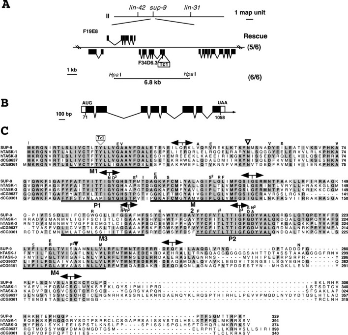
Molecular cloning, genomic structure, and sequence of the sup-9 gene. A, Schematic representation of the sup-9 genomic region and the Tc1 insertion found in the sup-9(n1428) strain. Cosmid F19E8 as well as the 6.8 kb HpaI subclone rescued a sup-9(n180);unc-93(e1500) mutant on the basis of the appearance of the rubberband Unc response. The numbers in parentheses indicate the number of rescued lines and total transgenic lines scored. B, Intron-exon structure of sup-9. The structure of the sup-9 gene was deduced by comparing its genomic sequence with the sequences of RT-PCR and RACE products. Black boxes indicate coding regions. White boxes indicate untranslated regions. The arrow indicates the direction of transcription. C, Alignment of SUP-9 with human TASK-1, human TASK-3, and Drosophila predicted proteins dCG9637 and dCG9361. Residues identical between SUP-9 and the other proteins are highlighted in gray. Black bars indicate proposed transmembrane domains (M1, M2, M3, and M4). White bars indicate P domains (P1 and P2). lf mutations in sup-9 are indicated by the amino acids above the aligned sequences. The superscript number indicates the number of alleles with that mutation. The black triangle indicates the site of all four gf alleles. The white triangle indicates a potential N-glycosylation site. The site of the Tc1 insertion from sup-9(n1428) is indicated. The intron-exon boundaries are indicated by a double arrow above the sequence. GenBank accession numbers are as follows: SUP-9, AY357729; TASK-1, NP_002237; TASK-3, NP_057685; dCG9637, AAF54970; and dCG9361, AAF54374.
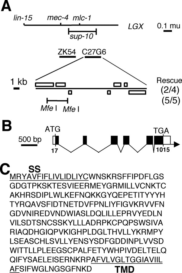
Molecular cloning and genomic structure of the sup-10 gene and sequence of the SUP-10 protein. A, Schematic representation of the sup-10 genomic region. Cosmid C27G6 as well as a 7.3 kb MfeI subclone of C27G6 suppressed the semidominant rubberband Unc paralysis of sup-10(n983) gf mutants as transgenes. Numbers in parentheses indicate the number of rescued lines and total transgenic lines scored. B, Intron-exon structure of sup-10. The structure of the sup-10 gene was deduced by comparing its genomic sequence with the sequence of cDNA GenBank accession number U43891. Black boxes indicate coding regions. White boxes indicate untranslated regions. The arrow indicates the direction of transcription. C, Amino acid sequence of SUP-10. The underlined residues represent the putative signal sequence (SS) and the transmembrane domain (TMD), which were determined using the PSORTII II program developed by Nakai and Horton (1999) (available at http://psort.nibb.ac.jp/ ).
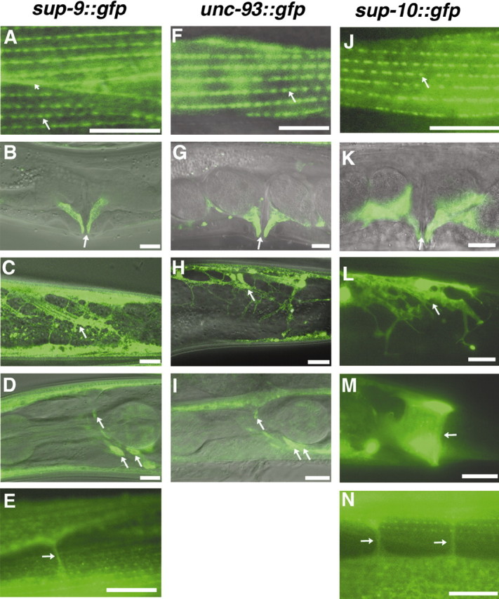
Expression of sup-9::gfp, unc-93::gfp, and sup-10::gfp fusions. A-N, Epifluorescence or merged Normarski and epifluorescence images of GFP expression in worms carrying a sup-9::gfp (A-E), unc-93::gfp (F-I), or sup-10:gfp (J-N) reporter transgenes. Scale bars, 10 μm. A, F, J, Body-wall muscle cells. The short arrow indicates the cell membranes of adjacent body-wall muscle cells; the long arrow indicates rows of dense bodies. B, G, K, Vulval muscle cells. Two muscle cells are visible in the focal planes shown. The arrow indicates the opening of the vulva. C, H, L, Intestinal muscle cells (arrows). D, I, Heads of adult animals expressing gfp in neurons (arrows). E, N, Muscle arms (arrows). M, Anal depressor muscle (arrow).
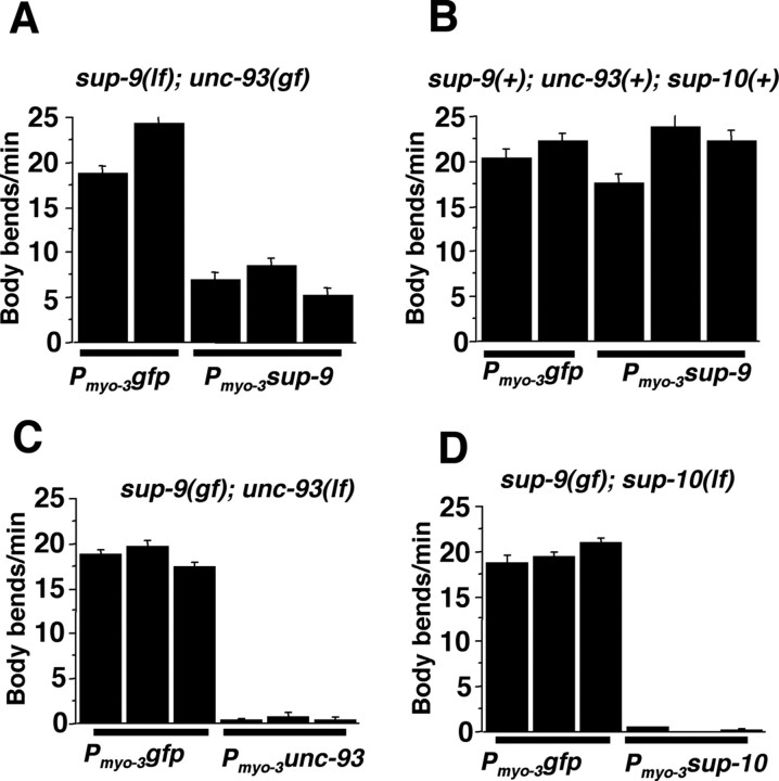
sup-9, sup-10, and unc-93 cDNA rescue of locomotory defects. Locomotory rates of young adult transgenic hermaphrodites were scored on a bacterial lawn during 60 sec intervals. A body-bend is defined as a 360o sine wave. Each bar represents the mean ± SEM of an independent transgenic line. All strains carry lin-15(n765) and the wild-type lin-15 gene as a transgene to aid in the identification of transgenic animals. A, Parental genotype: sup-9(n1913); unc-93(e1500); lin-15(n765); n = 20-24 worms per transgenic line. B, Parental genotype: lin-15(n765); n = 11-13. C, Parental genotype: sup-9(e2655); unc-93(e1500 n234); lin-15(n765); n = 16-17. D, Parental genotype: sup-9(n1550); sup-10(n183); lin-15(n765ts); n = 16.
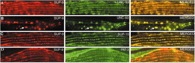
Subcellular colocalization of SUP-9 with UNC-93::GFP and SUP-10::GFP. Transgenic lines expressing sup-9 driven by the myo-3 promoter and an unc-93::gfp (A, B), sup-10::gfp (C), or pat-3::gfp (D) fusion was fixed and costained with anti-SUP-9 (red) and anti-GFP antisera (green). All panels display adult body-wall muscle cells analyzed by confocal microscopy for immunofluorescence. Scale bars, 10 μm.

Muscimol phenocopies the rubberband Unc phenotype of sup-9(gf), unc-93(gf), and sup-10(gf) mutants. A, Wild-type (N2) worms were scored for the rubberband Unc response at the indicated concentrations of muscimol, as defined in Materials and Methods (n = 170 for all concentrations except 1 mm , in which case n = 232). B, Rubberband mutants were scored for the rubberband Unc response in the absence of muscimol [n = 300 for all genotypes except sup-9(n1550I), in which case n = 200]. C, Wild-type and lf mutant worms were scored for the rubberband Unc response in 1 mm muscimol. The numbers of responses scored were n = 979, n = 360, n = 300, n = 490, and n = 300, respectively. D, Rubberband sup-9(n1550)/+ worms were scored for the rubberband Unc response in the absence and presence of 1 mm muscimol. The numbers of responses scored were n = 300 and n = 300.

Overexpression of SUP-9(gf) cannot bypass the need for UNC-93. A, Transgenic male animals were scored on a bacterial lawn for locomotory rate. Males, rather than hermaphrodites, were used because unc-93(lr12) heterozygous males can be unambiguously generated. All strains contained the lin-15(n765) mutation to enable the identification of transgenic animals and the unc-93(lr12) mutation as noted previously. The independently generated transgenic lines are indicated by the numbers below the graph. Each bar represents the mean ± SEM; n = 10 for all lines. B, Confocal images of a representative adult transgenic animal (top) and a nontransgenic sibling (bottom). unc-93(lr12) animals with or without the myo-3::sup-9(n1550) transgene were fixed and immunostained with anti-SUP-9 antisera. Epifluorescence images are on the left. Normarski images on the right allow visualization of dense bodies, in which SUP-9 is expected to localize. Scale bar, 10 μm.

Phylogenetic tree of UNC-93-like genes from C. elegans, Drosophila, mouse, and human. Protein alignments were generated using the ClustalW algorithm (Thompson et al., 1994). The horizontal length between each pair of branches represents the evolutionary distance between gene pairs as measured by the number of substitutions per residue. The predicted sequences of H.s. dJ366N23.1 and H.s. dJ366N23.2 were used to create H.s. dJ366N23.1/.2, because we believe that these sequences likely represent the N-terminal and C-terminal segments, respectively, of a single gene. M.m.AI615282 and M.m. 285761 are encoded by ESTs and likely represent partial sequences of their respective genes. B0554.9/.8/.7 contains the combined amino acid sequences of predicted genes B0554.9, B0554.8, and B0554.7, because we believe that these sequences are likely portions of one gene. The GenBank accession numbers of the genes are as follows: D.m. 30B8.6, CAA15704; D.m. CG2121, AAF59099; D.m. GH10120, AF145657; D.m. CG18549, AAF54824; H.s. ET10, AF015185; H.s. dJ366N23.1/.2, AL021331; H.s. ET22, AF015186; M.m.AI615282, AI615282; M.m. ET8, AF015191; M.m.AA285761, AA285761; UNC-93, S23352; ZK6.8, AAC17687; ZK6.6, AAC17693; Y39D8A.1, AAC69224; B0554.5, AAB37612; Y39B6B.L, CAB60917; Y39B6B.K, CAB60916; B0554.9/.8/.7, AAB37619, AAB37618, AAB37616; Y37A1A.2, CAB16469; Y11D7A.3, CAA21581; C27C12.4, CAA93741; Y52E8A.B, AAF59523; Y52E8A.A, AAF59522; F31D5.2, AAC71105; F31D5.1, AAC71106; M153.2, CAA91944; F36G9.3, CAB04342. H.s., Homo sapiens; M.m., Mus musculus; D.m., Drosophila melanogaster. All others are C. elegans.
Similar articles
-
The Caenorhabditis elegans iodotyrosine deiodinase ortholog SUP-18 functions through a conserved channel SC-box to regulate the muscle two-pore domain potassium channel SUP-9.PLoS Genet. 2014 Feb 20;10(2):e1004175. doi: 10.1371/journal.pgen.1004175. eCollection 2014 Feb. PLoS Genet. 2014. PMID: 24586202 Free PMC article.
-
The Caenorhabditis elegans unc-93 gene encodes a putative transmembrane protein that regulates muscle contraction.J Cell Biol. 1992 Apr;117(1):143-55. doi: 10.1083/jcb.117.1.143. J Cell Biol. 1992. PMID: 1313436 Free PMC article.
-
An abnormal ketamine response in mutants defective in the ryanodine receptor gene ryr-1 (unc-68) of Caenorhabditis elegans.J Mol Biol. 1997 Apr 11;267(4):849-64. doi: 10.1006/jmbi.1997.0910. J Mol Biol. 1997. PMID: 9135117
-
An evolutionarily conserved family of accessory subunits of K+ channels.Cell Biochem Biophys. 2006;46(1):91-9. doi: 10.1385/CBB:46:1:91. Cell Biochem Biophys. 2006. PMID: 16943626 Review.
-
Potassium channels in C. elegans.WormBook. 2005 Dec 30:1-15. doi: 10.1895/wormbook.1.42.1. WormBook. 2005. PMID: 18050399 Free PMC article. Review.
Cited by
-
Complex Locomotion Behavior Changes Are Induced in Caenorhabditis elegans by the Lack of the Regulatory Leak K+ Channel TWK-7.Genetics. 2016 Oct;204(2):683-701. doi: 10.1534/genetics.116.188896. Epub 2016 Aug 17. Genetics. 2016. PMID: 27535928 Free PMC article.
-
Differential modulation of C. elegans motor behavior by NALCN and two-pore domain potassium channels.PLoS Genet. 2022 Apr 28;18(4):e1010126. doi: 10.1371/journal.pgen.1010126. eCollection 2022 Apr. PLoS Genet. 2022. PMID: 35482723 Free PMC article.
-
UNC93B1 interacts with the calcium sensor STIM1 for efficient antigen cross-presentation in dendritic cells.Nat Commun. 2017 Nov 21;8(1):1640. doi: 10.1038/s41467-017-01601-5. Nat Commun. 2017. PMID: 29158474 Free PMC article.
-
Genome-wide view of cell fate specification: ladybird acts at multiple levels during diversification of muscle and heart precursors.Genes Dev. 2007 Dec 1;21(23):3163-80. doi: 10.1101/gad.437307. Genes Dev. 2007. PMID: 18056427 Free PMC article.
-
Natural Variation in plep-1 Causes Male-Male Copulatory Behavior in C. elegans.Curr Biol. 2015 Oct 19;25(20):2730-7. doi: 10.1016/j.cub.2015.09.019. Epub 2015 Oct 8. Curr Biol. 2015. PMID: 26455306 Free PMC article.
References
Publication types
MeSH terms
Substances
Grants and funding
LinkOut - more resources
Full Text Sources
Molecular Biology Databases