. 2003 Oct 8;23(27):9208-19.
doi: 10.1523/JNEUROSCI.23-27-09208.2003.

A receptor-like inositol lipid phosphatase is required for the maturation of developing cochlear hair bundles

Affiliations

A receptor-like inositol lipid phosphatase is required for the maturation of developing cochlear hair bundles

R J Goodyear et al. J Neurosci. .

Abstract

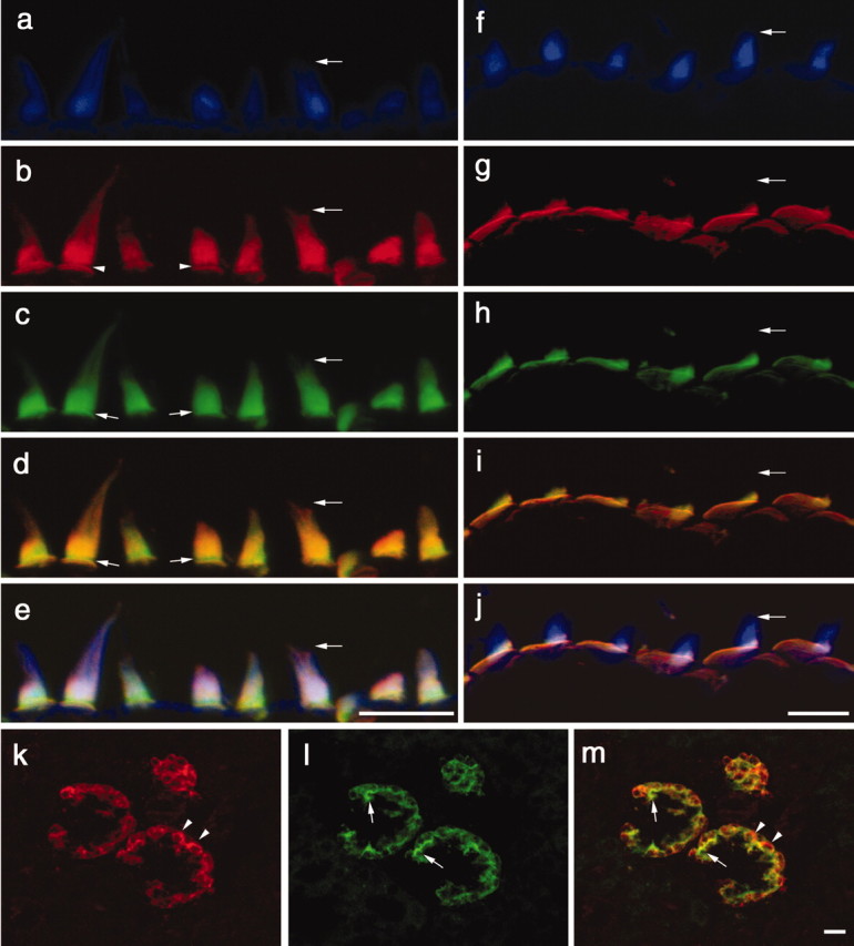
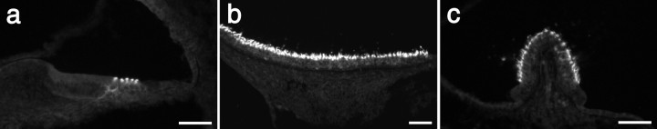
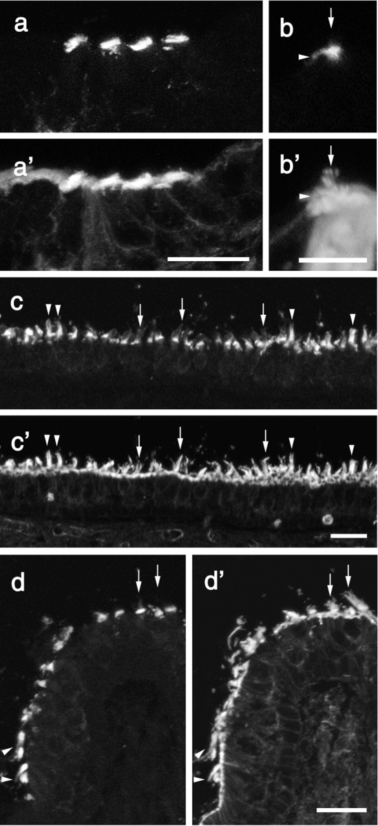
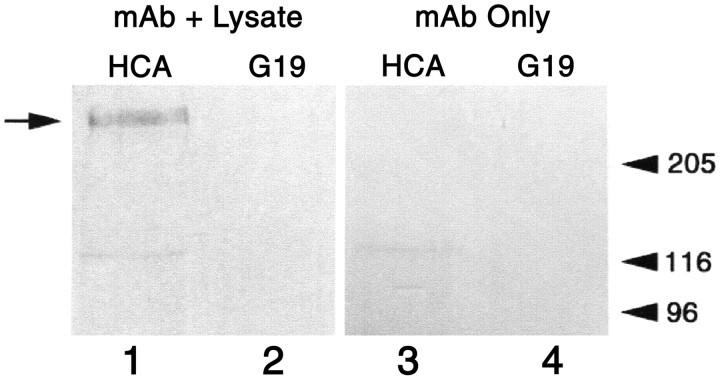
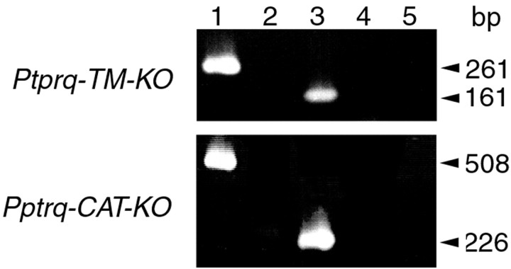
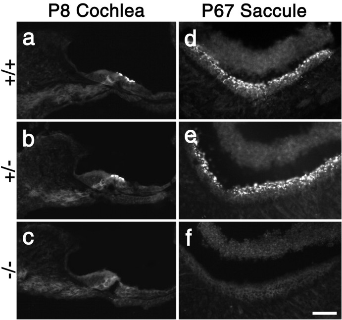
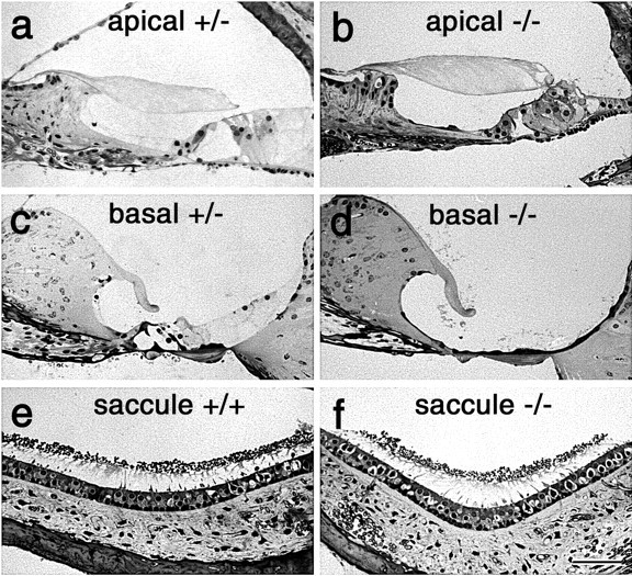
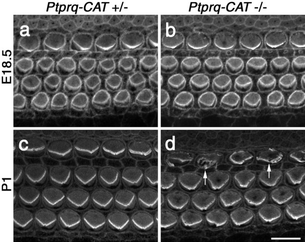
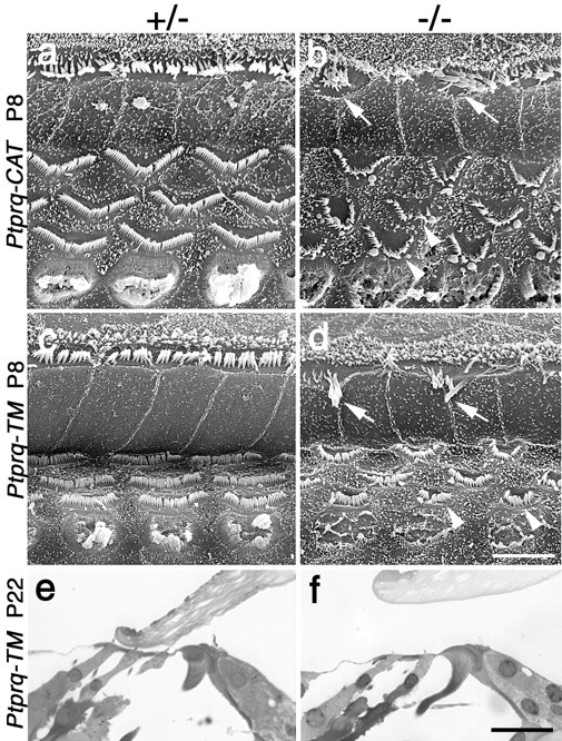
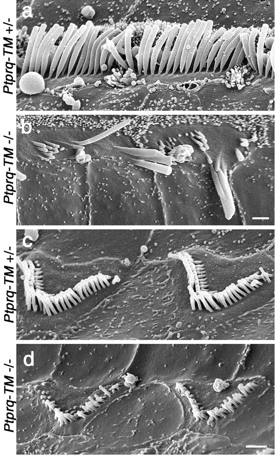
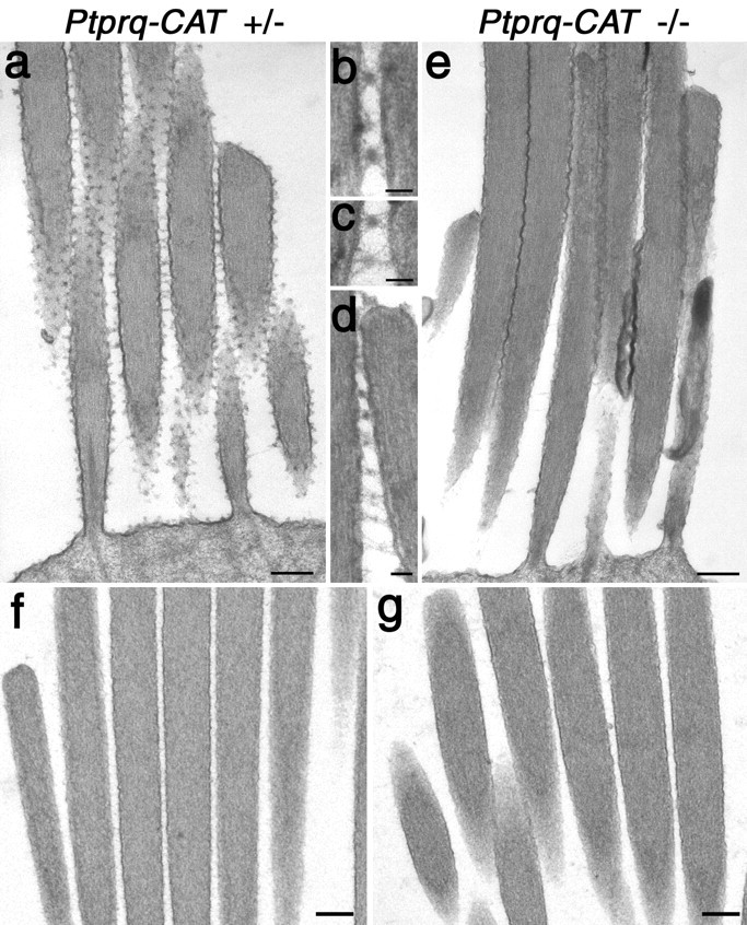
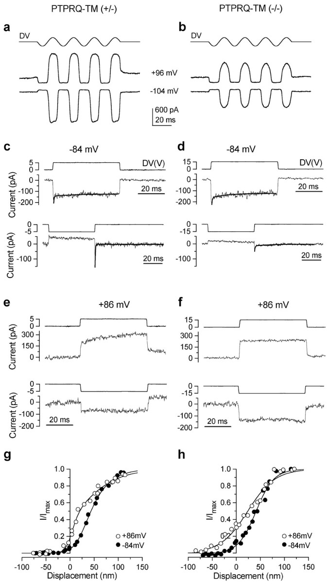
A screen for protein tyrosine phosphatases (PTPs) expressed in the chick inner ear yielded a high proportion of clones encoding an avian ortholog of protein tyrosine phosphatase receptor Q (Ptprq), a receptor-like PTP. Ptprq was first identified as a transcript upregulated in rat kidney in response to glomerular nephritis and has recently been shown to be active against inositol phospholipids. An antibody to the intracellular domain of Ptprq, anti-Ptprq, stains hair bundles in mice and chicks. In the chick ear, the distribution of Ptprq is almost identical to that of the 275 kDa hair-cell antigen (HCA), a component of hair-bundle shaft connectors recognized by a monoclonal antibody (mAb) that stains inner-ear hair bundles and kidney glomeruli. Furthermore, anti-Ptprq immunoblots a 275 kDa polypeptide immunoprecipitated by the anti-HCA mAb from the avian inner ear, indicating that the HCA and Ptprq are likely to be the same molecule. In two transgenic mouse strains with different mutations in Ptprq, anti-Ptprq immunoreactivity cannot be detected in the ear. Shaft connectors are absent from mutant vestibular hair bundles, but the stereocilia forming the hair bundle are not splayed, indicating that shaft connectors are not necessary to hold the stereocilia together; however, the mice show rapid postnatal deterioration in cochlear hair-bundle structure, associated with smaller than normal transducer currents with otherwise normal adaptation properties, a progressive loss of basal-coil cochlear hair cells, and deafness. These results reveal that Ptprq is required for formation of the shaft connectors of the hair bundle, the normal maturation of cochlear hair bundles, and the long-term survival of high-frequency auditory hair cells.


Figures

Figure 1.
Figure 1.
Distribution of anti-HCA and anti-Ptprq immunostaining in the inner ear and kidney of the early posthatch chick. a-j, Compressed z-stacks of confocal sections from the extrastriolar region of the utricular macula (a-e) and the basilar papilla (f-j), triple-labeled with Alexa 350 phalloidin (a, f), polyclonal anti-Ptprq (b, g), and monoclonal anti-HCA mAb (c, h). d, i, Merges of anti-Ptprq and anti-HCA mAb staining; e, j, merges of the images obtained through all three channels (anti-Ptprq, anti-HCA mAb, and F-actin). Horizontal arrows point to the tip of the same hair bundle in a-e and f-j. b, Arrowheads indicate the ankle-link region that is not stained by anti-Ptprq. c, d, Small arrows indicate that anti-HCA mAb staining is present in the ankle-link region. b-d, In the extrastriolar region of the macula, the hair bundle is stained to its distal tip by anti-Ptprq and the anti-HCA mAb. g-i, In the basilar papilla, staining with both antibodies is restricted to the proximal end of the hair bundle. g, h, The extensive, nonstereociliary apical membrane of the hair cell is clearly stained by both antibodies in the basilar papilla. k, l, Compressed z-stacks of confocal section from the chick kidney double labeled with anti-Ptprq (k) and the anti-HCA mAb (l). m, Merge of anti-Ptprq and anti-HCA mAb staining. Arrowheads indicate areas that stain only with anti-Ptprq; arrows indicate areas that stain only with the anti-HCA mAb. Scale bars, 10 μm.
Figure 2.
Figure 2.
Distribution of Ptprq in the mouse inner ear. Shown are sections of the cochlea at P2 (a), saccule at P15 (b), and crista at P21 (c) from the mouse inner ear stained with anti-Ptprq. Scale bars, 50 μm.
Figure 3.
Figure 3.
Fluorescence micrographs from mouse inner ear sections double labeled with affinity-purified antibodies to Ptprq (a-d) and rhodamine phalloidin (a′-d′).On cochlear inner hair cells (b,b′,arrows)and hair cells in the striolar regions of the utricular macula (c,c′,arrows) and at the apex of the crista (d, d′, arrows), anti-Ptprq staining is concentrated at the proximal end of the hair bundle. With hair bundles in the extrastriolar regions of the utricular macula (c, c′, arrowheads) and peripheral region of the crista (d, d′, arrowheads), the hair bundle is stained up to its tip. Sections are from mice at P2 (a), P21 (b, c), and P15 (d). Scale bars: a, 20 μm; b, 10 μm; c, d, 20 μm.
Figure 4.
Figure 4.
Immunoprecipitation of the HCA. An immunoblot stained with affinity-purified antibodies to the recombinant intracellular domain of Ptprq. Lysates prepared from the sensory epithelia of the chick inner ear (lanes 1, 2) or lysis buffer (lanes 3, 4) were incubated with an anti-HCA mAb (mAb D10; lanes 1, 3) or an irrelevant mAb of the same isotype (mAb G19; lanes 2, 4), and the mAbs were precipitated with rabbit anti-mouse Ig and collected with Protein A-Sepharose. The 275 kDa hair-cell antigen is indicated by the arrow on the left. Markers indicated on the right with molecular masses in kilodaltons are skeletal muscle myosin (205), β-galactosidase (116), and phosphorylase B (96)
Figure 5.
Figure 5.
Expression of Ptprq during mouse inner ear development. Pairs of fluorescence micrographs from adjacent sections of the vestibule at E13.5 (a, a′) and P21 (e, e′) and the cochlea at E18.5 (b, b′), P2 (c, c′), and P21 (d, d′) labeled with antibodies to myosin VIIa (a-e) or Ptprq (a′-e′). Ptprq first appears in the vestibule at E13.5 (a′) and is present on inner and outer hair cells in the base of the cochlea by E18.5 (b, b′). Ptprq expression on basal-coil outer hair cells increases by P2 (c, c′) but declines by P15 and can no longer be detected at P21 (d′). Inner hair cells (d′) and vestibular hair cells (e′) express Ptprq into maturity. Scale bars, 20 μm.
Figure 6.
Figure 6.
RT-PCR analysis of Ptprq mRNA expression in the cochlea of wild-type, Ptprq-TM-KO, and Ptprq-CAT-KO mice. Specific products spanning the exons deleted in the Ptprq-TM-KO and Ptprq-CAT-KO mutants were amplified from RT-PCR reactions performed on total RNA isolated from cochleas from wild-type (lanes 1, 2) and homozygous mutant (lanes 3, 4) mice. In the Ptprq-TM-KO line a product of 261 bp is amplified from wild-type mice (lane 1), and a product of 161 bp is amplified from homozygous mutant mice (lane 3). In the Ptprq-CAT-KO line a product of 508 bp is amplified from wild-type mice (lane 1), and a product of 226 bp is amplified from homozygous mutant mice (lane 3). Control reverse transcription reactions performed without reverse transcriptase are negative (lanes 2, 4) as are the water controls (lane 5).
Figure 7.
Figure 7.
Immunostaining with Ptprq intracellular domain antibodies in the cochlea of wild-type, Ptprq-TM-KO, and Ptprq-CAT-KO mice. Fluorescence micrographs of sections from the cochleas at P8 (a-c) and saccule at P67 (d-f) of wild-type (a, d), heterozygous (b, e), and homozygous (c, f) Ptprq-TM-KO (a-c) and Ptprq-CAT-KO (d-f) mice stained with affinity-purified antibodies to Ptprq. Ptprq cannot be detected with these antibodies in the inner ear of either homozygous mutant (c, f). Scale bar, 50 μm.
Figure 8.
Figure 8.
Structure of cochlea in wild-type, Ptprq-TM-KO, and Ptprq-CAT KO mice. Light micrographs of 1-μm-thick, toluidine blue-stained sections from the apical (a, b) and basal (c, d) regions of the cochlea, and the saccule(e,f),ofheterozygous(a,c),wild-type(e),andhomozygous(b, d,f)3-month-oldPtprq-CAT-KO mice. Note the complete loss of the organ of Corti in the basal end of the cochlea in the homozygous Ptprq-CAT-KO (d) mouse. Scale bar, 50 μm.
Figure 9.
Figure 9.
Phalloidin staining of hair bundles in Ptprq-CAT-KO mice. Confocal micrographs of the basal end of phalloidin-stained cochlear whole mounts from heterozygous (a, c) and homozygous Ptprq-CAT-KO(b,d)mice at E18.5(a,b)and P1(c,d). Defects in hair-bundle structure are first apparent in the inner hair cells (d, arrows) at P1. Scale bar, 10 μm.
Figure 10.
Figure 10.
Morphology of hair bundles and hair cells in Ptprq-TM-KO and Ptprq-CAT-KO mice. a-f, Survey scanning electron micrographs at P8 (a-d) and sections at P22 (e, f) of the organ of Corti in the basal coil of heterozygous (a, c, e) and homozygous (b, d, f) Ptprq-CAT-KO (a, b) and Ptprq-TM-KO (c-f) mice. The hair bundles of inner (arrows) and outer (arrowheads) hair cells are considerably affected in both the homozygous Ptprq-CAT-KO (b) and the Ptprq-TM-KO (d) mice by P8. Note the difference in the shape of the outer hair-cell hair bundles. Hair cells are still present in the organ of Corti in Ptprq-TM-KO mice at P22 (e, f). Scale bars: (in d) a-d, 5 μm; (in f) e, f, 20 μm.
Figure 11.
Figure 11.
Structure of hair bundles in heterozygous and homozygous Ptprq-TM-KO mice. High-magnification scanning electron micrographs of the hair bundles of inner (a, b) and outer (c, d) hair cells in the organ of Corti (basal coil) of heterozygous (a, c) and homozygous (b, d) Ptprq-TM-KO mice at P15. Note the loss of stereocilia from the hair bundles of homozygous mutant hair cells, the giant, fused stereocilia on the homozygous mutant inner hair cells (b), and how the stereocilia are shorter on the homozygous mutant outer hair cells (d). Scale bars, 1 μm.
Figure 12.
Figure 12.
TEM analysis of shaft connector structure in vestibular hair bundles of wild-type and Ptprq-CAT-KO mice. Transmission electron micrographs of extrastriolar hair bundles from the utricular macula of heterozygous (a-d, f) and homozygous (e, g) mutant Ptprq-CAT-KO mice at P21. Samples in a-e were fixed in fixatives containing ruthenium red; samples in f and g were fixed in the absence of any contrasting agents. Note the lack of shaft connectors in the homozygous mutant hair bundle (e) and the close apposition of the stereocilia. Details of shaft connector structure in heterozygous Ptprq-CAT-KO mice (b-d) reveal dense particles attached to the membrane by fine stalks. The spacing of stereocilia in the hair bundles of heterozygous(f) and homozygous (g) mutant Ptprq-CAT-KO mice is similar in the absence of ruthenium red. Scale bars: a, e-g, 200 nm; b-d, 50 nm.
Figure 13.
Figure 13.
Mechanotransducer currents in Ptprq-TM-KO outer hair cells. Transducer currents recorded in basal-coil OHCs from a heterozygous Ptprq-TM-KO mouse (a, c, e, g) and a homozygous Ptprq-TM-KO mouse (b, d, f, h). a, b, Transducer currents recorded at a holding potential of -104 and +96 mV by applying sinusoidal force stimuli of 45 Hz. The driver voltage signal (DV) (amplitude, 35 V) to the fluid jet is shown above each trace. Positive deflections are excitatory. c, d, The time course of onset adaptation to an excitatory step displacement at -84 mV (top panels). Note the difference in the driver voltages applied. Fitted time constants for onset adaptation are as follows: c, 0.34 and 16.1 msec; d, 0.39 and 12.3 msec. Bottom panels show the transducer current in response to a negative stimulus at -84 mV. After termination of the inhibitory stimulus the transducer current shows evidence of rebound adaptation. Fitted time constants for the rebound adaptation are as follows: c, 0.34 and 16.2 msec; d, 0.54 and 16.3 msec. e, f, The time course of the transducer current elicited by excitatory (top panels) and inhibitory (bottom panels) bundle displacements at +86 mV. Note the absence of onset and rebound adaptation. a, c, e, P7 OHC: Cm 6.9 pF; Rs 2.4 MΩ; b, d, f, P7 OHC: Cm 6.7 pF; Rs 3.0 MΩ. All responses shown are single traces. g, h, Normalized peak transducer current as a function of bundle displacement. Zero current is set as the holding current when the force stimulus closes most transducer channels. Smooth curves are second-order Boltzmann functions: I = Imax /(1 + exp(a2 (x2 - x))*(1 + exp(a1(x1 - x)))). g, At -84 mV, Imax = -732 pA, a1 = 0.06 nm-1, a2 = 0.04 nm-1, x1 = 26 nm, x2 = 35 nm; at +86 mV, Imax = 810 pA, a1 = 0.12 nm-1, a2 = 0.02 nm -1, x1 = 7 nm, x2 = 14 nm. P7 OHC: Cm 7.0 pF; Rs 1.4 MΩ. Bundle height 4 μm; bundle width 8 μm. h, At -84 mV, Imax = -211 pA, a1 = 0.10 nm-1, a2 = 0.054 nm -1, x1 = -32nm, x2 = 41 nm; at +86 mV, Imax = 316 pA, a1 = 0.15nm -1, a2 = 0.035 nm -1, x1 = -76 nm, x2 = 24 nm. P7 OHC: Cm 6.8 pF; Rs 2.5 MΩ. Bundle height 3.5 μm; bundle width 6 μm.

References

    1. Alagraman KN, Murcia CL, Kwon HY, Pawlowski KS, Wright CG, Woychik RP ( 2001) The mouse Ames waltzer hearing-loss mutant is caused by a mutation of Pdch15, a novel protocadherin gene. Nat Genet 27: 99-102. - PubMed
    1. Altschul F, Gish W, Miller W, Myers EW, Lipman DJ ( 1990) Basic local alignment research tool. J Mol Biol 215: 403-410. - PubMed
    1. Anniko M ( 1983) Cytodifferentiation of cochlear hair cells. Am J Otolaryngol 4: 375-388. - PubMed
    1. Aricescu AR, McKinnell IW, Halfter W, Stoker AW ( 2002) Heparan sulphate proteoglycans are ligands for receptor protein tyrosine phosphatase sigma. Mol Cell Biol 22: 1881-1892. - PMC - PubMed
    1. Assad JA, Hacohen N, Corey DP ( 1989) Voltage dependence of adaptation and active bundle movement in bullfrog saccular hair cells. Proc Natl Acad Sci USA 86: 2918-2922. - PMC - PubMed

Publication types

MeSH terms