. 2003 Oct 10:4:25.
doi: 10.1186/1471-2202-4-25.

Serial pathways from primate prefrontal cortex to autonomic areas may influence emotional expression

Affiliations

Serial pathways from primate prefrontal cortex to autonomic areas may influence emotional expression

Helen Barbas et al. BMC Neurosci. .

Abstract

Background: Experiencing emotions engages high-order orbitofrontal and medial prefrontal areas, and expressing emotions involves low-level autonomic structures and peripheral organs. How is information from the cortex transmitted to the periphery? We used two parallel approaches to map simultaneously multiple pathways to determine if hypothalamic autonomic centres are a key link for orbitofrontal areas and medial prefrontal areas, which have been associated with emotional processes, as well as low-level spinal and brainstem autonomic structures. The latter innervate peripheral autonomic organs, whose activity is markedly increased during emotional arousal.

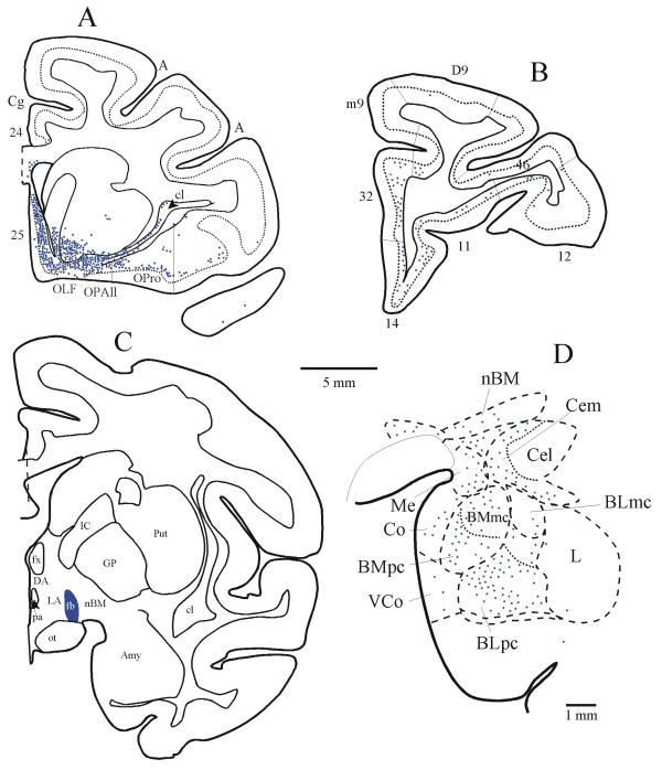
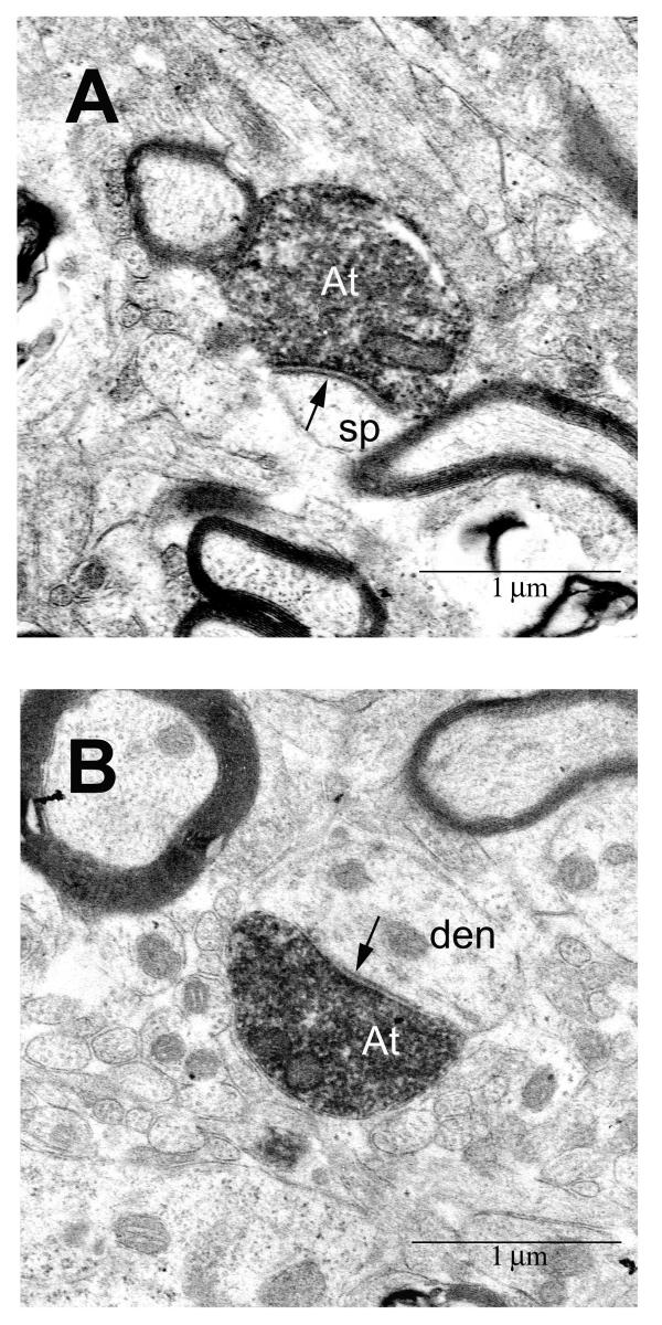
Results: We first determined if pathways linking the orbitofrontal cortex with the hypothalamus overlapped with projection neurons directed to the intermediolateral column of the spinal cord, with the aid of neural tracers injected in these disparate structures. We found that axons from orbitofrontal and medial prefrontal cortices converged in the hypothalamus with neurons projecting to brainstem and spinal autonomic centers, linking the highest with the lowest levels of the neuraxis. Using a parallel approach, we injected bidirectional tracers in the lateral hypothalamic area, an autonomic center, to label simultaneously cortical pathways leading to the hypothalamus, as well as hypothalamic axons projecting to low-level brainstem and spinal autonomic centers. We found densely distributed projection neurons in medial prefrontal and orbitofrontal cortices leading to the hypothalamus, as well as hypothalamic axonal terminations in several brainstem structures and the intermediolateral column of the spinal cord, which innervate peripheral autonomic organs. We then provided direct evidence that axons from medial prefrontal cortex synapse with hypothalamic neurons, terminating as large boutons, comparable in size to the highly efficient thalamocortical system. The interlinked orbitofrontal, medial prefrontal areas and hypothalamic autonomic centers were also connected with the amygdala.

Conclusions: Descending pathways from orbitofrontal and medial prefrontal cortices, which are also linked with the amygdala, provide the means for speedy influence of the prefrontal cortex on the autonomic system, in processes underlying appreciation and expression of emotions.


Figures

Figure 1
Figure 1
Overlap in hypothalamus of serial pathways from prefrontal area 12 and the intermediolateral spinal column. (A) The bidirectional tracer fluororuby (fr) was injected in prefrontal area 12 (red area). (B) The injection of the retrograde tracer fast blue (fb) was in the spinal cord, covering the intermediolateral cell column, the lowest central autonomic center (blue area). Fast blue labeled neurons projecting to the spinal cord (blue dots) and labeled axons (brown lines) originating in area 12 were intermingled in the following hypothalamic areas: (C) dorsal hypothalamic area (DA) and tuberomammillary nucleus (TM); (D) perifornical nucleus (Pef); (E) fields of Forel (FF). (F) In the same case, labeled neurons in the basolateral (BL) and lateral (L) nuclei of the amygdala projected to area 12 (red dots).
Figure 2
Figure 2
Serial pathways from the prefrontal cortex reach central autonomic structures. Pathways were mapped after injection of the bidirectional tracer BDA in the lateral (LA) hypothalamic area. (A) The first pathway is marked by projection neurons (blue dots) originating most densely from orbitofrontal and medial prefrontal cortices leading to the injection site (brown area in B). (B) The second pathway is marked by labelled axons emanating from the injection site and terminating in several autonomic nuclei (brown lines): (C, D) brainstem nuclei; (E) the thoracic spinal cord; (F) A bidirectional pathway links the amygdala with the same hypothalamic nuclei. The shaded areas in the amygdala show the specific termination zones of axons from orbitofrontal cortex (yellow), and the diffuse termination zone by axons from medial prefrontal cortex (light brown), as described by Ghashghaei and Barbas [24]. The dotted line in A indicates the upper border of cortical layer V, and brown lines show the terminations of hypothalamic axons.
Figure 3
Figure 3
Simultaneous labelling of pathways from orbitofrontal cortex and the amygdala to the lateral hypothalamic area. (A) Densely distributed projection neurons in the posterior orbitofrontal cortex (areas OPAll, OPro), and in medial area 25 projected to the lateral hypothalamic area (LA in C). (B) Projection neurons in prefrontal areas 32 and 14 and in other prefrontal areas directed to LA; (C) The injection site of fast blue (fb) was in LA of the hypothalamus. (D) Projection neurons from the amygdala directed to the same area (LA) of the hypothalamus. The dotted line in A and B indicates the upper border of cortical layer V.
Figure 4
Figure 4
Axons from prefrontal area 32 synapse in the lateral hypothalamic area. Axon terminals (At) were identified by BDA label injected in area 32. (A) synapse (arrow) with dendritic spine (sp); (B) synapse (arrow) with dendrite (den). Scale bar = 1 μm.
Figure 5
Figure 5
Summary of pathways linking prefrontal cortex with structures associated with perception and expression of emotions. Line thickness represents the density of projections. Direct and indirect sensory input to the orbitofrontal cortex: The orbitofrontal cortex and the amygdala receive projections from every sensory modality through the cortex (pathways so, s), and are robustly linked with each other, providing the structural basis for direct (so), and indirect (s and s') sensory input to the orbitofrontal cortex. The orbitofrontal cortex disinhibits hypothalamic autonomic centers: Orbitofrontal axons terminate heavily in the intercalated masses of the amygdala (IM, pathway a), which project to the central nucleus (a'), which projects to hypothalamic autonomic centers (pathway b). Activation of pathways (a, a') leads to disinhibition of hypothalamic autonomic centers, which innervate brainstem and spinal autonomic centers (pathways c', o'). Direct and indirect pathways from medial prefrontal areas to hypothalamic autonomic centers: The direct pathway courses from medial prefrontal cortex to hypothalamic autonomic centers (c), forming asymmetric (and presumed excitatory) synapses in the lateral and posterior hypothalamic areas. The indirect pathway courses from medial prefrontal cortices to the parvicellular sector of the basolateral nucleus of the amygdala (BLpc, pathway d), which projects to hypothalamic autonomic centers (d'), and is presumed to be excitatory. Activation of the direct or indirect pathways ultimately activates brainstem and spinal autonomic nuclei (pathways c, c', o'), which innervate peripheral organs. Red, inhibitory pathways; green, excitatory pathways.

References

    1. Kling A, Steklis HD. A neural substrate for affiliative behavior in nonhuman primates. Brain Behav Evol. 1976;13:216–238. - PubMed
    1. Damasio AR. Descarte's Error: Emotion, Reason, and the Human Brain. 1. New York: G. P. Putnam's Sons; 1994.
    1. Damasio AR, Tranel D, Damasio H. Individuals with sociopathic behavior caused by frontal damage fail to respond autonomically to social stimuli. Behav Brain Res. 1990;41:81–94. - PubMed
    1. Bechara A, Tranel D, Damasio H, Damasio AR. Failure to respond autonomically to anticipated future outcomes following damage to prefrontal cortex. Cereb Cortex. 1996;6:215–225. - PubMed
    1. Rempel-Clower NL, Barbas H. Topographic organization of connections between the hypothalamus and prefrontal cortex in the rhesus monkey. J Comp Neurol. 1998;398:393–419. - PubMed

Publication types

MeSH terms

Substances

LinkOut - more resources