

Transmural left ventricular mechanics underlying torsional recoil during relaxation
- PMID: 14551052
- PMCID: PMC2954110
- DOI: 10.1152/ajpheart.00575.2003
Transmural left ventricular mechanics underlying torsional recoil during relaxation
Abstract
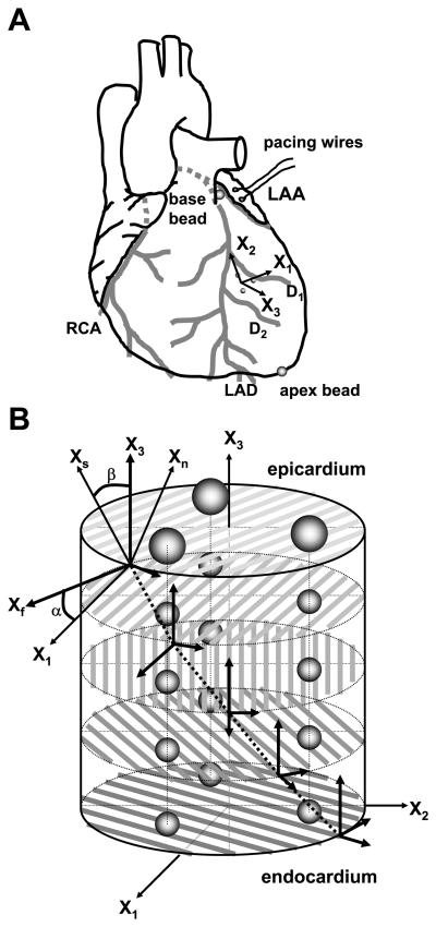
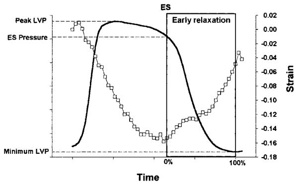
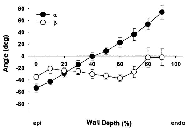
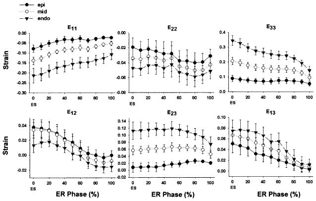
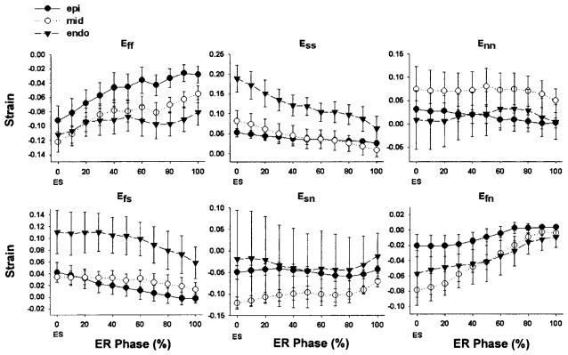
Early relaxation in the cardiac cycle is characterized by rapid torsional recoil of the left ventricular (LV) wall. To elucidate the contribution of the transmural arrangement of the myofiber to relaxation, we determined the time course of three-dimensional fiber-sheet strains in the anterior wall of five adult mongrel dogs in vivo during early relaxation with biplane cineangiography (125 Hz) of implanted transmural markers. Fiber-sheet strains were found from transmural fiber and sheet orientations directly measured in the heart tissue. The strain time course was determined during early relaxation in the epicardial, midwall, and endocardial layers referenced to the end-diastolic configuration. During early relaxation, significant circumferential stretch, wall thinning, and in-plane and transverse shear were observed (P < 0.05). We also observed significant stretch along myofibers in the epicardial layers and sheet shortening and shear in the endocardial layers (P < 0.01). Importantly, predominant epicardial stretch along the fiber direction and endocardial sheet shortening occurred during isovolumic relaxation (P < 0.05). We conclude that the LV mechanics during early relaxation involves substantial deformation of fiber and sheet structures with significant transmural heterogeneity. Predominant epicardial stretch along myofibers during isovolumic relaxation appears to drive global torsional recoil to aid early diastolic filling.
Figures
References
-
- Arts T, Costa KD, Covell JW, McCulloch AD. Relating myocardial laminar architecture to shear strain and muscle fiber orientation. Am J Physiol Heart Circ Physiol. 2001;280:H2222–H2229. - PubMed
-
- Arts T, Reneman RS. Dynamics of left ventricular wall and mitral valve mechanics—a model study. J Biomech. 1989;22:261–271. - PubMed
-
- Bell SP, Nyland L, Tischler MD, McNabb M, Granzier H, LeWinter MM. Alterations in the determinants of diastolic suction during pacing tachycardia. Circ Res. 2000;87:235–240. - PubMed
-
- Beyar R, Sideman S. The dynamic twisting of the left ventricle: a computer study. Ann Biomed Eng. 1986;14:547–562. - PubMed
-
- Beyar R, Yin FC, Hausknecht M, Weisfeldt ML, Kass DA. Dependence of left ventricular twist-radial shortening relations on cardiac cycle phase. Am J Physiol Heart Circ Physiol. 1989;257:H1119–H1126. - PubMed
Publication types
MeSH terms
Grants and funding
LinkOut - more resources
Full Text Sources