. 2003 Nov;133(3):1170-80.
doi: 10.1104/pp.103.028266. Epub 2003 Oct 9.

Microspore separation in the quartet 3 mutants of Arabidopsis is impaired by a defect in a developmentally regulated polygalacturonase required for pollen mother cell wall degradation

Affiliations

Microspore separation in the quartet 3 mutants of Arabidopsis is impaired by a defect in a developmentally regulated polygalacturonase required for pollen mother cell wall degradation

Seung Y Rhee et al. Plant Physiol. 2003 Nov.

Abstract

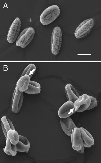
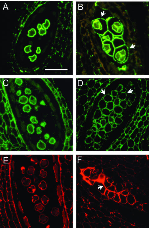
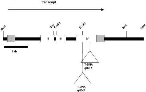
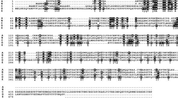
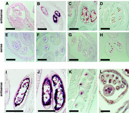
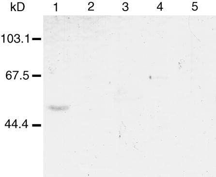
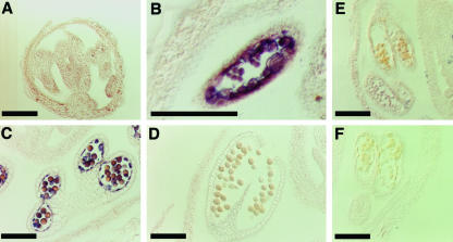
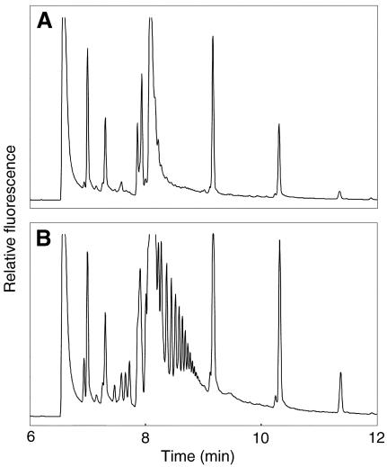
Mutations in the QUARTET loci in Arabidopsis result in failure of microspore separation during pollen development due to a defect in degradation of the pollen mother cell wall during late stages of pollen development. Mutations in a new locus required for microspore separation, QRT3, were isolated, and the corresponding gene was cloned by T-DNA tagging. QRT3 encodes a protein that is approximately 30% similar to an endopolygalacturonase from peach (Prunus persica). The QRT3 protein was expressed in yeast (Saccharomyces cerevisiae) and found to exhibit polygalacturonase activity. In situ hybridization experiments showed that QRT3 is specifically and transiently expressed in the tapetum during the phase when microspores separate from their meiotic siblings. Immunohistochemical localization of QRT3 indicated that the protein is secreted from tapetal cells during the early microspore stage. Thus, QRT3 plays a direct role in degrading the pollen mother cell wall during microspore development.


Figures

Figure 1.
Figure 1.
Scanning electron micrograph of pollen from the wild type (A) and the qrt3-3 mutant (B). The arrows indicate material deposited on the pollen that appears to derive from the pollen mother cell wall. Bar = 10 μm.
Figure 2.
Figure 2.
Immunohistochemical staining of pectin associated with microspores of wild type (A, C, and E) and the qrt3-1 mutant (B, D, and F). Sections were stained with antibodies against unesterified pectin (A and B), methyl-esterified pectin (C and D), and RGII (E and F). In the wild type, the developing pollen grains are separated from each other. In the qrt3 mutant, the developing pollen are enclosed by residual pollen mother cell wall (arrows). Scale bar = 50 μm. The wild-type images were previously published by Rhee and Somerville (1998).
Figure 3.
Figure 3.
Structure of the QRT3 locus At4G20050. The regions of the primary transcript present in a putative full-length cDNA are shown as boxes. The 5′ and 3′ non-translated regions are shown as gray boxes. The primary transcript is processed to remove three introns, one of which is in the 5′-non-translated region. The site of insertion of the TDNA inserts corresponding to the qrt3-1 and qrt3-3 mutations are indicated below the figure.
Figure 4.
Figure 4.
Alignment of QRT3 with sequences of polygalacturonases. A, Tap2g (GenBank accession no. U70480) was isolated from the abscission zone of tomato (Lycopersicon esculentum). B, PGase (GenBank accession no. X76735) was from peach. C, CRYJ2 was from Japanese cedar (Cryptomeria japonica). D, QRT3. E, Consensus sequence.
Figure 5.
Figure 5.
In situ hybridization of antisense and sense QRT3 transcripts to sections of flowers at various stages of microsporogenesis. A to D, Hybridization of QRT3 antisense probe to sections of developing flowers at various stages. E to H, Hybridization of QRT3 sense probes to sections of flowers at the same stages shown in A to E. I and J, Enlarged images of hybridization of antisense QRT3 probe to developing anthers at the tetrad stage. K, Hybridization of QRT3 antisense to ovules of mature flowers. L, Hybridization of antisense eIF4A transcript to section of developing flower at tetrad stage was used as a positive control. The following developmental stages were characterized: A and E, PMC stage buds; B, F, I, and J, tetrad stage buds; C and G, early microspore stage buds; and D and H, late microspore stage buds. Scale bars = 100 μm.
Figure 6.
Figure 6.
Western blot probed with antibody against QRT3. Approximately 20 μg of extracted protein from various tissues of 4-week old plants was loaded in each lane, blotted to nitrocellulose, and detected with an antibody against QRT3 at a dilution of 1:10,000 (v/v). Lane 1, Wild-type unopened flower buds; lane 2, wild type flowers; lane 3, wild-type expanded leaves; lane 4, wild-type roots; lane 5, qrt3-1 unopened flower buds. The positions of Mr standards are indicated on the left.
Figure 7.
Figure 7.
Immunohistochemical localization of QRT3 in wild-type flower buds. Ten-micrometer sections of developing anthers were incubated with anti-QRT3 sera at a dilution of 1:1,000 (v/v). A, Tetrad stage; B, early microspore stage; C, late microspore stage; D, mature pollen; E, early microspore stage probed with pre-immune sera; F, early microspore stage probed with secondary antibody. Bars = 100 μm.
Figure 8.
Figure 8.
Electropherograms of reaction products from QRT3-mediated hydrolysis of poly-GalUA. Poly-GalUA was incubated with protein extracts from control yeast cells (A) or cells expressing the QRT3 enzyme (B) for 12 h, and then the reaction products were resolved by capillary electrophoresis. The series of peaks between approximately 8 and 9 min in the QRT3 reaction products correspond to the elution times of oligo-GalUA standards containing two to eight residues. Other peaks correspond to contaminants in the dye and by-products of the derivatization reactions.

References

    1. Albani D, Altosaar I, Arnison PG, Fabijanski SF (1991) A gene showing sequence similarity to pectin esterase is specifically expressed in developing pollen of Brassica napus: sequences in its 5′ flanking region are conserved in other pollen-specific promoters. Plant Mol Biol 16: 501–513 - PubMed
    1. Amagai M, Ariizumi T, Endo M, Hatakeyama K, Kuwata C, Shibata D, Toriyama K, Watanabe M (2003) Identification of anther-specific genes in a cruciferous model plant, Arabidopsis thaliana, by using a combination of Arabidopsis macroarray and mRNA derived from Brassica oleracea. Sex Plant Reprod 15: 213–220
    1. Ariizumi T, Amagai M, Shibata D, Hatakeyama K, Watanabe M, Toriyama K (2002) Comparative study of promoter activity of three anther-specific genes encoding lipid transfer protein, xyloglucan endotransglucosylase/hydrolase and polygalacturonase in transgenic Arabidopsis thaliana. Plant Cell Rep 21: 90–96
    1. Ausubel FM, Brent R, Kingston RE, Moore DD, Seidman JG, Smith JA, Struhl K (1989) Preparation of genomic DNA from plant tissue. In FM Ausubel, R Bent, RE Kingston, DD Moore, JG Seidman, JA Smith, K Struhl, eds, Current Protocols in Molecular Biology, Vol. 1. Wiley, New York, pp 2.3.1–2.3.3
    1. Barnett MJ, Long SR (1997) Identification and characterization of a gene on Rhizobium meliloti pSyma, syrB, that negatively affects syrM expression. Mol Plant-Microbe Interact 10: 550–559 - PubMed

Publication types

MeSH terms

Associated data