. 2003 Nov 15;553(Pt 1):13-9.
doi: 10.1113/jphysiol.2003.054551. Epub 2003 Oct 10.

Small conductance Ca2+-activated K+ channels formed by the expression of rat SK1 and SK2 genes in HEK 293 cells

Affiliations

Small conductance Ca2+-activated K+ channels formed by the expression of rat SK1 and SK2 genes in HEK 293 cells

David C H Benton et al. J Physiol. .

Abstract

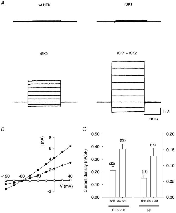
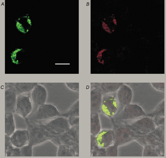
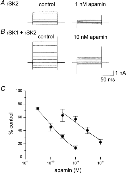
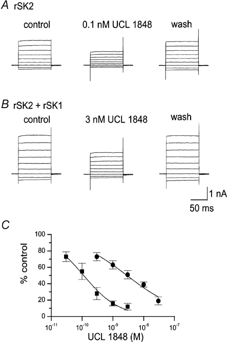
The rat SK1 gene (rSK1) does not form functional Ca2+-activated potassium channels when expressed alone in mammalian cell lines. Using a selective antibody to the rSK1 subunit and a yellow fluorescent protein (YFP) tag we have discovered that rSK1 expression produces protein that remains largely at intracellular locations. We tested the idea that rSK1 may need an expression partner, rSK2, in order to form functional channels. When rSK1 was co-expressed with rSK2 in HEK 293 cells it increased the current magnitude by 77 +/- 34% (as compared with cells expressing rSK2 alone). Co-expression of rSK1 with rSK2 also changed the channel pharmacology. The sensitivity of SK current to block by apamin was reduced approximately 16-fold from an IC50 of 94 pM (for SK2 alone) to 1.4 nM (for SK2 and SK1 together). The sensitivity to block by UCL 1848 (a potent small molecule blocker of SK channels) was similarly reduced, approximately 26-fold, from an IC50 of 110 pM to 2.9 nM. These data clearly demonstrate that rSK1 and rSK2 subunits interact. The most likely explanation for this is that the subunits are able to form heteromeric assemblies.


Figures

Figure 1
Figure 1. Expression of rSK1 and rSK2 alone or together in HEK 293 cells
A, typical whole-cell current records obtained from an untransfected (wild-type, wt) HEK 293 cell (top left) and from cells transfected with rSK1 (top right), rSK2 (bottom left) or rSK1 together with rSK2 (bottom right). The cells were held at −80 mV and 100 ms steps applied from −120 to +40 mV. In all cases, the pipette solution contained 1 µm free calcium to activate SK currents. Currents were recorded 1-2 min after patch rupture. B, current-voltage relationships for the currents shown in A. ○, wild-type HEK; ▴, rSK1 (obscured beneath wt data); •, rSK2; ▪, rSK1 and rSK2. Currents recorded from wild-type cells or cells transfected with rSK1 were essentially identical and reversed between −20 and −10 mV. In contrast cells transfected with rSK2 or a combination of rSK1 and rSK2 exhibited large currents with an approximately linear current-voltage relationship, which reversed at approximately −80 mV, close to the predicted value of EK (−85 mV). C, comparison of SK current density in HEK 293 (left) or H4 (right) cells transfected with rSK2 either alone or with a 2-fold excess of rSK1. Histograms represent the mean current density at −20 mV, with the number of cells for each condition shown in parentheses. In both cases co-transfection with rSK1 caused a significant (P < 0.05) increase in current. Error bars indicate s.e.m.
Figure 2
Figure 2. Distribution of rSK1 protein in HEK 293 cells
A, confocal image of HEK 293 cells transiently transfected with a YFP-tagged rSK1 construct (green filter) showing that the expressed protein is unevenly distributed around the cell and has a punctate staining pattern. B, the staining seen with rSK1-specific antibody UCL 56. Comparison with A shows that it recognizes the channel protein because the staining coincides well with YFP fluorescence. C, bright field image of cells depicted in A and B. D, overlay of images A, B and C. Scale bar is 20 µm.
Figure 3
Figure 3. Apamin block of current in cells expressing rSK2 alone or rSK1 and rSK2
A, typical current traces showing the effect of 1 nm apamin on homomeric rSK2 currents. B, typical current traces showing the effect of 10 nm apamin on a cell co-transfected with rSK1 and rSK2. In both A and B, currents were recorded immediately before and after 4 min exposure to apamin. The scale bar applies to both A and B. C, apamin concentration-inhibition curves for cells transfected with rSK2 alone (▪) or rSK1 and rSK2 (•). Continuous lines are fits of the Hill equation to the data, yielding estimates of IC50 of 95 ± 8 pm (nH = 0.80 ± 0.06) and 1.4 ± 0.3 nm (nH = 0.6 ± 0.1) for rSK2 and rSK1 with rSK2, respectively. Each point is the mean of 4–9 observations and the error bars indicate s.e.m.
Figure 4
Figure 4. UCL 1848 block of current in cells expressing rSK2 alone or rSK1 and rSK2
A, effect of 0.1 nm UCL 1848 on rSK2 currents. B, effect of 3 nm UCL 1848 on a cell co-transfected with rSK1 and rSK2. In A and B currents were recorded before and after 3 min exposure to UCL 1848, and 3 min after washout. The scale bar applies to both A and B. C, UCL 1848 concentration-inhibition curves for cells transfected with rSK2 alone (▪) or rSK1 and rSK2 (•). Continuous lines show fits of the Hill equation to the data, yielding estimates of IC50 of 110 ± 26 pm (nH = 0.7 ± 0.1) and 2.9 ± 0.3 nm (nH = 0.49 ± 0.04) for rSK2 and rSK1 with rSK2, respectively. Each point is the mean of 3-5 observations. Error bars indicate s.e.m.

References

    1. Benton DCH, Dunn PM, Chen JQ, Galanakis D, Ganellin CR, Malik-Hall M, Shah M, Haylett DG, Jenkinson DH. UCL 1848: a novel bis-quinolinium cyclophane which blocks apamin-sensitive K+ channels with nanomolar affinity. Br J Pharmacol. 1999;128:39P.
    1. Benton DCH, Monaghan AS, Hosseini R, Bahia PK, Shah Y, Haylett DG, Moss GWJ. Interactions between rat SK channel subunits expressed in HEK 293 cells. J Physiol. 2003;551.P:C43. - PMC - PubMed
    1. Bowden SE, Fletcher S, Loane DJ, Marrion NV. Somatic colocalization of rat SK1 and D class (Cav1. 2) L-type calcium channels in rat CA1 hippocampal pyramidal neurons. J Neurosci. 2001;21:RC175. - PMC - PubMed
    1. Chandy KG, Fantino E, Wittekindt O, Kalman K, Tong LL, Ho TH, Gutman GA, Crocq MA, Ganguli R, Nimgaonkar V, Morris-Rosendahl DJ, Gargus JJ. Isolation of a novel potassium channel gene hSKCa3 containing a polymorphic CAG repeat: a candidate for schizophrenia and bipolar disorder? Mol Psychiatry. 1998;3:32–37. - PubMed
    1. Chen JQ, Galanakis D, Ganellin CR, Dunn PM, Jenkinson DH. bis-Quinolinium cyclophanes: 8, 14-diaza-1,7 (1,4)-diquinolinacyclotetradecaphane (UCL 1848), a highly potent and selective, nonpeptidic blocker of the apamin-sensitive Ca2+-activated K+ channel. J Med Chem. 2000;43:3478–3481. - PubMed

Publication types

MeSH terms

LinkOut - more resources