. 2003 Nov 15;553(Pt 1):183-9.
doi: 10.1113/jphysiol.2003.051896. Epub 2003 Oct 10.

Small- and intermediate-conductance calcium-activated K+ channels provide different facets of endothelium-dependent hyperpolarization in rat mesenteric artery

Affiliations

Small- and intermediate-conductance calcium-activated K+ channels provide different facets of endothelium-dependent hyperpolarization in rat mesenteric artery

G J Crane et al. J Physiol. .

Abstract

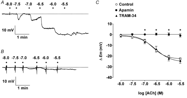
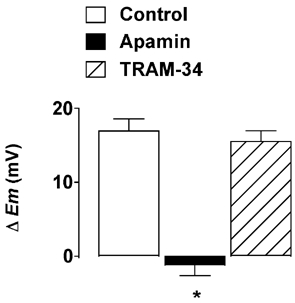
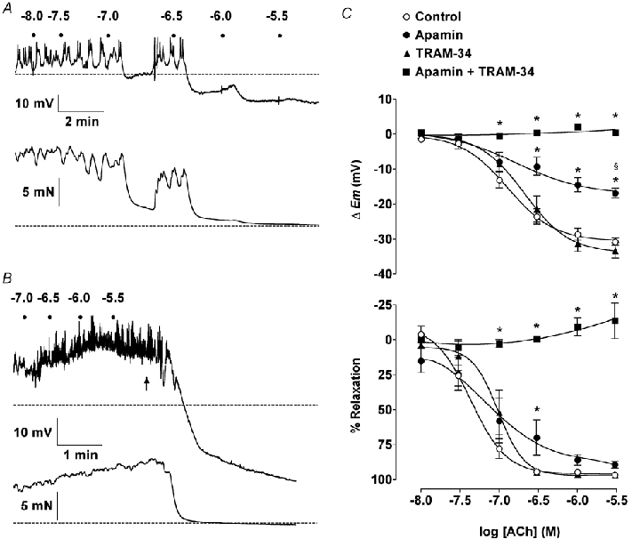
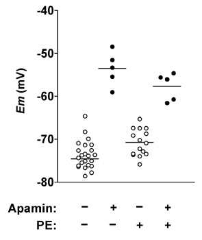
Activation of both small-conductance (SKCa) and intermediate-conductance (IKCa) Ca2+-activated K+ channels in endothelial cells leads to vascular smooth muscle hyperpolarization and relaxation in rat mesenteric arteries. The contribution that each endothelial K+ channel type makes to the smooth muscle hyperpolarization is unknown. In the presence of a nitric oxide (NO) synthase inhibitor, ACh evoked endothelium and concentration-dependent smooth muscle hyperpolarization, increasing the resting potential (approx. -53 mV) by around 20 mV at 3 microM. Similar hyperpolarization was evoked with cyclopiazonic acid (10 microM, an inhibitor of sarcoplasmic endoplasmic reticulum calcium ATPase (SERCA)) while 1-EBIO (300 microM, an IKCa activator) only increased the potential by a few millivolts. Hyperpolarization in response to either ACh or CPA was abolished with apamin (50 nM, an SKCa blocker) but was unaltered by 1-[(2-chlorophenyl) diphenylmethyl]-1H-pyrazole (1 microM TRAM-34, an IKCa blocker). During depolarization and contraction in response to phenylephrine (PE), ACh still increased the membrane potential to around -70 mV, but with apamin present the membrane potential only increased just beyond the original resting potential (circa -58 mV). TRAM-34 alone did not affect hyperpolarization to ACh but, in combination with apamin, ACh-evoked hyperpolarization was completely abolished. These data suggest that true endothelium-dependent hyperpolarization of smooth muscle cells in response to ACh is attributable to SKCa channels, whereas IKCa channels play an important role during the ACh-mediated repolarization phase only observed following depolarization.


Figures

Figure 1
Figure 1. Effect of apamin or TRAM-34 on smooth muscle hyperpolarization in response to ACh
A and B, representative original traces show ACh cumulative concentration-increases in smooth muscle cell membrane potential in rat isolated mesenteric arteries. Each dot represents the addition of ACh (log molar concentration). A, in the presence of 100 µml-NAME (Control in C) the impaled cell hyperpolarized from a resting membrane potential (rmp) of − 53.5 mV (dashed line) to a maximum of − 78.1 mV. In the presence of 50 nm apamin (B) the rmp (−52.6 mV, dashed line) was not significantly increased. C, summarized data showing the average change in membrane potential (▵Em) to cumulative increases in [ACh] under control conditions (rmp = −51.7 ± 0.6 mV, n = 17–25), and in the presence of 50 nm apamin (rmp = −52.8 ± 1.0 mV, n = 5) or 1 µm TRAM-34 (rmp = −51.1 ± 1.0 mV, n = 6–7). Apamin alone was fully able to abolish hyperpolarization to ACh, whereas TRAM-34 had no effect. * P < 0.05vs. control.
Figure 2
Figure 2. Effect of apamin or TRAM-34 on smooth muscle hyperpolarization in response to CPA
Under control conditions, cyclopiazonic acid (CPA, 10 µm) hyperpolarized smooth muscle cells by an average of 16.9 ± 1.7 mV (n = 9). This hyperpolarization was abolished in the presence of 50 nm apamin (n = 5), but unaffected by 1 µm TRAM-34 (n = 3). * P < 0.05vs. control. ▵Em, change in membrane potential.
Figure 3
Figure 3. Effect of apamin and TRAM-34 on smooth muscle hyperpolarization in response to ACh in the presence of phenylephrine
A and B, representative records show simultaneous smooth muscle cell hyperpolarization and relaxation in response to ACh in rat isolated mesenteric arteries contracted with phenylephrine. Dots represent addition of ACh (log molar concentrations). A, in the presence of 100 µml-NAME (Control) 0.6 µm phenylephrine caused depolarization from −52.5 mV (rmp, dashed line) to − 44.6 mV and contraction from 2.3 mN (resting tension, dashed line) to 16.0 mN. ACh stimulated hyperpolarization to levels beyond rmp (maximum −70.1 mV), associated with almost complete relaxation. B, in the combined presence of 50 nm apamin and 1 µm TRAM-34, 1.3 µm phenylephrine stimulated depolarization from −51.5 mV to −39.3 mV and contraction from 3.2 mN to 15.4 mN. Under these conditions, ACh was unable to stimulate hyperpolarization or relaxation, but the subsequent addition of 3 µm levcromakalim (indicated by arrow) was able to stimulate hyperpolarization (to − 81.3 mV) and relaxation. C, summarized data showing the average change in smooth muscle membrane potential (▵Em, top panel) and tension (% Relaxation, bottom panel) to cumulative increases in [ACh]. TRAM-34 had no effect on ACh responses, whereas apamin prevented hyperpolarization beyond rmp with slightly reduced relaxation. The combination of apamin plus TRAM-34 abolished hyperpolarization or relaxation to ACh.
Figure 4
Figure 4. Effect of apamin on smooth muscle hyperpolarization in response to ACh
Scatter of individual impalements, showing the maximum steady-state membrane potential in response to 3 µm ACh with or without 50 nm apamin present, and in both uncontracted arteries or arteries contracted with phenylephrine (PE). Each horizontal line represents the mean for that data set. Membrane potentials achieved with ACh were independent of resting potential in the absence but not the presence of apamin.

References

    1. Barriere E, Tazi KA, Pessione F, Heller J, Poirel O, Lebrec D, Moreau R. Role of small-conductance Ca2+-dependent K+ channels in in vitro nitric oxide-mediated aortic hyporeactivity to alpha-adrenergic vasoconstriction in rats with cirrhosis. J Hepatol. 2001;35:350–357. - PubMed
    1. Bolz SS. Endothelium-derived hyperpolarizing factor but not NO reduces smooth muscle Ca2+ during acetylcholine-induced dilation of microvessels. Br J Pharmacol. 1999;128:124–134. - PMC - PubMed
    1. Burdyga T, Shmygol A, Eisner DA, Wray S. A new technique for simultaneous and in situ measurements of Ca2+ signals in arteriolar smooth muscle and endothelial cells. Cell Calcium. 2003;34:27–33. - PubMed
    1. Burnham MP, Bychkov R, Feletou M, Richards GR, Vanhoutte PM, Weston AH, Edwards G. Characterization of an apamin-sensitive small-conductance Ca2+-activated K+ channel in porcine coronary artery endothelium: relevance to EDHF. Br J Pharmacol. 2002;135:1133–1143. - PMC - PubMed
    1. Busse R, Edwards G, Feletou M, Fleming I, Vanhoutte PM, Weston AH. EDHF: bringing the concepts together. Trends Pharmacol Sci. 2002;23:374–380. - PubMed

Publication types

MeSH terms

LinkOut - more resources