Comparative Study
. 2004 Jan 15;554(Pt 2):335-52.
doi: 10.1113/jphysiol.2003.051748. Epub 2003 Oct 10.

The mechanism of the force response to stretch in human skinned muscle fibres with different myosin isoforms

Affiliations
Comparative Study

The mechanism of the force response to stretch in human skinned muscle fibres with different myosin isoforms

Marco Linari et al. J Physiol. .

Erratum in

  • J Physiol. 2004 Mar 16;555(Pt 3):851

Abstract

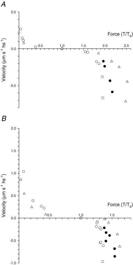
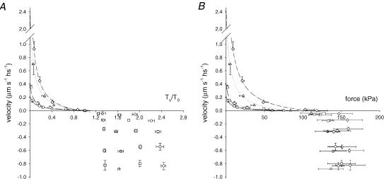
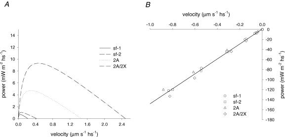
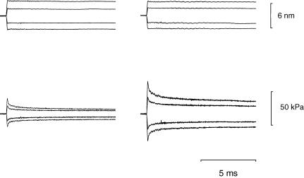
Force enhancement during lengthening of an active muscle, a condition that normally occurs during locomotion in vivo, is attributed to recruitment of myosin heads that exhibit fast attachment to and detachment from actin in a cycle that does not imply ATP splitting. We investigated the kinetic and mechanical features of this cycle in Ca(2+) activated single skinned fibres from human skeletal muscles containing different myosin heavy chain (MHC) isoforms, identified with single-fibre gel electrophoresis. Fibres were activated by using a new set-up that allows development of most of the tension following a temperature jump from 0-1 degrees C to the test temperature (approximately 12 degrees C). In this way we could prevent the development of sarcomere non-uniformity and record sarcomere length changes with a striation follower in any phase of the mechanical protocol. We found that: (i) fibres with fast MHC isoforms develop 40-70% larger isometric forces than those with slow isoforms, as a result of both a larger fraction of force-generating myosin heads and a higher force per head; (ii) in both slow and fast fibres, force enhancement by stretch is due to recruitment of myosin head attachments, without increase in strain per head above the value generated by the isometric heads; and (iii) the extent of recruitment is larger in slow fibres than in fast fibres, so that the steady force and power output elicited by lengthening become similar, indicating that mechanical and kinetic properties of the actin-myosin interactions under stretch become independent of the MHC isoform.


Figures

Figure 1
Figure 1. Slow-time-base record from a fast fibre to show the experimental protocol
Upper trace, force; lower trace, motor position. The dashed lines mark the time when the solution was changed; the dotted line marks the time of transition from low temperature (LT) to test temperature (TT). Arrows indicate the time during which a ramp lengthening (8% of fibre length) at a velocity of 0.09 μm s−1 hs−1 was imposed on the fibre before returning to relaxing solution. A large release (10% of fibre length) is imposed on the fibre to measure the force baseline (not resolved in the force record since the trace is from the chart recorder). Fibre length, 3.03 mm; average sarcomere length, 2.53 μm; CSA, 8900 μm2. Low temperature (LT), 1.0°C; test temperature (TT), 11.5°C. Fibre characterization indicates that the MHC isoform is 2A/2X type.
Figure 2
Figure 2. Force development following a T-jump (from 1 to 12°C) in slow (left column) and fast (right column) fibres and identification of fibre types
A, traces indicate segment length change (upper panel), force response and zero force (lower panel). The artefact before the striation follower signal is due to the fibre travelling in air and within the shadow zone. Slow fibre (type 1 isoform): fibre length, 2.10 mm; segment length under the striation follower, 1.00 mm; average segment sarcomere length, 2.58 μm; CSA, 5200 μm2. Fast fibre (type 2A/2X isoform): fibre length, 2.45 mm; segment length under the striation follower, 0.87 mm; average segment sarcomere length, 2.52 μm; CSA, 9000 μm2. B, MHC isoform identification in single fibres by SDS-PAGE. The areas of migration of MHC-1, MHC-2A and MHC-2X are indicated on the left. Lane a: a single fibre containing MHC-2A and MHC-2X, i.e. a fast, type 2AX, fibre; lane b: a sample from vastus lateralis muscle containing all three adult MHC isoforms; lane c: a single fibre containing MHC-1, i.e. a slow, type 1, fibre.
Figure 3
Figure 3. Force response to steady shortening (A and B) and lengthening (CF) imposed on activated fast fibres
Left column: 2A type; right column: 2A/2X type. Figures close to the records indicate the velocity (μm s−1 hs−1) during the steady-state force response. In each row, force responses obtained at similar velocities are compared. In each frame, from top to bottom, traces indicate: sarcomere length change, force response and zero force. The time-scale is 400 ms (AD) or 100 ms (EF). 2A fibre: fibre length, 2.25 mm; segment length, 0.98 mm; average sarcomere length in the segment, 2.71 μm; CSA, 6700 μm2, temperature, 11.8°C. 2A/2X fibre: fibre length, 3.03 mm; segment length, 0.85 mm; average sarcomere length in the segment, 2.52 μm; CSA, 8900 μm2, temperature, 11.5°C. The two fibres are from vastus lateralis muscle of two different subjects.
Figure 4
Figure 4. Force response to steady shortening (A and B) and lengthening (CF) imposed on activated slow fibres. Left column: sf-1 type; right column: sf-2 type
Figures close to the records indicate the velocity (μm s−1 hs−1) at the steady state of force response. In each row, force responses obtained for similar velocities are compared. In each frame, from top to bottom, traces indicate: sarcomere length change, force response and zero force. The time-scale is 400 ms (AD) or 100 ms (EF). sf-1 fibre: fibre length, 1.96 mm; segment length, 1.05 mm; average sarcomere length in the segment, 2.51 μm; CSA, 7000 μm2, temperature, 11.8°C. sf-2 fibre: fibre length, 2.10 mm; segment length, 1.0 mm; average sarcomere length in the segment, 2.58 μm; CSA, 5200 μm2, temperature, 11.8°C. The two fibres are from vastus lateralis muscle of the same subject.
Figure 5
Figure 5. Force–velocity relations in two slow fibres (A) and in two fast fibres (B). Negative velocity refers to lengthening
Open symbols, steady force; filled circles, peak of force during lengthening. A, triangles (sf-1) and circles (sf-2) refer to the same slow fibres as in Fig. 4. B, triangles: 2A fast fibre (same fibre as in Fig. 3); circles: 2A/2X fast fibre (fibre length, 3.40 mm; segment length under the striation follower, 1.20 mm; average segment sarcomere length, 2.64 μm; CSA, 12 400 μm2; temperature, 11.6°C; isometric force, 170 kPa). Filled circles show the relation between the peak of force and the lengthening velocity during the corresponding phase of force response for the sf-2 and 2A/2X fibres.
Figure 6
Figure 6. Mean force–velocity relations in slow and fast fibres
A, force relative to the isometric value. B, force in kPa. Negative velocities refer to lengthening. Circles: sf-1, n = 8; squares, sf-2, n = 5; triangles: 2A, n = 6; diamonds: 2A/2X, n = 11. Bars are ± s.e.m.. Data are grouped in classes of velocity. The sample number in each class varies between 2 and 14. Lines are Hill's equations fitted to the pooled force–shortening velocity data. Continuous lines, sf-1; dashed lines, sf-2; dotted lines, 2A; dot–dashed lines, 2A/2X.
Figure 7
Figure 7. Relations between power and velocity during steady shortening (A) and lengthening (B)
Lines in A were obtained from Hill's equation fitted to the relative force–shortening velocity curves of Fig. 6. The points in B represent the mean values obtained from the points for lengthening in Fig. 6, and the line represents the linear regression (slope, 148.9 ± 2.9 kPa; ordinate intercept, 0.81 ± 1.31 mW m−2 hs−1) fitted to the pooled data.
Figure 8
Figure 8. Superimposed tension responses to length steps of different size in a slow fibre activated at different pCa
Left column, pCa = 5.63; right column, pCa = 4.50. Upper panels, length change per hs; lower panels, force response. Fibre length, 3.15 mm; segment length, 1.05 mm; average sarcomere length in the segment, 2.40 μm; CSA, 5800 μm2; temperature, 12.1°C; isometric force at pCa = 4.50, 66 kPa.
Figure 9
Figure 9. Isometric force–pCa relation (A and B) and T1 relations at different pCa (C and D) in a slow fibre (A and C) and a fast fibre (B and D)
The continuous lines in A and B are calculated from Hill's equation: T0/T0,4.50= 1/[1 + 10n(pK–pCa)], where n, the Hill coefficient, expresses the slope and pK is pCa at which T0= 0.5T0,4.50. The best-fitting parameters, obtained with the nonlinear least-squares method (SigmaPlot, Jandel Scientific), are listed in the figure close to the graphs. T1 curves in C and D are obtained by plotting the extreme tension attained at the end of the length step (T1, relative to T0 at pCa = 4.50, T0,S) against the step amplitude. Different symbols refer to different pCa as indicated by the figure close to the corresponding relation. Circles refer to full activation before (open circles) and after (filled circles) the series at larger pCa. The lines represent the linear regressions fitted to the experimental points at each pCa. A and C, same fibre as in Fig. 8. B and D, fast (2A/2X) fibre: fibre length, 3.51 mm; segment length, 1.03 mm; average sarcomere length in the segment, 2.45 μm; CSA, 9500 μm2; temperature, 12.1°C; T0,S, 109 kPa. Mean values (± s.e.m.) of the isometric force (T0) and stiffness (e0) at full activation are as follows. Slow fibres (3 fibres): T0= 72 ± 5 kPa; e0= 13.97 ± 2.36 kPa nm−1; fast fibres (2 fibres): T0= 106 ± 4 kPa; e0= 16.15 ± 0.93 kPa nm−1.
Figure 10
Figure 10. Relation of hs extension (Y0) versus T0 at different pCa values
Open symbols: slow fibres; filled symbols: fast 2A/2X fibres. Different symbols refer to different fibres. The continuous lines were obtained by linear regression on the pooled data for either slow or fast fibres.
Figure 11
Figure 11. Stiffness during steady lengthening in slow and fast fibres
A, sample records of force responses to step length changes imposed on the active fibre either in isometric conditions (upper row) or during steady force response to lengthening at about 0.65 μm s−1 hs−1 (lower row) in a slow fibre (left column) and in a fast 2A/2X fibre (right column). In each panel, the upper trace is length change per hs; the lower trace is the force response. Slow fibre: fibre length, 3.35 mm; segment length, 1.07 mm; average sarcomere length in the segment, 2.51 μm; CSA, 4500 μm2; temperature, 12.1°C; T0, 67 kPa; Tv/T0, 2.48. 2A/2X fibre: fibre length, 3.4 mm; segment length, 1.2 mm; average sarcomere length in the segment, 2.64 μm; CSA, 12 400 μm2; temperature, 11.6°C; T0, 170 kPa; Tv/T0, 1.32. B, relation of stiffness versus lengthening velocity. Stiffness is relative to that measured in isometric conditions. Filled circles are mean values (± s.e.m.) for slow fibres (nine fibres; data contributing to each class between 5 and 15). Open symbols are for fast fibres (four fibres; each symbol refers to a different fibre). C, relation of the stiffness measured in kPa nm−1 hs−1versus lengthening velocity for the same data as in B.

Similar articles

Cited by

References

    1. Bagni MA, Cecchi G, Colombini B, Colomo F. A non-cross-bridge stiffness in activated frog muscle fibers. Biophys J. 2002;82:3118–3127. - PMC - PubMed
    1. Blangé T, Stienen GJM, Treijtel BW. Active stiffness in frog skinned muscle fibres at different Ca concentrations. J Physiol. 1985;366:65P.
    1. Bottinelli R. Functional heterogeneity of mammalian single muscle fibres: do myosin isoforms tell the whole story? Pflugers Arch. 2001;443:6–17. - PubMed
    1. Bottinelli R, Betto R, Schiaffino S, Reggiani C. Unloaded shortening velocity and myosin heavy chain and alkali light chain isoform composition in rat skeletal muscle fibres. J Physiol. 1994;478:341–349. - PMC - PubMed
    1. Bottinelli R, Canepari M, Pellegrino MA, Reggiani C. Force-velocity properties of human skeletal muscle fibres: myosin heavy chain isoform and temperature dependence. J Physiol. 1996;495:573–586. - PMC - PubMed

Publication types