. 2003 Oct 28;100(22):12601-6.
doi: 10.1073/pnas.1635129100. Epub 2003 Oct 10.

Processive phosphorylation of alternative splicing factor/splicing factor 2

Affiliations

Processive phosphorylation of alternative splicing factor/splicing factor 2

Brandon E Aubol et al. Proc Natl Acad Sci U S A. .

Abstract

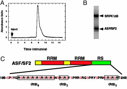
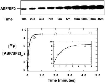
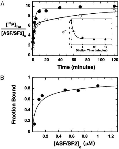
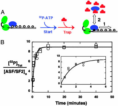
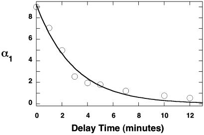
SR proteins, named for their multiple arginine/serine (RS) dipeptide repeats, are critical components of the spliceosome, influencing both constitutive and alternative splicing of pre-mRNA. SR protein function is regulated through phosphorylation of their RS domains by multiple kinases, including a family of evolutionarily conserved SR protein-specific kinases (SRPKs). The SRPK family of kinases is unique in that they are capable of phosphorylating repetitive RS domains with remarkable specificity and efficiency. Here, we carried out kinetic experiments specially developed to investigate how SRPK1 phosphorylates the model human SR protein, ASF/SF2. By using the start-trap strategy, we monitored the progress curve for ASF/SF2 phosphorylation in the absence and presence of an inhibitor peptide directed at the active site of SRPK1. ASF/SF2 modification is not altered when the inhibitor peptide (trap) is added with ATP (start). However, when the trap is added first and allowed to incubate for a specific delay time, the decrease in phosphate content of the enzyme-substrate complex follows a simple exponential decline corresponding to the release rate of SRPK1. These data demonstrate that SRPK1 phosphorylates a specific region within the RS domain of ASF/SF2 by using a fully processive catalytic mechanism, in which the splicing factor remains "locked" onto SRPK1 during RS domain modification.


Figures

Fig. 1.
Fig. 1.
Isolation of an SRPK1ΔS–ASF/SF2 complex. (A) Size-exclusion chromatogram of refolded complex. (B) SDS/12% PAGE of size-exclusion peak. (C) Domain structure of ASF/SF2. The two RRM domains are shown in red, whereas the C-terminal RS domain is shown in green. All the potential serine phosphorylation sites are shown in red.
Fig. 2.
Fig. 2.
Progress curve for the phosphorylation of ASF/SF2. SRPK1ΔS–ASF/SF2 is diluted to 0.6 μM in a buffer containing 200 μM [32P]ATP, and phosphate incorporation was measured at various times from 10 s to 45 min by using autoradiography. The data were fit to an exponential function with a forward rate constant (kf) of 0.80 ± 0.08 min–1 and an amplitude of 9 ± 0.5 phosphates.
Fig. 3.
Fig. 3.
Complex stability. (A) Progress curves under dissociating conditions. SRPK1ΔS–ASF/SF2 was diluted from 4 to 0.1 μM in a buffer containing 200 μM [32P]ATP, and phosphorylation was monitored by using autoradiography (•). In a separate experiment, the complex was diluted to 0.1 μM and allowed to incubate for 5 min before addition of 200 μM [32P]ATP (○). The data at zero delay time were fit to a single-exponential function with an amplitude of 9.5 ± 0.40 phosphates. The data at the 5-min dilution were fit to a double-exponential function with amplitudes of α1 = 6.1 ± 0.4 and α2 = 3.4 ± 0.4 phosphates. (Inset) Values for α1 at a series of dilution times (0–15 min). The line drawn through the data was obtained from kinetic simulations (see text). (B) Dissociation constant for SRPK1ΔS–ASF/SF2. Progress curves for ASF/SF2 phosphorylation were measured a several complex concentrations. The fraction bound was determined from α1 after 10-min dilution times and plotted against total ASF/SF2 concentration. The data are fit to Eq. 1 to obtain a Kd of 50 ± 25 nM.
Fig. 4.
Fig. 4.
Start–trap experiment. (A) Experimental design. SRPK1ΔS–ASF/SF2 is mixed simultaneously with [32P]ATP (start) and AlaPep (trap). In this experimental scheme, the enzyme and AlaPep are shown in blue and red, respectively. The two RRMs of ASF/SF2 are shown in green. The unphosphorylated RS domain is designated with open circles. One phosphorylated serine is shown with a filled circle. Pathways 1 and 2 describe processive and distributive mechanisms, respectively. (B) Progress curves with and without trap. SRPK1ΔS–ASF/SF2 (1 μM) is mixed with 200 μM[32P]ATP, and time-dependent phosphorylation is monitored by using the autoradiographic assay in the absence (○) and presence (□) of 20 mM AlaPep. The data are fit to a single-exponential function with a common rate constant of 0.80 ± 0.10 min –1 and amplitude of 9.0 ± 0.5 phosphates.
Fig. 5.
Fig. 5.
Trap–delay–start experiment. SRPK1ΔS–ASF/SF2 complex (1 μM) is mixed with AlaPep (20 mM) and allowed to equilibrate for various delay times (0–12 min) before 200 μM[32P]ATP is added (start phase). The amplitude of the first kinetic phase (α1) in the progress curves is plotted as a function of the delay time and then fit to a single-exponential function with a rate constant of 0.30 ± 0.02 min–1.

References

    1. Fu, X. D. (1995) RNA 1 663–680. - PMC - PubMed
    1. Zahler, A. M., Lane, W. S., Stolk, J. A. & Roth, M. B. (1992) Genes Dev. 6 837–847. - PubMed
    1. Gui, J. F., Tronchere, H., Chandler, S. D. & Fu, X. D. (1994) Proc. Natl. Acad. Sci. USA 91 10824–10828. - PMC - PubMed
    1. Wang, H. Y., Lin, W., Dyck, J. A., Yeakley, J. M., Songyang, Z., Cantley, L. C. & Fu, X. D. (1998) J. Cell Biol. 140 737–750. - PMC - PubMed
    1. Stojdl, D. F. & Bell, J. C. (1999) Biochem. Cell Biol. 77 293–298. - PubMed

Publication types

MeSH terms

LinkOut - more resources