Comparative Study
. 2003 Oct 28;100(22):13087-92.
doi: 10.1073/pnas.2132313100. Epub 2003 Oct 10.

Trace but not delay fear conditioning requires attention and the anterior cingulate cortex

Affiliations
Comparative Study

Trace but not delay fear conditioning requires attention and the anterior cingulate cortex

C J Han et al. Proc Natl Acad Sci U S A. .

Abstract

Higher cognitive functions such as attention have been difficult to model in genetically tractable organisms. In humans, attention-distracting stimuli interfere with trace but not delay conditioning, two forms of associative learning. Attention has also been correlated with activation of anterior cingulate cortex (ACC), but its functional significance is unclear. Here we show that a visual distractor interferes selectively with trace but not delay auditory fear conditioning in mice. Trace conditioning is associated with increased neuronal activity in ACC, as assayed by relative levels of c-fos expression, and is selectively impaired by lesions of this structure. The effects of the ACC lesions are unlikely to be caused by indirect impairment of the hippocampus, which is required for mnemonic aspects of trace conditioning. These data suggest that trace conditioning may be useful for studying neural substrates of attention in mice, and implicate the ACC as one such substrate.


Figures

Fig. 1.
Fig. 1.
The conditioning and testing procedures. See Materials and Methods for parameters. (A, B, and E) The training box, the conditioning paradigms, and the testing paradigm. (C) A light presentation served as the distractor. (D) For animals in the distraction conditions, the distractor is presented with a random interstimulus interval of 5, 10, 15, or 20 sec.
Fig. 2.
Fig. 2.
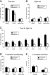
Animals received either a distractor or no distractor during training. On the testing day, animals were presented with the tone and the light, or vice versa. (A) Percent time spent freezing during tone testing. The distractor during conditioning selectively disrupts trace learning, without affecting delay learning. An asterisk indicates significant reduction in time spent freezing for the distractor group compared with the nondistractor group of trace conditioning training (P < 0.05). Both delay and trace conditioning are significantly different from the shock-only conditioning for the nondistracted animals (black bars, P < 0.05). Error bars indicate SEM. (B) For light testing, there is no difference in percent time spent freezing between the animals that did and did not receive the distractor during training. (C) Six groups of mice received one to six tone–shock pairings and the flashing light as the distractor during trace conditioning. One group of mice received standard six tone–shock pairings and no distractor during trace conditioning. There is no difference in freezing to the flashing light for any of the six tone–shock pairings, compared with mice that received no flashing light. As a positive control, the ND group shows a high level of freezing in the tone test. TS, tone–shock pairing; ND, no distractor. (D) No difference was observed between the distractor and no distractor groups, indicating that the distractor did not affect contextual fear conditioning. (E) No difference was found in locomotor activity, as assessed by the number of crossing and rearing events between the distractor and no distractor groups.
Fig. 3.
Fig. 3.
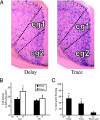
c-fos-positive cell counts after trace or delay conditioning. (A) Representative c-fos in situ hybridization expression in the ACC during delay and trace conditioning. Darker dots are c-fos-positive cells stained by BCIP/NBT. (B) c-fos-positive cell density in the Cg1 and M1. Mice that received trace conditioning have significantly more c-fos-positive cells in the ACC compared with mice that received delay conditioning (*P < 0.05). There is no such difference in M1. (C) Percent time spent freezing during the tone test of the mice trained simultaneously with the mice killed for c-fos mRNA in situ hybridization. Mice trained on delay and trace conditioning exhibit significantly more freezing than the shock-only group (*P < 0.05).
Fig. 4.
Fig. 4.
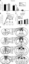
The effects of the pretraining ACC lesion on trace and delay conditioning. (A) Tone test. The ACC lesion group show impaired freezing performance in trace conditioning, but not in delay conditioning, compared with the sham control group. There is no difference between the V1 lesion group and the sham group in either delay or trace group. An asterisk identifies signifi-cantly less time spent freezing of ACC lesions compared with sham operation and V1 lesions in trace conditioning (P < 0.05). An “x” means the corresponding V1 and ACC groups did not exist. (B) Percent time spent freezing as a function of trial number for trace conditioning. The impairment of the ACC lesions can be seen as early as the first trial. (C) Locomotor activities. No difference is found between the ACC, V1, and sham lesion groups trained in trace, assessed by crossing and rearing activities. (D) Schematic representations of the ACC lesions. The dark gray area indicates the smallest and the light gray area indicates the greatest extent of the lesions. Cg, ACC; DP, dorsal peduncular cortex; IL, infralimbic cortex; M2, secondary motor cortex; MO, medial orbital cortex; RS, retrosplenial cortex (posterior cingulate cortex); RSA, retrosplenial cortex agranular; RSG, retrosplenial cortex granular; PrL, prelimbic cortex.

References

    1. Parasuraman, R. (1998) The Attentive Brain (MIT Press, Cambridge, MA).
    1. Braun, J., Koch, C. & Davis, J. L. (2001) Visual Attention and Cortical Circuits (MIT Press, Cambridge, MA).
    1. Robbins, T. W. (2002) Psychopharmacology 163 362-380. - PubMed
    1. Clark, R. E., Manns, J. R. & Squire, L. R. (2002) Trends Cognit. Sci. 6 524-531. - PubMed
    1. Balogh, S. A., Radcliffe, R. A., Logue, S. F. & Wehner, J. M. (2002) Behav. Neurosci. 116 947-957. - PubMed

Publication types