. 2003 Oct 13;163(1):45-56.
doi: 10.1083/jcb.200305051.

AIP is a mitochondrial import mediator that binds to both import receptor Tom20 and preproteins

Affiliations

AIP is a mitochondrial import mediator that binds to both import receptor Tom20 and preproteins

Masato Yano et al. J Cell Biol. .

Abstract

Most mitochondrial preproteins are maintained in a loosely folded import-competent conformation by cytosolic chaperones, and are imported into mitochondria by translocator complexes containing a preprotein receptor, termed translocase of the outer membrane of mitochondria (Tom) 20. Using two-hybrid screening, we identified arylhydrocarbon receptor-interacting protein (AIP), an FK506-binding protein homologue, interacting with Tom20. The extreme COOH-terminal acidic segment of Tom20 was required for interaction with tetratricopeptide repeats of AIP. An in vitro import assay indicated that AIP prevents preornithine transcarbamylase from the loss of import competency. In cultured cells, overexpression of AIP enhanced preornithine transcarbamylase import, and depletion of AIP by RNA interference impaired the import. An in vitro binding assay revealed that AIP specifically binds to mitochondrial preproteins. Formation of a ternary complex of Tom20, AIP, and preprotein was observed. Hsc70 was also found to bind to AIP. An aggregation suppression assay indicated that AIP has a chaperone-like activity to prevent substrate proteins from aggregation. These results suggest that AIP functions as a cytosolic factor that mediates preprotein import into mitochondria.


Figures
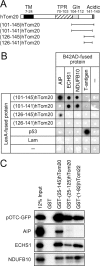
Figure 1.
Figure 1.
AIP binds to the extreme COOH-terminal segment of hTom20. (A) The schematic structures of hTom20 and its deletion mutants fused with LexA are shown. TM, predicted transmembrane domain; TPR, tetratricopeptide repeat motif; Gln, glutamine-rich segment; Acidic, the segment rich in acidic residues. (B) Plasmids expressing LexA-fused hTom20s and B42AD-fused proteins were cotransformed into yeast EGY48[p8op-lacZ] cells. The yeast cells were cultured on indicator medium containing 5-bromo-4-chloro-3-indolyl-d-galactoside. Blue color (seen here in black) indicates a specific interaction in the two-hybrid system. Interaction between p53 and T-antigen was also monitored as positive control. Human lamin C [66–230] (Lam) was used as negative control. ECHS1, mitochondrial short-chain enoyl-coenzyme A hydratase 1 precursor; NDUFB10, mitochondrial NADH:ubiquinone oxidoreductase 1β subcomplex 10. (C) Translation product (10 μl) containing 35S-labeled pOTC-GFP, AIP, ECHS1, or NDUFB10 was incubated with glutathione-Sepharose beads prebound with GST, GST-(25–145)hTom20, GST-(25–125)hTom20, or GST-(1–82)hTom22 (0.6 nmol). After washing, GST derivatives and bound proteins were eluted and analyzed by SDS-PAGE and fluorography. 12% Input: 12% of input 35S-labeled proteins.
Figure 2.
Figure 2.
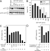
AIP maintains the import competency of pOTC in vitro. (A) 35S-labeled human pOTC or pOTC-GFP translated in 10 μl reticulocyte lysate was mixed with 25 μg isolated mitochondria in the presence of indicated amounts of purified GST-AIP in the import reaction mixture (total, 50 μl). After a 20-min incubation at 25°C, the import reaction was stopped, and reisolated mitochondria were subjected to SDS-PAGE and fluorography. 20% Input; 20% of input 35S-labeled preproteins; p and m, precursor and mature forms, respectively. (B) The radioactive cleaved mature proteins shown in A were quantified. The percentage of import represents the amount of mature proteins compared with controls without GST-AIP. (C) 35S-labeled human pOTC translated in 5 μl reticulocyte lysate was mixed with indicated amounts of GST-AIP in the import reaction mixture without mitochondria (total, 90 μl), and the mixture was incubated at 25°C for 20 min (preincubation). The mixture was then mixed with isolated mitochondria (total, 100 μl). The import reaction and quantification were done as in A and B. The percentage of import represents the amount of mature proteins compared with control without preincubation in the absence of GST-AIP. (D) AIP was translated in reticulocyte lysates in the presence of cold methionine. Mock translation without mRNA was also done. After stopping the translation by adding cycloheximide, the AIP- and mock-translated mixtures were combined as indicated (total, 5 μl), and mixed with 35S-labeled human pOTC in 5 μl reticulocyte lysate. The 10-μl mixture was preincubated at 25°C for 20 min in the import reaction mixture without mitochondria (total, 90 μl), and then mixed with isolated mitochondria (total, 100 μl). The import reaction and quantification were done as in A and B. The percentage of import represents the amount of mature proteins compared with control without preincubation in the presence of mock-translated mixture.
Figure 3.
Figure 3.
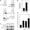
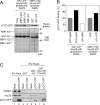
AIP enhances the import of pOTC in cultured cells. (A) Both pCAGGS-pOTC (a plasmid expressing pOTC; 5 μg) and pCAGGS-GFP (a plasmid expressing GFP; 5 μg) were cotransfected with pEGFP-C1 (a plasmid expressing EGFP-C1; 5 μg) or pEGFP-AIP (a plasmid expressing EGFP-AIP; 5 μg) into COS-7 cells on 100-mm dishes. After culturing for 36 h, cells were harvested, and the cell extracts (16 μg protein) were subjected to SDS-PAGE and immunoblot analysis for the indicated proteins; p and m, precursor and mature forms, respectively; AIP, endogenous AIP in COS-7; GAPDH, glyceraldehyde-3-phosphate dehydrogenase. (B) GFP and the processed mature OTC (mOTC) in A were quantified. The ratios of mOTC versus GFP were calculated (mean ± SD, n = 3). The mean of the values for EGFP-C1–expressing cells was set as 1. (C) 5 μg pCAGGS-pOTC, 5 μg pCAGGS-R23GpOTC (a plasmid expressing R23GpOTC), or 5 μg pCAGGS-GFP were cotransfected with 5 μg pEGFP-C1 or 5 μg pEGFP-AIP into COS-7 cells on 100-mm dishes. After culturing for 36 h, cells were radiolabeled for 20 min and then chased using cold methionine. After a 0- and 40-min chase, proteins were immunoprecipitated with anti-OTC or -GFP antiserum and analyzed by SDS-PAGE and fluorography. (D) The radiolabeled proteins chased for 0 min in C were quantified. The ratios of the indicated radiolabeled proteins in EGFP-AIP– expressing cells (EGFP-AIP) versus those in EGFP-C1–expressing cells (EGFP-C1) are shown as “Fold increase” (mean ± SD, n = 3). (E) COS-7 cells on 35-mm dishes were cotransfected with 1 μg pCAGGS-pOTC-GFP and 2.8 μg siRNA. After 3.5 d, the cells were harvested and the cell extract (8 μg protein) was subjected to immunoblot analysis. Control, siRNA for Discosoma sp. red fluorescent protein; (205–225)AIP, siRNA for AIP. (F) The extracts (20 μg protein) of HeLa/pSN and HeLa/pSN-AIP cells were subjected to SDS-PAGE and immunoblot analysis for the indicated proteins. (G) HeLa/pSN and HeLa/pSN-AIP cells on 35-mm dishes were cotransfected with 2 μg pCAGGS-pOTC and 2 μg pCAGGS-GFP. After 48 h, the cells were harvested and the cell extract (10 μg protein) was subjected to immunoblot analysis. (H) GFP and mOTC in G were quantified. The ratios of mOTC versus GFP were calculated (mean ± SD, n = 3). The mean of the values for HeLa/pSN cells was set as 1.
Figure 3.
Figure 3.
AIP enhances the import of pOTC in cultured cells. (A) Both pCAGGS-pOTC (a plasmid expressing pOTC; 5 μg) and pCAGGS-GFP (a plasmid expressing GFP; 5 μg) were cotransfected with pEGFP-C1 (a plasmid expressing EGFP-C1; 5 μg) or pEGFP-AIP (a plasmid expressing EGFP-AIP; 5 μg) into COS-7 cells on 100-mm dishes. After culturing for 36 h, cells were harvested, and the cell extracts (16 μg protein) were subjected to SDS-PAGE and immunoblot analysis for the indicated proteins; p and m, precursor and mature forms, respectively; AIP, endogenous AIP in COS-7; GAPDH, glyceraldehyde-3-phosphate dehydrogenase. (B) GFP and the processed mature OTC (mOTC) in A were quantified. The ratios of mOTC versus GFP were calculated (mean ± SD, n = 3). The mean of the values for EGFP-C1–expressing cells was set as 1. (C) 5 μg pCAGGS-pOTC, 5 μg pCAGGS-R23GpOTC (a plasmid expressing R23GpOTC), or 5 μg pCAGGS-GFP were cotransfected with 5 μg pEGFP-C1 or 5 μg pEGFP-AIP into COS-7 cells on 100-mm dishes. After culturing for 36 h, cells were radiolabeled for 20 min and then chased using cold methionine. After a 0- and 40-min chase, proteins were immunoprecipitated with anti-OTC or -GFP antiserum and analyzed by SDS-PAGE and fluorography. (D) The radiolabeled proteins chased for 0 min in C were quantified. The ratios of the indicated radiolabeled proteins in EGFP-AIP– expressing cells (EGFP-AIP) versus those in EGFP-C1–expressing cells (EGFP-C1) are shown as “Fold increase” (mean ± SD, n = 3). (E) COS-7 cells on 35-mm dishes were cotransfected with 1 μg pCAGGS-pOTC-GFP and 2.8 μg siRNA. After 3.5 d, the cells were harvested and the cell extract (8 μg protein) was subjected to immunoblot analysis. Control, siRNA for Discosoma sp. red fluorescent protein; (205–225)AIP, siRNA for AIP. (F) The extracts (20 μg protein) of HeLa/pSN and HeLa/pSN-AIP cells were subjected to SDS-PAGE and immunoblot analysis for the indicated proteins. (G) HeLa/pSN and HeLa/pSN-AIP cells on 35-mm dishes were cotransfected with 2 μg pCAGGS-pOTC and 2 μg pCAGGS-GFP. After 48 h, the cells were harvested and the cell extract (10 μg protein) was subjected to immunoblot analysis. (H) GFP and mOTC in G were quantified. The ratios of mOTC versus GFP were calculated (mean ± SD, n = 3). The mean of the values for HeLa/pSN cells was set as 1.
Figure 4.
Figure 4.
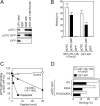
AIP binds to the presequence peptide of pOTC in the same manner as does Tom20. (A) Translation mixture containing 35S-labeled pOTC, pOTC-GFP, or GFP (10 μl) was incubated with glutathione-Sepharose beads prebound with GST, GST-AIP, or GST-(25–145)hTom20 (0.6 nmol) in the binding buffer (total, 300 μl). After washing, GST derivatives and bound proteins were eluted and subjected to SDS-PAGE and fluorography. 12% Input: 12% of input 35S-labeled proteins. (B) The radioactive proteins in A were quantified. The binding was expressed as a percentage of input proteins (mean ± SD, n = 3). (C) Binding of 35S-labeled pOTC-GFP to GST-AIP (0.6 nmol) was assessed in the binding buffer (total, 300 μl) containing 0, 1.2, 6, and 30 nmol of synthetic presequence peptide of human pOTC (Prepeptide), Tom20C[131–145], or PTH[69–84] (Control) as in A, and quantified as in B. Binding was expressed as a percentage of controls without peptides. (D) Binding of 35S-labeled pOTC-GFP derivatives to GST-AIP or GST-(25–145)hTom20 was assessed and quantified as in A and B. WT, pOTC-GFP; R23A, R23ApOTC-GFP; R15/23/26A, R15/23/26ApOTC-GFP.
Figure 5.
Figure 5.
AIP and Tom20 bind to mitochondrial preproteins in a similar manner. (A) Binding of various 35S-labeled mitochondrial and nonmitochondrial proteins to GST-AIP or GST-(25–145)-hTom20 was assessed and quantified. pAAT, preaspartate aminotransferase; pSPT, preserine:pyruvate aminotransferase; AAC, ATP/ADP carrier protein; EYFP-ER, EYFP fused with ER targeting and retrieval signals of calreticulin; NDK-B, nucleoside diphosphate kinase B; TAF9, TATA-binding protein–associated factor. (B) The radioactive proteins in A were quantified. The binding is expressed as a percentage of the input proteins.
Figure 6.
Figure 6.
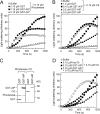
AIP has a chaperone-like activity. (A) Aggregation of 0.15 μM rhodanese was monitored at 55°C in the absence (Buffer) or presence of indicated mounts of GST fusions by measuring the turbidity of the solution at 360 nm. (B) Aggregation of 0.15 μM citrate synthase (CS) was monitored at 43°C as in A. (C) Aggregation of 0.15 μM rhodanese and 0.15 μM citrate synthase was done in the presence of 0.5 μM GST or 0.5 μM GST-AIP. Aggregation of rhodanese was tested at 55°C for 4 min, and that of citrate synthase was tested at 43°C for 10 min. Aggregated proteins were recovered by centrifugation, subjected to SDS-PAGE, and stained with Coomassie brilliant blue R-250. (D) Aggregation of 0.15 μM citrate synthase was monitored in the solution containing 1 mM ATP-MgCl2 at 43°C.
Figure 7.
Figure 7.
Complex formation among preprotein, AIP, Tom20, and other chaperones. (A) Ternary complex formation of preprotein, AIP, and Tom20 was examined. The translation mixture containing 10 μl 35S-labeled pOTC-GFP was incubated with GST or MBP derivatives (0.2 nmol) bound to glutathione beads or amylose beads in the presence of purified MBP or GST derivatives (0.2 nmol). After washing, GST and MBP derivatives with bound proteins were eluted and subjected to SDS-PAGE, followed by staining with Coomassie brilliant blue R-250 (CBB) and fluorography. (B) The radioactive preproteins in A were quantified. The binding is expressed as percentage of controls with MBP-LacZ or GST. (C) The involvement of Hsc70 and Hsp90s in preprotein binding to AIP was examined. The translation mixture containing 10 μl nonlabeled pOTC-GFP was subjected to a binding assay, as in Fig. 4 A. The eluate was subjected to SDS-PAGE followed by immunoblot analysis.
Figure 8.
Figure 8.
Two-hybrid analysis of the binding sites for Tom20 and preproteins in AIP. (A) The schematic structures of AIP and its deletion mutants fused with LexA are shown. PPIase, a domain homologous to that of peptidyl-prolyl cis/trans isomerase; TPRs, three tetratricopeptide-repeat motifs. (B and C) Interaction between LexA-fused hTom20s and B42AD-fused AIPs (B) and interaction between LexA-fused AIPs and B42AD-fused mitochondrial preproteins (C) were analyzed as in Fig. 1 B.

Similar articles

Cited by

References

    1. Abe, Y., T. Shodai, T. Muto, K. Mihara, H. Torii, S. Nishikawa, T. Endo, and D. Kohda. 2000. Structural basis of presequence recognition by the mitochondrial protein import receptor Tom20. Cell. 100:551–560. - PubMed
    1. Artigues, A., A. Iriarte, and M. Martinez-Carrion. 2002. Binding to chaperones allows import of a purified mitochondrial precursor into mitochondria. J. Biol. Chem. 277:25047–25055. - PubMed
    1. Bell, D.R., and A. Poland. 2000. Binding of aryl hydrocarbon receptor (AhR) to AhR-interacting protein. The role of hsp90. J. Biol. Chem. 275:36407–36414. - PubMed
    1. Bose, S., T. Weikl, H. Bugl, and J. Buchner. 1996. Chaperone function of Hsp90-associated proteins. Science. 274:1715–1717. - PubMed
    1. Carver, L.A., and C.A. Bradfield. 1997. Ligand-dependent interaction of the aryl hydrocarbon receptor with a novel immunophilin homolog in vivo. J. Biol. Chem. 272:11452–11456. - PubMed

Publication types

MeSH terms

Substances