. 2003 Oct 13;163(1):57-69.
doi: 10.1083/jcb.200306118.

A role for Yip1p in COPII vesicle biogenesis

Affiliations

A role for Yip1p in COPII vesicle biogenesis

Matthew Heidtman et al. J Cell Biol. .

Abstract

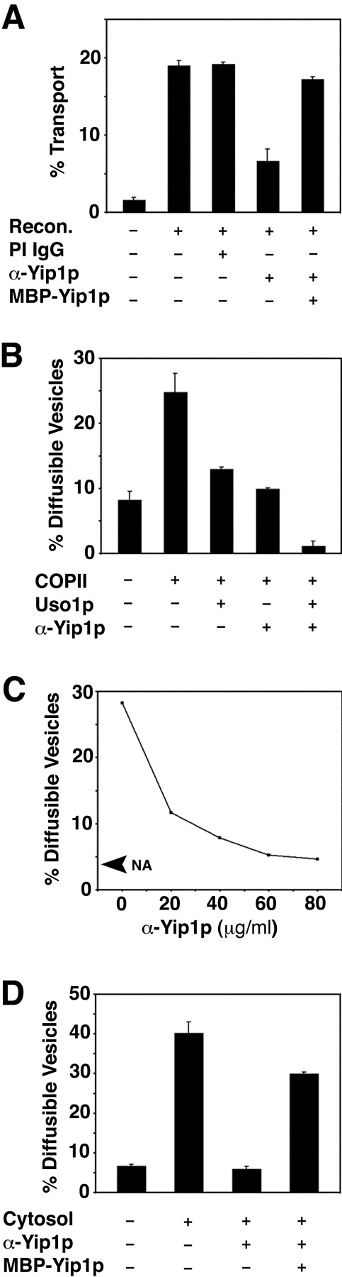
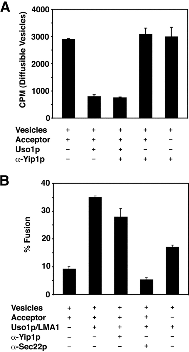
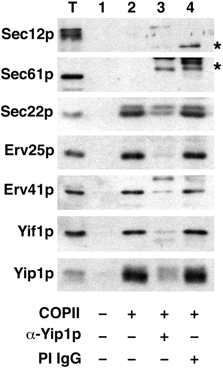
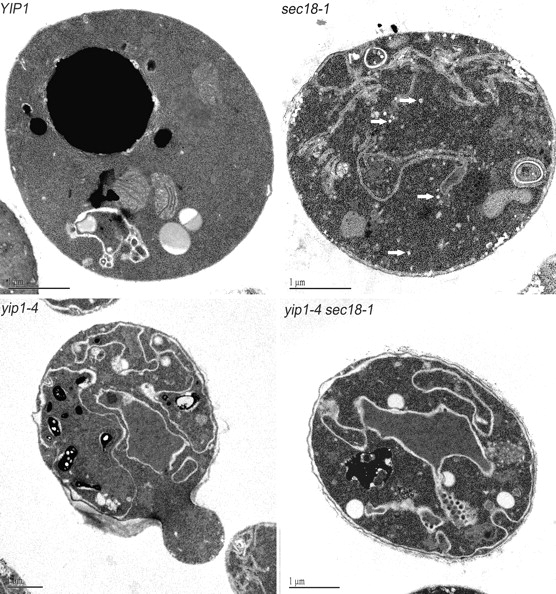
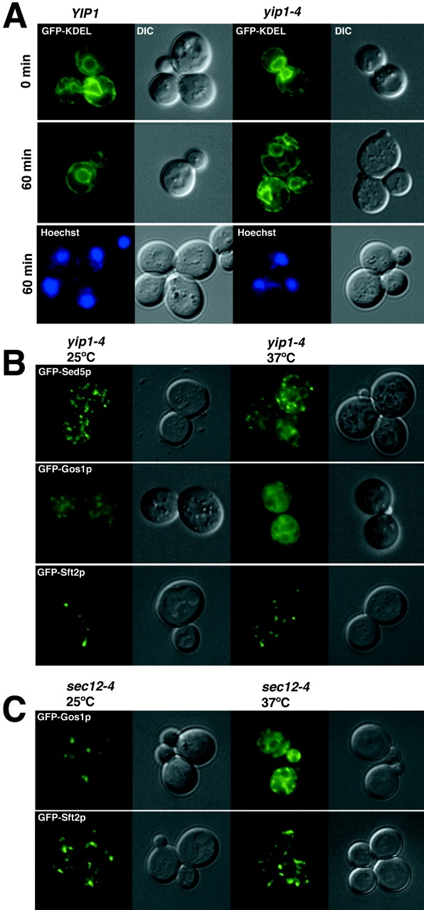
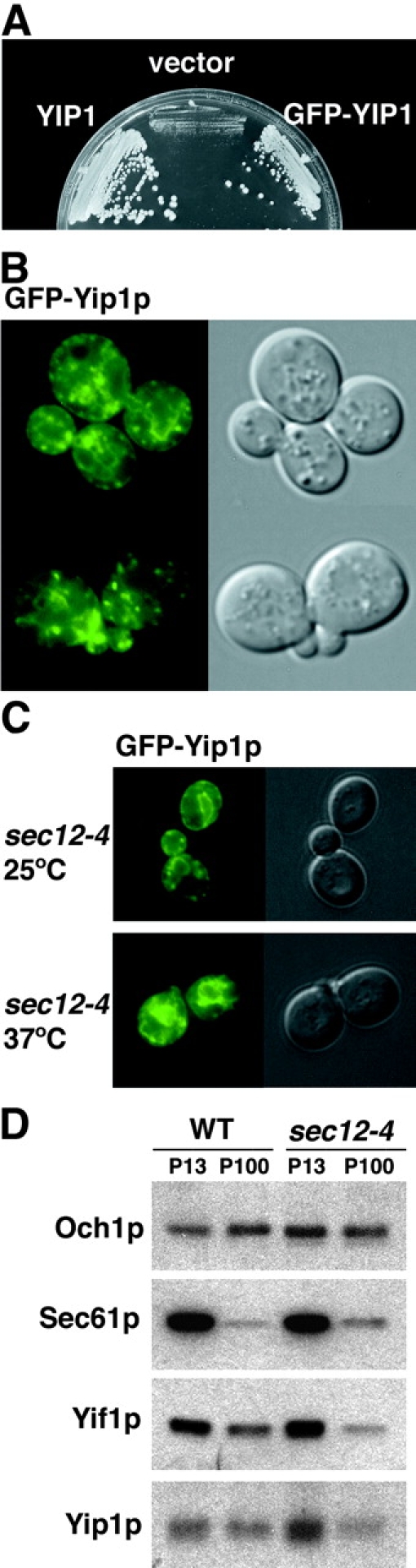
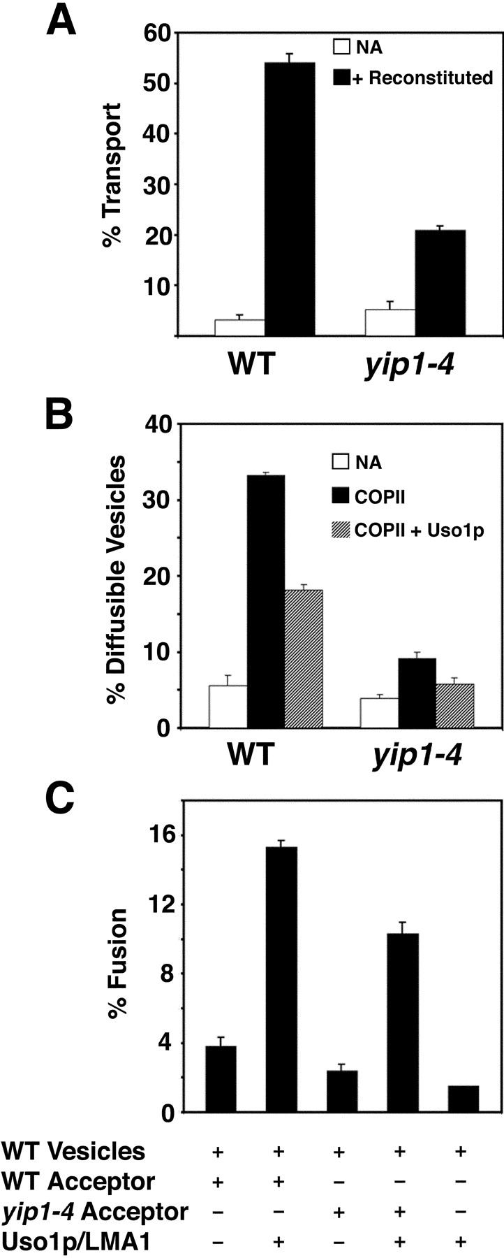
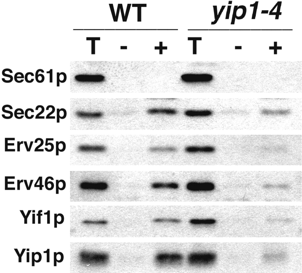
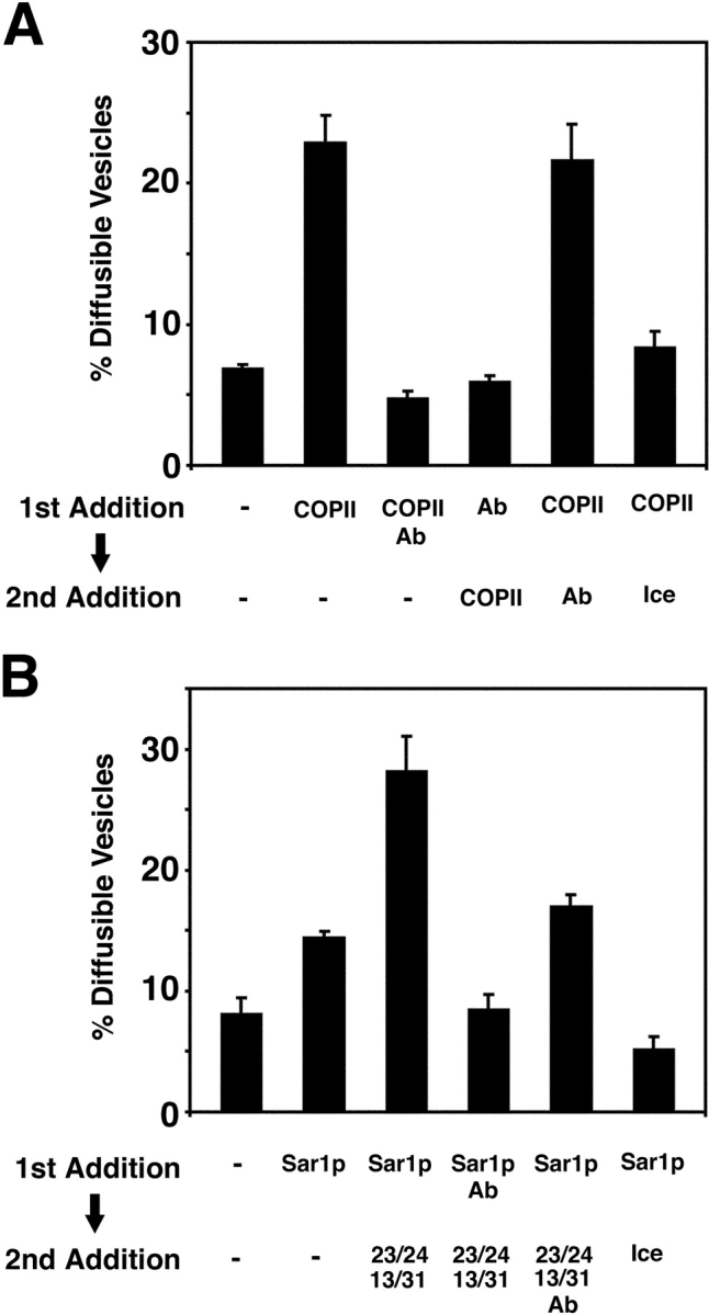
Yeast Ypt1p-interacting protein (Yip1p) belongs to a conserved family of transmembrane proteins that interact with Rab GTPases. We encountered Yip1p as a constituent of ER-derived transport vesicles, leading us to hypothesize a direct role for this protein in transport through the early secretory pathway. Using a cell-free assay that recapitulates protein transport from the ER to the Golgi complex, we find that affinity-purified antibodies directed against the hydrophilic amino terminus of Yip1p potently inhibit transport. Surprisingly, inhibition is specific to the COPII-dependent budding stage. In support of this in vitro observation, strains bearing the temperature-sensitive yip1-4 allele accumulate ER membranes at a nonpermissive temperature, with no apparent accumulation of vesicle intermediates. Genetic interaction analyses of the yip1-4 mutation corroborate a function in ER budding. Finally, ordering experiments show that preincubation of ER membranes with COPII proteins decreases sensitivity to anti-Yip1p antibodies, indicating an early requirement for Yip1p in vesicle formation. We propose that Yip1p has a previously unappreciated role in COPII vesicle biogenesis.


Figures

Figure 1.
Figure 1.
Anti-Yip1p antibodies inhibit in vitro transport between the ER and the Golgi complex at the budding stage. (A) Washed wild-type (FY834) semi-intact cells containing [35S]gpαf were incubated with Recon proteins (COPII, Uso1p, and LMA1) and an ATP regeneration system. After 75 min at 23°C, the amount of Golgi-modified [35S]gpαf was measured to determine transport efficiency. Where indicated, anti-Yip1p antibodies (40 μg/ml), preimmune IgGs (40 μg/ml), or MBP-Yip1p (144 μg/ml) were added to reactions. (B) Semi-intact cells prepared as in A were incubated with COPII or COPII plus Uso1p to measure budding and tethering in the presence or absence of anti-Yip1p antibodies (20 μg/ml). After 30 min at 23°C, freely diffusible vesicles containing [35S]gpαf were separated from semi-intact cell membranes by centrifugation at 18,000 g and [35S]gpαf quantified by Con A precipitation. (C) Vesicle budding as in B with increasing amounts of anti-Yip1p antibodies (20–80 μg/ml). No addition (NA) shows level of budding minus COPII. (D) Vesicle budding as in B, except cytosol was used to drive reactions. Where indicated, anti-Yip1p antibodies (40 μg/ml) and MBP-Yip1p (144 μg/ml) were added.
Figure 2.
Figure 2.
Anti-Yip1p antibodies do not inhibit vesicle tethering or vesicle fusion in a two-stage reaction. (A) COPII vesicles containing [35S]gpαf were synthesized from wild-type (FY834) membranes and mixed with wild-type semi-intact cell acceptor membranes. Reactions were incubated with Uso1p in the presence or absence of anti-Yip1p antibodies (120 μg/ml) where indicated. Freely diffusible vesicles containing [35S]gpαf were quantified by Con A precipitation. (B) COPII vesicles prepared as in A were mixed with wild-type semi-intact cell acceptor membranes. Reactions contained Uso1p and LMA1 in the presence or absence of anti-Yip1p antibodies (120 μg/ml) or anti-Sec22p antibodies (60 μg/ml) where indicated. After 75 min at 23°C, the amount of Golgi-modified [35S]gpαf was measured to determine fusion efficiency.
Figure 3.
Figure 3.
Anti-Yip1p antibodies block COPII-dependent budding of vesicle proteins. COPII-budding reactions were performed from wild-type semi-intact cells (FY834) in the presence or absence of anti-Yip1p antibodies (40 μg/ml) or preimmune IgGs (40 μg/ml) as indicated. One tenth of a total reaction (T), or budding reactions without COPII proteins (lane 1), with COPII proteins (lane 2), with COPII plus anti-Yip1p antibodies (lane 3), and with COPII plus preimmune antibodies (lane 4) were separated on a 12.5% polyacrylamide gel. ER resident proteins (Sec61p and Sec12p) and vesicle proteins (Sec22p, Erv25p, Erv41p, Yif1p, and Yip1p) were detected using immunoblot. Asterisks indicate antibody heavy chain cross-reactivity with secondary antibodies.
Figure 4.
Figure 4.
Themosensitive yip1-4 mutants display morphological phenotypes characteristic of an early block in the secretory pathway. Wild-type and mutant cells were shifted to 37°C for 40 min and then fixed and prepared for EM as described under the Materials and methods section. Representative thin sections are shown for each condition. The white arrows in the sec18-1 panel point to transport vesicles that have accumulated in this strain. Note that the accumulation of vesicles was absent in the yip1-4 and yip1-4 sec18-1 strains. Bars, 1 μm.
Figure 5.
Figure 5.
Distribution of GFP-tagged proteins in wild-type and yip1-4 strains. (A) Fluorescence images of GFP-KDEL showing the outline of ER membranes in wild-type (RCY1768) and yip1-4 (RCY1764) strains after 60 min at 37°C. Hoechst stain for DNA indicates nuclei remain intact in yip1-4 cells after a 60-min shift to the restrictive temperature. (B) GFP-tagged versions of Sed5p, Gos1p, and Sft2p were expressed in the yip1-4 strain and monitored by fluorescence microscopy at 25°C or after shift to 37°C for 30 min. Note the partial localization to perinuclear structures for GFP-Sed5p and GFP-Gos1p upon shift to the restrictive temperature. (C) GFP-Gos1p shifts to a perinuclear distribution when ER export is blocked in a sec12-4 strain, whereas GFP-Sft2p remains in a punctate pattern as in the yip1-4 strain.
Figure 6.
Figure 6.
Yip1p cycles between the ER and Golgi compartments. (A) GFP fused to the amino terminus of Yip1p (GFP-Yip1p) complements a yip1Δ strain. (B) Cells expressing GFP-Yip1p were analyzed by fluorescence imaging. A punctate fluorescence pattern typical of Golgi-localized proteins in addition to ring-like perinuclear structures of the ER were observed. (C) GFP-Yip1p redistributes to perinuclear structures when transport from the ER is blocked in a sec12 strain shifted to 37°C for 30 min (D) ER and Golgi membrane fractions from WT and sec12-4. Cells were shifted to 37°C for 45 min before lysis and collection of membrane fractions. The contents of ER (P13) and Golgi (P100) fractions were monitored by immunoblot. Sec61p serves as an ER marker, whereas Och1p is a Golgi-localized protein.
Figure 7.
Figure 7.
yip1-4 membranes display a defect in COPII vesicle budding in vitro. (A) Washed semi-intact cells containing [35S]gpαf were prepared from wild-type (RCY1768) and yip1-4 (RCY1764) strains. Semi-intact cells were incubated with COPII proteins, Uso1p, LMA1, and an ATP regeneration system. After 75 min at 23°C, the amount of Golgi-modified [35S]gpαf was measured to determine transport efficiency. (B) Semi-intact cells from wild-type and mutant strains were prepared as in A and incubated with COPII or COPII plus Uso1p to measure vesicle budding and tethering. (C) COPII vesicles containing [35S]gpαf were synthesized from wild-type microsomes and purified on density gradients. Purified vesicles were mixed with wild-type or yip1-4 semi-intact cell acceptor membranes in second-stage assays in the presence or absence of Uso1p and LMA1. After 75 min at 23°C, the amount of Golgi-modified [35S]gpαf was measured to determine transport efficiency.
Figure 8.
Figure 8.
COPII-dependent budding of vesicle proteins is reduced in the yip1-4 strain. COPII-budding reactions in wild-type (RCY1768) or yip1-4 (RCY1764) semi-intact cells. One tenth of a total reaction (T), budded vesicles isolated after incubation with COPII proteins (+), or a mock reaction without COPII proteins (−) were separated on a 12.5% polyacrylamide gel. Sec61p (ER resident protein) and Sec22p, Erv25p, Erv46p, Yif1p, and Yip1p (vesicle proteins) were detected by immunoblot.
Figure 9.
Figure 9.
Genetic interaction analysis of yip1-4. Strains carrying a thermosensitive mutation in genes involved in ER/Golgi transport were combined with a yip1Δ allele together with a wild-type YIP1 on a URA3 plasmid. Strains also contained vector only, wild-type YIP1 LEU2 vector, or a yip1-4 LEU vector. All transformants were assessed by plasmid shuffling on 5-fluorouracil at 25°C. Plates containing yip1-4 sec17ts double mutants are shown as an example where no genetic interaction was observed. In contrast, yip1-4 sec12-4 double mutants are shown as an example of complete lethality between two mutants. A summary of genetic interaction results is shown in Table I.
Figure 10.
Figure 10.
Preincubation of donor membranes with COPII produces a decrease in sensitivity to anti-Yip1p antibodies. (A) Washed semi-intact cells containing [35S]gpαf were prepared from a wild-type strain (FY834) and treated with either COPII or anti-Yip1p antibodies (40 μg/ml) for 5 min at 15°C. Secondary factors were then added as indicated, and the reactions proceeded for an additional 25 min. Freely diffusible vesicles containing [35S]gpαf were then separated from semi-intact cell membranes by centrifugation and quantified by Con A precipitation. (B) Semi-intact cells prepared as in A were treated with Sar1p or anti-Yip1p antibodies alone (40 μg/ml) for 5 min at 15°C. Secondary factors were then added and diffusible vesicles were quantified as in A.

Similar articles

Cited by

References

    1. Allan, B.A., B.D. Moyer, and W.E. Balch. 2000. Rab1 recruitment of p115 into a cis-SNARE complex: programming budding of COPII vesicles for fusion. Science. 289:444–448. - PubMed
    1. Ausubel, R.M., R. Brent, R.E. Kingston, D.D. Moore, J.G. Seidman, J.A. Smith, and K. Struhl. 1987. Current Protocols in Molecular Biology. Greene Publishing Associates and Wiley-InterScience, New York. 3.0.1–3.14.3.
    1. Baker, D., L. Hicke, M. Rexach, M. Schleyer, and R. Schekman. 1988. Reconstitution of SEC gene product-dependent intercompartmental protein transport. Cell. 54:335–344. - PubMed
    1. Barlowe, C. 1997. Coupled ER to Golgi transport reconstituted with purified cytosolic proteins. J. Cell Biol. 139:1097–1108. - PMC - PubMed
    1. Barlowe, C., L. Orci, T. Yeung, M. Hosobuchi, S. Hamamoto, N. Salama, M. Rexach, M. Ravazzola, M. Amherdt, and R. Schekman. 1994. COPII: a membrane coat formed by Sec proteins that drive vesicle budding from the ER. Cell. 77:895–907. - PubMed

Publication types

MeSH terms