Review
. 2003 Oct;16(4):698-712.
doi: 10.1128/CMR.16.4.698-712.2003.

Antigens and alternatives for control of Anaplasma marginale infection in cattle

Affiliations
Review

Antigens and alternatives for control of Anaplasma marginale infection in cattle

Katherine M Kocan et al. Clin Microbiol Rev. 2003 Oct.

Abstract

Anaplasmosis, a tick-borne cattle disease caused by the rickettsia Anaplasma marginale, is endemic in tropical and subtropical areas of the world. The disease causes considerable economic loss to both the dairy and beef industries worldwide. Analyses of 16S rRNA, groESL, and surface proteins have resulted in the recent reclassification of the order Rickettsiales. The genus Anaplasma, of which A. marginale is the type species, now also includes A. bovis, A. platys, and A. phagocytophilum, which were previously known as Ehrlichia bovis, E. platys, and the E. phagocytophila group (which causes human granulocytic ehrlichiosis), respectively. Live and killed vaccines have been used for control of anaplasmosis, and both types of vaccines have advantages and disadvantages. These vaccines have been effective in preventing clinical anaplasmosis in cattle but have not blocked A. marginale infection. Thus, persistently infected cattle serve as a reservoir of infective blood for both mechanical transmission and infection of ticks. Advances in biochemical, immunologic, and molecular technologies during the last decade have been applied to research of A. marginale and related organisms. The recent development of a cell culture system for A. marginale provides a potential source of antigen for the development of improved killed and live vaccines, and the availability of cell culture-derived antigen would eliminate the use of cattle in vaccine production. Increased knowledge of A. marginale antigen repertoires and an improved understanding of bovine cellular and humoral immune responses to A. marginale, combined with the new technologies, should contribute to the development of more effective vaccines for control and prevention of anaplasmosis.


Figures

FIG. 1.
FIG. 1.
Bovine erythrocytes infected with A. marginale. (A) Inclusion bodies (arrowheads) are located at the periphery of the erythrocyte in a stained blood film. (B) Electron micrograph of an A. marginale inclusion that contains three organisms. Bar, 10 μm (A) and 0.5 μm (B).
FIG. 2.
FIG. 2.
The high A. marginale levels in acute rickettsemia (>109 ml−1) are resolved after the development of a primary immune response, but the emergence of antigenic variants results in persistent infection. Persistence is characterized by sequential rickettsemic cycles, occurring at approximately 5-week intervals, in which new MSP2 variants replicate to a peak of >106 ml−1 and are then controlled by a variant-specific immune response. Variants arising in three sequential rickettsemic cycles are shown and are designated V1, V2, and V3. The points of variant emergence and variant control are designated for V2. (Reprinted from reference 125 with permission of the publisher.)
FIG. 3.
FIG. 3.
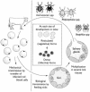
Schematic of the development cycle of A. marginale in cattle and ticks. Infected erythrocytes are ingested by ticks (Dermacentor spp., Rhipicephalus spp., or Boophilus spp.) with the blood meal. The first site of infection of A. marginale in ticks is the gut cells. When the ticks feed a second time, many tick tissues become infected, including salivary gland cells, from where the rickettsia is transmitted back to cattle. Two forms of A. marginale, reticulated and dense forms, are found in infected tick cells. Reticulated forms appear first and are the vegetative stage that divides by binary fission. The reticulated form changes into the dense form, which is the infective form and can survive extracellularly. (Reprinted from reference 125 with permission from the publisher.)
FIG. 4.
FIG. 4.
Micrographs of colonies of A. marginale in tick gut cells. (A) Light micrograph of a large colony (C) in a tick gut cell. (B) Electron micrograph of a colony in a tick gut cell. Bar, 10 μm (A) and 5 μm (B).
FIG. 5.
FIG. 5.
Micrographs of colonies of A. marginale in tick salivary gland cells. (A) Light micrograph of two colonies (C) in salivary gland cells. (B) Electron micrograph of a tick salivary gland cell that contains several A. marginale colonies (arrowheads). Bar, 10 μm (A) and 5 μm (B).
FIG. 6.
FIG. 6.
Electron micrographs of the two developmental stages of A. marginale within colonies in tick cells. (A) Reticulated forms within a colony, dividing by binary fission (arrowhead). (B) Dense forms within a colony in an infected tick cell. Bars, 1 μm.
FIG. 7.
FIG. 7.
Maximum-parsimony analysis of MSP4 sequences. The topology of 1 of 11 equally most-parsimonious trees based on DNA sequence variation in the msp4 gene is shown. The tree was rooted with A. centrale and A. ovis. The solid portion of each branch represents the minimum branch length; the dashed portion of each branch represents the maximum branch length as determined by PAUP*4.0b4a. Numbers below the branches represent the percentage of 500 bootstrap iterations in which each clade was detected, and numbers with asterisks indicate bootstrap support based on a phylogenetic analysis of deduced amino acid residues. Vertical lines show synapomorphic amino acid changes documenting the monophyly of A. marginale and the Latin American clade of A. marginale isolates along with the amino acid residue and its position in the sequence. (Adapted from reference 52 with permission of the publisher.)
FIG. 8.
FIG. 8.
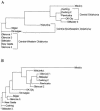
Phylogenetic tree constructed from analysis of the msp4 (A) and msp1α (B) coding sequences based on a sequence distance method utilizing the neighbor-joining algorithm (147). Sequences derived from the Mexico isolate of A. marginale (52) were used as outgroup. The geographic distribution of the Oklahoma isolates of A. marginale is shown for the msp4 analysis in panel A.

Similar articles

Cited by

References

    1. Abdala, A. A., E. Pipano, D. H. Aguirre, A. B. Gaido, M. A. Zurbriggen, A. J. Mangold, and A. A. Guglielmone. 1990. Frozen and fresh Anaplasma centrale vaccines in the protection of cattle against Anaplasma marginale infection. Rev. Elev. Med. Vet. Pays Trop. 43:155-158. - PubMed
    1. Aguirre, D. H., A. C. Bermúdez, A. J. Mangold, and A. A. Guglielmone. 1988. Infección natural con Anaplasma marginale en bovines de raza Hereford, Criolla and Nelore en Tucumán, Argentina. Rev. Latinoam. Microbiol. 30:37-42. - PubMed
    1. Aguirre, D. H., A. B. Gaido, A. A. Abdala, L. G. de Ríos, A. J. Mangold, and A. A. Guglielmone. 1988. Evaluación de la protección conferida contra Anaplasma marginale por una vacuna de A. marginale muerto, una vacuna de Anaplasma centrale vivo y una combinación de ambas en bovinos Holando Argentino. Rev. Med. Vet. (Buenos Aires) 69:13-19.
    1. Aguirre, D. H., A. J. Mangold, L. G. de Ríos, and A. A. Guglielmone. 1991. Respuesta clínica y evolución del peso corporal (Bos taurus) vacunadas simultáneamente contra babesiosis y anaplasmosis con inmunógenos vivos. Med. Vet. (Barcelona) 8:95-101.
    1. Alleman, A. R., and A. F. Barbet. 1996. Evaluation of Anaplasma marginale major surface protein 3 (MSP3) as a diagnostic test antigen. J. Clin. Microbiol. 34:270-276. - PMC - PubMed

Publication types

MeSH terms

LinkOut - more resources