

Current perspectives on ophthalmic mycoses
- PMID: 14557297
- PMCID: PMC207127
- DOI: 10.1128/CMR.16.4.730-797.2003
Current perspectives on ophthalmic mycoses
Abstract
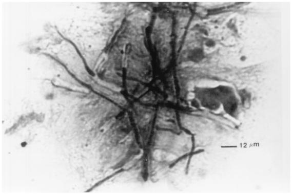
Fungi may infect the cornea, orbit and other ocular structures. Species of Fusarium, Aspergillus, Candida, dematiaceous fungi, and Scedosporium predominate. Diagnosis is aided by recognition of typical clinical features and by direct microscopic detection of fungi in scrapes, biopsy specimens, and other samples. Culture confirms the diagnosis. Histopathological, immunohistochemical, or DNA-based tests may also be needed. Pathogenesis involves agent (invasiveness, toxigenicity) and host factors. Specific antifungal therapy is instituted as soon as the diagnosis is made. Amphotericin B by various routes is the mainstay of treatment for life-threatening and severe ophthalmic mycoses. Topical natamycin is usually the first choice for filamentous fungal keratitis, and topical amphotericin B is the first choice for yeast keratitis. Increasingly, the triazoles itraconazole and fluconazole are being evaluated as therapeutic options in ophthalmic mycoses. Medical therapy alone does not usually suffice for invasive fungal orbital infections, scleritis, and keratitis due to Fusarium spp., Lasiodiplodia theobromae, and Pythium insidiosum. Surgical debridement is essential in orbital infections, while various surgical procedures may be required for other infections not responding to medical therapy. Corticosteroids are contraindicated in most ophthalmic mycoses; therefore, other methods are being sought to control inflammatory tissue damage. Fungal infections following ophthalmic surgical procedures, in patients with AIDS, and due to use of various ocular biomaterials are unique subsets of ophthalmic mycoses. Future research needs to focus on the development of rapid, species-specific diagnostic aids, broad-spectrum fungicidal compounds that are active by various routes, and therapeutic modalities which curtail the harmful effects of fungus- and host tissue-derived factors.
Figures


References
-
- Adler, D. E., T. H. Milhorat, and J. I. Miller. 1998. Treatment of rhinocerebral mucormycosis with intravenous, interstitial and cerebrospinal fluid administration of amphotericin B: case report. Neurosurgery 42:644-648. - PubMed
-
- Agarwal, A., A. Gupta, V. Sakhuja, P. Talwar, K. Joshi, and K. S. Chugh. 1991. Retinitis following disseminated cryptococcosis in a renal allograft recipient. Efficacy of oral fluconazole. Acta Ophthalmol. Copenh. 69:402-405. - PubMed
-
- Agrawal, V., J. Biswas, H. N. Madhavan, G. Mangat, M. K. Reddy, J. S. Saini, S. Sharma, and M. Srinivasan. 1994. Current perspectives in infectious keratitis. Indian J. Ophthalmol. 42:171-191. - PubMed
-
- Ainbinder, D. J., V. C. Parmley, T. H. Mader, and M. L. Nelson. 1998. Infectious crystalline keratopathy caused by Candida guilliermondii. Am. J. Ophthalmol. 125:723-725. - PubMed
-
- Ajayi, B. G., B. Osuntokun, O. Olurin, O. O. Kale, and T. A. Junaid. 1986. Orbital histoplasmosis due to Histoplasma capsulatum var. duboisii: successful treatment with septrin. J. Trop. Med. Hyg. 89:179-187. - PubMed
Publication types
MeSH terms
Substances
LinkOut - more resources
Full Text Sources
Other Literature Sources
Medical
Miscellaneous
