. 2003 Oct 28;100(22):12735-40.
doi: 10.1073/pnas.2135500100. Epub 2003 Oct 13.

A subset of membrane-associated proteins is ubiquitinated in response to mutations in the endoplasmic reticulum degradation machinery

Affiliations

A subset of membrane-associated proteins is ubiquitinated in response to mutations in the endoplasmic reticulum degradation machinery

Amy L Hitchcock et al. Proc Natl Acad Sci U S A. .

Abstract

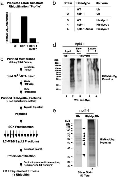
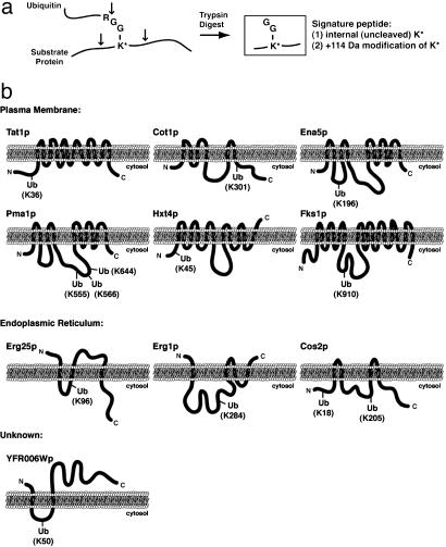
Ubiquitination of membrane-associated proteins can direct their proteasome-mediated degradation or activation at the endoplasmic reticulum (ER), as well as their endocytosis and intracellular sorting. However, the full spectrum of ubiquitinated membrane proteins has not been determined. Here we combined proteomic analysis with yeast genetics to identify 211 ubiquitinated membrane-associated proteins in Saccharomyces cerevisiae and map >30 precise sites of ubiquitination. Major classes of identified ubiquitinated proteins include ER-resident membrane proteins, plasma membrane-localized permeases, receptors, and enzymes, and surprisingly, components of the actin cytoskeleton. By determining the differential abundance of ubiquitinated proteins in yeast mutated for NPL4 and UBC7, which are major components of ER-associated degradation (ERAD), we furthermore were able to classify 83 of these identified ubiquitinated membrane proteins as potential endogenous substrates of the ERAD pathway. These substrates are highly enriched for proteins that localize to or transit through the ER. Interestingly, we also identified novel membrane-bound transcription factors that may be subject to ubiquitin/proteasome-mediated cleavage and activation at the ER membrane.


Figures

Fig. 1.
Fig. 1.
NPL4-dependent ubiquitinated ER protein degradation. (a) Simplified model of ERAD. Unfolded and/or damaged ER proteins (a membrane protein is depicted, soluble lumenal proteins are also subject to ERAD) are ubiquitinated by the E2 ubiquitin-conjugating enzyme Ubc7p (recruited to the ER membrane by Cue1p) and the E3 ubiquitin ligases Hrd1p and Doa10p. The E2s Ubc6p and Ubc1p also make minor contributions to ER protein ubiquitination. Once ubiquitinated, an ERAD substrate requires the activity of the Npl4p–Ufd1p–Cdc48p chaperone to be fully extracted from the membrane and degraded by the proteasome. (b) Accumulation of ubiquitinated proteins in npl4-1 membranes. WT and npl4-1 cells expressing 6×His–Myc-tagged ubiquitin were subjected to subcellular fractionation. Equal amounts of total protein (10 μg) from total cell extract (lanes 1 and 2), soluble (lanes 3 and 4), and membrane (lanes 5 and 6) fractions were separated by SDS/PAGE and immunoblotted with anti-Myc antibodies. (c) Extragenic suppression of npl4-1 by deletion of genes encoding ERAD ubiquitination machinery. Yeast strains of the indicated genetic backgrounds were serially diluted, spotted onto rich-media plates, and incubated for 2–3 days at the indicated temperatures. The number of cells spotted in each dilution is indicated on the bottom. (d) Loss of UBC7 abrogates accumulation of ubiquitinated proteins in npl4-1 membranes. WT and npl4-1 cells ±Δubc7 expressing 6×His–Myc-tagged ubiquitin were subjected to subcellular fractionation. Equal amounts of total protein (10 μg) from total cell extract (lanes 1–4), soluble (lanes 5–8), and membrane (lanes 9–12) fractions were separated by SDS/PAGE and immunoblotted with anti-Myc antibodies.
Fig. 2.
Fig. 2.
A genetic/proteomic screen for endogenous ERAD substrates in S. cerevisiae. (a) Predicted “profile” of ubiquitination for a typical ERAD substrate in WT vs. npl4-1 vs. npl4-1Δubc7 yeast. (b) Yeast strains from which membrane-associated 6×His–Myc-tagged ubiquitinated proteins were purified and identified. WT and npl4-1 strains expressing untagged ubiquitin (strains 1 and 2) served as negative controls for the experimental WT, npl4-1, and npl4-1Δubc7 strains expressing 6×His–Myc-tagged ubiquitin (strains 3–5, respectively). (c) Strategy used for ubiquitinated protein purification and identification. For each of the five strains listed in b, a membrane fraction containing 20 mg of total protein was solubilized under native conditions, and 6×His–Myc-ubiquitin-conjugated proteins were purified by immobilized metal (nickel) affinity chromatography. Columns were washed extensively under highly denaturing conditions (8 M urea) to eliminate copurification of proteins associated with ubiquitin-conjugated proteins. Bound proteins were eluted with imidazole and directly digested with trypsin. The resulting peptide mixture was resolved by SCX chromatography, and 12 peptide-containing SCX fractions were collected. Each SCX fraction was individually subjected to nanoscale microcapillary reverse-phase (RP) chromatography. The RP chromatography was coupled directly to an ion trap tandem mass (MS/MS) spectrometer. Each MS/MS spectrum was searched independently against a database of predicted spectra derived from S. cerevisiae-encoded proteins, leading to the identification of >6,000 peptides corresponding to 527 unique proteins among the five data sets. These identified proteins were subjected to a stringent quality evaluation (see Materials and Methods), and only 211 unambiguously identified candidate ubiquitinated proteins were kept. (d) Example of a representative purification of 6×His–Myc-tagged ubiquitinated proteins (from npl4-1 membranes) as monitored by anti-Myc Western blot. Equivalent amounts (0.025% total) of input (lane 2), flow-through (lane 3), and two protein-containing elution (lanes 4 and 5) fractions were separated by SDS/PAGE and immunoblotted with anti-Myc antibodies to detect ubiquitin conjugates. An equivalent amount of input from the npl4-1-negative control strain expressing untagged ubiquitin (lane 1) was included to indicate the specificity of the anti-Myc antibodies. (e) Example of a representative purification of 6×His–Myc-tagged ubiquitinated proteins (from npl4-1 membranes) as monitored by silver staining. Equivalent amounts (1% total) of purified proteins from npl4-1 cells expressing either untagged (lane 1) or 6×His–Myc-tagged (lane 2) ubiquitin were subjected to SDS/PAGE and were detected by silver staining. Arrowheads indicate the presence of “false positive” proteins purified in the absence of tagged ubiquitin. Many of these false positive proteins contain endogenous histidine-rich sequences and/or have metal binding capabilities.
Fig. 3.
Fig. 3.
Identification of precise sites of substrate protein ubiquitination. (a) Schematic diagram indicating the ability to detect ubiquitinated lysine residues by tandem mass spectrometry. After trypsin digestion, a diglycine ubiquitin remnant remains covalently attached by an isopeptide linkage to the ε-amino side chain of a lysine residue within the substrate protein. The modified lysine, which is resistant to trypsin cleavage, can be identified by tandem mass spectrometry as a 114-Da-modified internal lysine residue. (b) Schematic representations of the predicted membrane topology and identified site(s) of ubiquitination for 15 integral membrane proteins localized to the plasma membrane, ER, and unknown membranes as indicated.
Fig. 4.
Fig. 4.
Subcellular localization and functional classes of identified ubiquitinated membrane-associated proteins, including candidate ERAD pathway substrates. (a) Venn diagram depiction of the distribution of 211 ubiquitinated proteins among the WT, npl4-1, and npl4-1 Δubc7 data sets. (b) Venn diagram depiction of the overlap between the 108 NPL4-(npl4-1 up-regulated) and 79 UBC7-responsive (Δubc7 down-regulated) ubiquitinated proteins [the small class (<10%) of npl4-1 down-regulated and Δubc7 up-regulated ubiquitinated proteins are not included]. The 68 NPL4- and UBC7-responsive proteins were classified as “strong” candidate endogenous ERAD substrates. Manual inspection of the remaining 51 NPL4-or UBC7-responsive proteins led to the classification of an additional 15 “weak” candidate ERAD substrates (see Materials and Methods). (c) Histogram representation of the membrane associations of 211 identified ubiquitinated proteins (white bars plus black bars) and the ERAD-candidate subpopulation (83 proteins, white bars). A total of 80 proteins (34 ERAD candidates) have known TMDs, and an additional 63 proteins (22 ERAD candidates) contain predicted TMDs. Of the non-TMD containing population, 43 (17 ERAD substrates) have known peripheral associations with cellular membranes, and 25 proteins (10 ERAD substrates) are not known or predicted to associate with membranes. (d) Histogram representation of the distribution of 186 membrane-associated (integral or peripheral) ubiquitinated proteins (73 of which are candidate ERAD substrates) among subcellular membranes. Asterisks indicate cellular membranes that contain an enriched number of ERAD candidate proteins. (e) Histogram representation of the distribution of 211 ubiquitinated proteins (including 83 candidate ERAD substrates) among eight functional classes of proteins. Asterisks indicate functional classes that contain an enriched number of ERAD candidate proteins.

References

    1. Kostova, Z. & Wolf, D. H. (2003) EMBO J. 22 2309–2317. - PMC - PubMed
    1. Plemper, R. K. & Wolf, D. H. (1999) Trends Biochem. Sci. 24 266–270. - PubMed
    1. Deak, P. M. & Wolf, D. H. (2001) J. Biol. Chem. 276 10663–10669. - PubMed
    1. Biederer, T., Volkwein, C. & Sommer, T. (1997) Science 278 1806–1809. - PubMed
    1. Bays, N. W., Gardner, R. G., Seelig, L. P., Joazeiro, C. A. & Hampton, R. Y. (2001) Nat. Cell Biol. 3 24–29. - PubMed

Publication types

MeSH terms