. 2003 Oct 14;100(21):12420-5.
doi: 10.1073/pnas.1635213100. Epub 2003 Oct 13.

The primary mechanism of attenuation of bacillus Calmette-Guerin is a loss of secreted lytic function required for invasion of lung interstitial tissue

Affiliations

The primary mechanism of attenuation of bacillus Calmette-Guerin is a loss of secreted lytic function required for invasion of lung interstitial tissue

Tsungda Hsu et al. Proc Natl Acad Sci U S A. .

Abstract

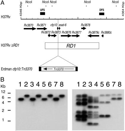
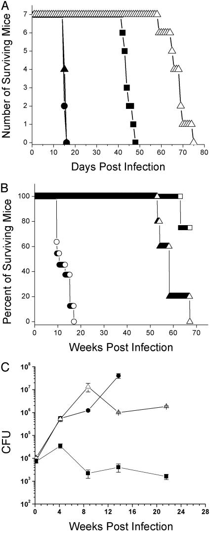
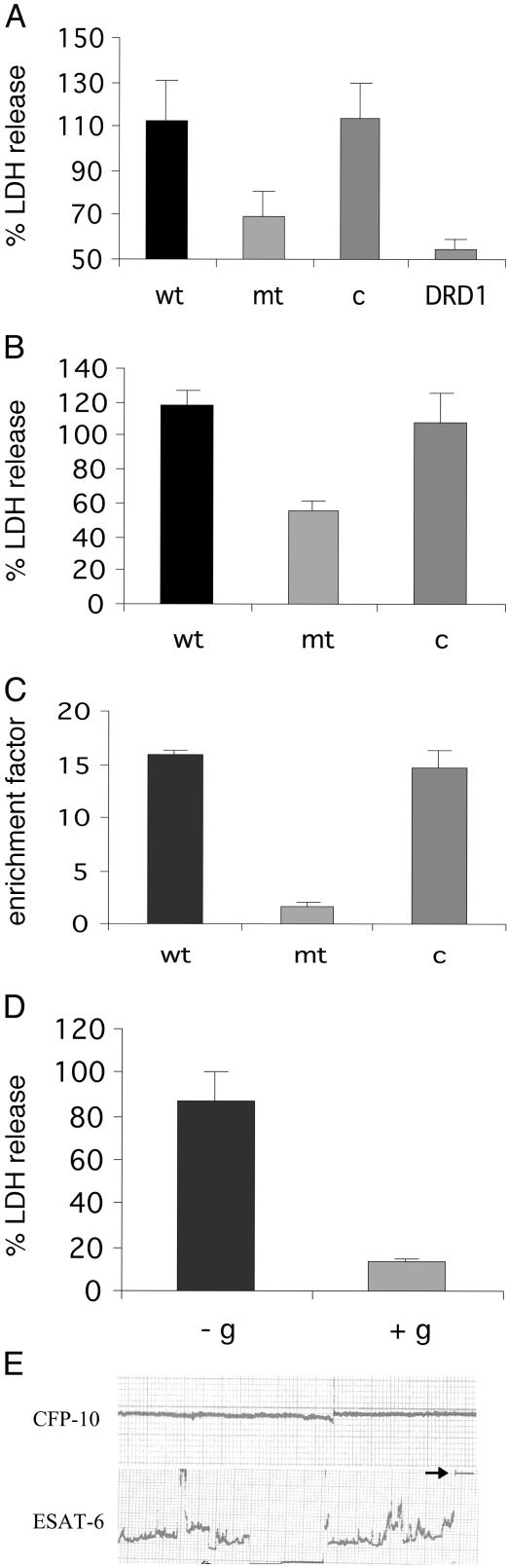
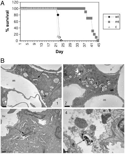
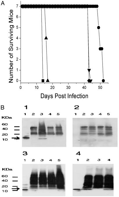
Tuberculosis remains a leading cause of death worldwide, despite the availability of effective chemotherapy and a vaccine. Bacillus Calmette-Guérin (BCG), the tuberculosis vaccine, is an attenuated mutant of Mycobacterium bovis that was isolated after serial subcultures, yet the functional basis for this attenuation has never been elucidated. A single region (RD1), which is absent in all BCG substrains, was deleted from virulent M. bovis and Mycobacterium tuberculosis strains, and the resulting DeltaRD1 mutants were significantly attenuated for virulence in both immunocompromised and immunocompetent mice. The M. tuberculosis DeltaRD1 mutants were also shown to protect mice against aerosol challenge, in a similar manner to BCG. Interestingly, the DeltaRD1 mutants failed to cause cytolysis of pneumocytes, a phenotype that had been previously used to distinguish virulent M. tuberculosis from BCG. A specific transposon mutation, which disrupts the Rv3874 Rv3875 (cfp-10 esat-6) operon of RD1, also caused loss of the cytolytic phenotype in both pneumocytes and macrophages. This mutation resulted in the attenuation of virulence in mice, as the result of reduced tissue invasiveness. Moreover, specific deletion of each transcriptional unit of RD1 revealed that three independent transcriptional units are required for virulence, two of which are involved in the secretion of ESAT-6 (6-kDa early secretory antigenic target). We conclude that the primary attenuating mechanism of bacillus Calmette-Guérin is the loss of cytolytic activity mediated by secreted ESAT-6, which results in reduced tissue invasiveness.


Figures

Fig. 1.
Fig. 1.
Generation of mutants in the RD1 region of M. tuberculosis and M. bovis. (A) Schematic of M. tuberculosis H37Rv RD1 region showing predicted NcoI sites. Arrows at the top represent the genes in this region. UFSs and DFSs used to generate the knockout are indicated as filled bars above the grid line. Each increment in the grid line represents 1 kbp. The RD1 sequence deleted from M. bovis BCG is represented by an open bar spanning from Rv3871 to Rv3879c. The site of the insertion of transposon Tn5370 is also indicated. (B) Southern analysis of the NcoI-digested genomic DNA isolated from the wild type and the ΔRD1 mutants generated by using specialized transduction in M. tuberculosis and M. bovis. Lane 1, M. tuberculosis H37Rv; lane 2, M. tuberculosis H37Rv ΔRD1; lane 3, M. tuberculosis Erdman; lane 4, M. tuberculosis Erdman ΔRD1; lane 5, M. tuberculosis CDC1551; lane 6, M. tuberculosis CDC1551 ΔRD1; lane 7, M. bovis Ravenel; and lane 8, M. bovis Ravenel ΔRD1. The probe used in the Southern analysis was either DFS (Left), demonstrating the deletion of RD1, or IS6110-specific (Right). The IS6110 probe is used to characterize the four different strains (described in ref. 31).
Fig. 2.
Fig. 2.
Deletion of RD1 in M. tuberculosis confers an attenuation of virulence in vivo. (A) Survival experiment using SCID mice i.v. infected with 2 × 10⁶ cfu per mouse. Infection was carried out as described in Materials and Methods. Strains used were M. tuberculosis H37Rv (•), M. tuberculosis ΔRD1 (▪), M. tuberculosis ΔRD1 (pYUB412::Rv3860Rv3885c)(▴), and M. bovis BCG (▵). (B) Survival experiment with BALB/c mice i.v. infected with M. tuberculosis H37Rv (○), M. tuberculosis ΔRD1 (▵), and M. bovis BCG (□), at a dose of 2 × 10⁶ cfu (C) Growth kinetics in the lungs of BALB/c mice i.v. infected with M. bovis BCG (▪), M. tuberculosis H37Rv (•), and M. tuberculosis H37Rv ΔRD1 (▵). The infecting dose per mouse was 2 × 10⁶ cfu. Data represent the mean of cfus from three mice per time point.
Fig. 3.
Fig. 3.
The cfp-10 esat-6 (Rv3874/Rv3875) operon of RD1 is required for host cell lysis. (A) LDH release assay of infected lung epithelial cells, at 72 h postinfection. Cells were infected at a multiplicity of infection of 10:1, and supernatants were analyzed for LDH release. wt, M. tuberculosis Erdman + pYUB412 (empty vector); mt, mc24513 = cfp10::Tn5370 + pYUB412; c, mc24513 + pYUB412::Rv3860Rv3885c; DRD1, M. tuberculosis Erdman ΔRD1. Values are means ± SD of triplicate measurements. (B) LDH release assay of bone marrow-derived macrophages at day 7 postinfection. Labels wt, mt, and c are as in A. Values are means ± SD of triplicate measurements. (C) Necrosis-determining assay, measuring histone-associated DNA fragments in the supernatant. Labels wt, mt, and c are as in A and B. (D) The M. tuberculosis necrosis phenotype is inhibited by 5 mM exogenous glycine (+g), compared with the control, in which there is no exogenous glycine (-g). Values are means ± SD of triplicate measurements. (E) ESAT-6, but not CFP-10, is sufficient to induce disruption, leading to total destruction (see arrowhead) of an artificial lipid bilayer. Artificial planal bilayers were constructed by using the lipid diphytanoylphosphatidylcholine as described in ref. . Voltages of 20–50 mV were applied, and the conductance across the bilayer was measured on the addition of 1 μg of each protein. The addition of ESAT-6 and CFP-10 in combination resulted in similar disruption to ESAT-6 alone. These studies were carried out in triplicate. For further details, see Materials and Methods.
Fig. 4.
Fig. 4.
The loss of cfp-10/esat-6 (Rv3874/Rv3875) confers attenuation of virulence. (A) Survival in SCID mice i.v. infected with 2 × 10⁶ cfu of M. tuberculosis Erdman + pYUB412 (wt), mc24513 = cfp10::Tn5370 + pYUB412 (mt), and mc24513 + pYUB412::Rv3860Rv3885c (c). (B) Electron micrographs of early lesions after high-dose aerosol infection of BALB/c mice with M. tuberculosis Erdman wild type + pYUB412, compared with mc24513 + pYUB412, at 3 weeks postinfection (×5,000). (B1) Mutant-infected macrophage in the alveolar airspace. This result was not observed in the wild-type-infected mice at this time point. (B2) A macrophage in the lumen of the airspace with large numbers of intracellular mutant bacilli. (B3) Wild-type bacillus residing within a macrophage, located interstitially. This result was not observed in mutant-infected mice at this time point. (B4) Cell within the alveolar space with ingested wild-type bacillus is lysed, resulting in the release of the organism. This result was not observed in mice infected with the mutant bacilli. as, alveolar space; bv, blood vessel; lc, lytic cell; m, macrophage; p, type I pneumocyte. Arrows denote mycobacteria.
Fig. 5.
Fig. 5.
The genes Rv3871 and Rv3876/Rv3877 of RD1 are required for virulence and for the secretion of ESAT-6. (A) Survival time of SCID mice infected i.v. with 2 × 10⁶ cfu of M. tuberculosis H37Rv (▪), ΔRv3871 (•), ΔRv3872/3 (▴), ΔRv3874/5 (▾), and ΔRv3876/7 (⋄). (B) Western analyses of the whole cell extracts and culture filtrates. Arrow indicates the ESAT-6-specific band. (B1) Culture filtrates probed with both anti-ESAT-6 primary antibody HYB76-8 and goat anti-mouse Ig secondary antibody. (B2) Culture filtrates probed only with secondary antibody. (B3) Whole-cell lysates probed with both primary and secondary antibodies. In B1B3, lanes are as follows: lane 1, purified ESAT-6 protein (50 ng); lane 2, ΔRv3871; lane 3, ΔRv3872/3; lane 4, ΔRv3874/5; and lane 5, ΔRv3876/7.(B4) Western analysis of culture filtrates of M. tuberculosis H37Rv, H37Rv ΔRD1, and complemented strain H37Rv ΔRD1 (pYUB412::Rv3860Rv3885c), reacted with both primary and secondary antibodies. Lane 1, purified ESAT-6 protein (50 ng); lane 2, H37Rv; lane 3, H37Rv ΔRD1; and lane 4, H37Rv ΔRD1 (pYUB412::Rv3860Rv3885c).

References

    1. Bloom, B. R. & Fine, P. (1994) in Tuberculosis Pathogenesis, Protection, and Control, ed. Bloom, B. R. (Am. Soc. Microbiol., Washington, DC).
    1. Calmette, A. & Guérin, C. (1909) C. R. Acad. Sci. 149, 716-718.
    1. Gheorghiu, M. (1996) in Vaccinia, Vaccination, and Vaccinology: Jenner, Pasteur and Their Successors, eds. Plotkin, S. A. & Fantini, B. (Elsevier, Paris), pp. 87-94.
    1. Calmette, A. & Guérin, C. (1905) Ann. Institut Pasteur 19, 601-618.
    1. Calmette, A. & Guérin, C. (1920) Ann. Inst. Pasteur 34, 553-561.

Publication types

MeSH terms

LinkOut - more resources