. 2003 Sep-Oct;10(5):337-45.
doi: 10.1101/lm.63703.

Developmental changes in eyeblink conditioning and neuronal activity in the pontine nuclei

Affiliations

Developmental changes in eyeblink conditioning and neuronal activity in the pontine nuclei

John H Freeman Jr et al. Learn Mem. 2003 Sep-Oct.

Abstract

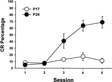
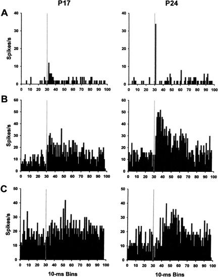
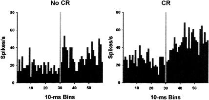
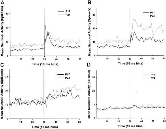
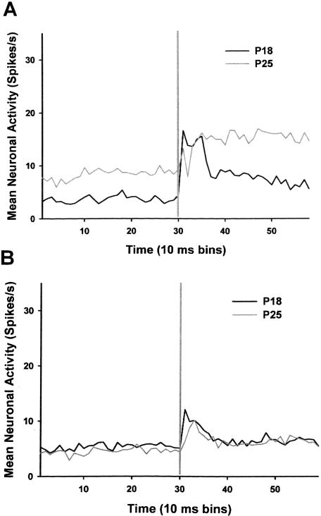
Neuronal activity was recorded in the pontine nuclei of developing rats during eyeblink conditioning on postnatal days 17-18 (P17-P18) or P24-P25. A pretraining session consisted of unpaired presentations of a 300-msec tone conditioned stimulus (CS) and a 10-msec periorbital shock unconditioned stimulus (US). Five paired training sessions followed the unpaired session, consisting of 100 trials of the CS paired with the US. The rats trained on P24-P25 exhibited significantly more conditioned responses (CRs) than the rats trained on P17-P18, although both groups produced CRs by the end of training. Ontogenetic increases in pre-CS and stimulus-elicited activity in the pontine nuclei were observed during the pretraining session and after paired training. The activity of pontine units was greater on trials with CRs relative to trials without CRs in rats trained on P24-P25, but almost no CR-related modulation was observed in the pontine units of rats trained on P17-P18. The findings indicate that pontine neuronal responses to the CS and modulation of pontine activity by the cerebellum and red nucleus undergo substantial postnatal maturation. The developmental changes in pontine neuronal activity might play a significant role in the ontogeny of eyeblink conditioning.


Figures

Figure 1
Figure 1
Mean (±SEM) conditioned response (CR) percentage from rat pups trained on postnatal days 17–18 (open circles) and postnatal days 24–25 (filled circles) across five paired training sessions.
Figure 2
Figure 2
Drawing of three coronal sections of the infant rat brain with labels indicating the placement of the tips of the electrode bundles in the pontine nuclei (PN) for rats trained on postnatal days 17–18 (gray dots) or postnatal days 24–25 (black dots).
Figure 3
Figure 3
Drawings of three coronal sections of the infant rat brain with labels indicating the placement of the tips of the electrode bundles with phasic (left), sustained (middle), and late (right) unit response profiles (see text for definitions) in the pontine nuclei (PN) for rats trained on postnatal days 17–18 (gray dots) or postnatal days 24–25 (black dots).
Figure 4
Figure 4
Mean firing rate (spikes/sec) of representative single units recorded from the pontine nuclei during the pretraining session. The firing frequencies of phasic (A), sustained (B), and late (C) units recorded from rats trained on postnatal day 17 (P17; left column) and P24 (right column) are displayed. The gray lines indicate the onset of the conditioned stimulus.
Figure 5
Figure 5
Mean firing rate (spikes/sec) of a pontine unit with significantly greater activity during trials with a conditioned response (CR) relative to trials with no CR.
Figure 6
Figure 6
Mean firing rate (spikes/sec) of phasic (A), sustained (B), late (C), and nonsignificant (D) response units before and during the tone conditioned stimulus (CS). The units were recorded during the pretraining session on P17 (black line) and P24 (gray line). The vertical gray line in each graph indicates the onset of the CS.
Figure 7
Figure 7
Mean firing rate (spikes/sec) of sustained (A) and phasic (B) response units before and during the tone conditioned stimulus (CS). The units were recorded during the last paired training session on P18 (black line) and P25 (gray line). The vertical gray line in each graph indicates the onset of the CS.
Figure 8
Figure 8
Overlays of all of the digitized waveforms recorded for two units during the last training session (A) and individual waveforms from the overlays (B). The duration of each event is 1.3 msec.

References

    1. Aitkin, L.M. and Boyd, J. 1978. Acoustic input to the lateral pontine nuclei. Hear. Res. 1: 67–77. - PubMed
    1. Andersson, G., Garwicz, M., and Hesslow, G. 1988. Evidence for a GABA-mediated cerebellar inhibition of the inferior olive in the cat. Exp. Brain Res. 72: 450–456. - PubMed
    1. Attwell, P.J., Rahman, S., and Yeo C.H. 2001. Acquisition of eyeblink conditioning is critically dependent on normal function in cerebellar cortical lobule HVI. J. Neurosci. 21: 5715–5722. - PMC - PubMed
    1. Attwell, P.J., Cooke, S.F., and Yeo, C.H. 2002. Cerebellar function in consolidation of a motor memory. Neuron 34: 1011–1020. - PubMed
    1. Bao S., Chen, L., and Thompson, R.F. 2000. Learning- and cerebellum-dependent neuronal activity in the lateral pontine nucleus. Behav. Neurosci. 114: 254–261. - PubMed

Publication types

LinkOut - more resources