Review
. 2003 Sep-Oct;10(5):373-86.
doi: 10.1101/lm.66103.

Multiple serotonergic mechanisms contributing to sensitization in aplysia: evidence of diverse serotonin receptor subtypes

Affiliations
Review

Multiple serotonergic mechanisms contributing to sensitization in aplysia: evidence of diverse serotonin receptor subtypes

Demian Barbas et al. Learn Mem. 2003 Sep-Oct.

Abstract

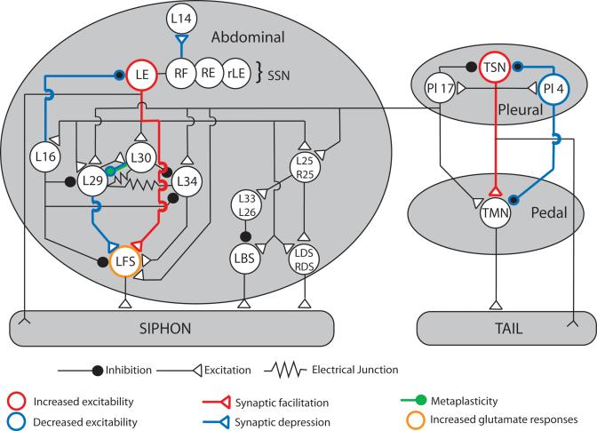
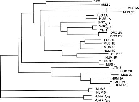
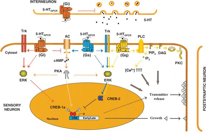
The neurotransmitter serotonin (5-HT) plays an important role in memory encoding in Aplysia. Early evidence showed that during sensitization, 5-HT activates a cyclic AMP-protein kinase A (cAMP-PKA)-dependent pathway within specific sensory neurons (SNs), which increases their excitability and facilitates synaptic transmission onto their follower motor neurons (MNs). However, recent data suggest that serotonergic modulation during sensitization is more complex and diverse. The neuronal circuits mediating defensive reflexes contain a number of interneurons that respond to 5-HT in ways opposite to those of the SNs, showing a decrease in excitability and/or synaptic depression. Moreover, in addition to acting through a cAMP-PKA pathway within SNs, 5-HT is also capable of activating a variety of other protein kinases such as protein kinase C, extracellular signal-regulated kinases, and tyrosine kinases. This diversity of 5-HT responses during sensitization suggests the presence of multiple 5-HT receptor subtypes within the Aplysia central nervous system. Four 5-HT receptors have been cloned and characterized to date. Although several others probably remain to be characterized in molecular terms, especially the Gs-coupled 5-HT receptor capable of activating cAMP-PKA pathways, the multiplicity of serotonergic mechanisms recruited into action during learning in Aplysia can now be addressed from a molecular point of view.


Figures

Figure 1
Figure 1
Schematic diagram of the neuronal circuit mediating tail and siphon withdrawal reflex. Neuronal cell bodies are located in three central ganglia (pleural, pedal, and abdominal). A tactile stimulus applied to the tail or siphon activates tail (TSN) or siphon sensory neurons (SSN), which excite MNs through monosynaptic (SN–MN synapses) and polysynaptic pathways depending on interneurons. 5-HT exerts multiple actions on the circuit: It usually induces synaptic depression and/or decreases in excitability in interneurons (blue) and synaptic facilitation and increase in excitability in SNs (red). L14, an ink gland MN, illustrates 5-HT-induced synaptic depression in RF SNs. 5-HT also induces metaplasticity at L30-L29 synapses (green) and increases glutamate responses in LFS MNs (orange). Adapted from Cleary et al. 1995.
Figure 2
Figure 2
Dendrogram analysis of different members of the 5-HT receptor superfamily. Sequences that were used for phylogenetic analysis were retrieved from the GenBank database. The sequences of serotonin receptors were compared and aligned using ClustalW (Thompson et al. 1994), which was executed from GDE (Genetic Data Environment; J. Felsenstein 1993, PHYLIP, Phylogeny Inference Package, version 3.5. 1c and 3.6, University of Washington, Seattle, WA). Only amino acid positions that could be aligned without ambiguity were used for the analysis. The alignment was then used for phylogenetic comparisons using the PHYLIP package. Analysis was performed with a bootstrap procedure that computes the probability of occurrence of the branches for 1000 possible trees. Branching order was determined using the Fitch-Margoliash algorithm included in the PHYLIP package. Only branches occurring in >800 trees are represented. DRO, Drosophila; FUG, Fugu rubripes (pufferfish); LYM, Lymnaea; HUM, human; MUS, mouse.
Figure 3
Figure 3
Schematic representation of putative roles of 5-HT receptor modulation of neuronal properties in Aplysia californica. The sensory neuron to motor neuron synapse involved in withdrawal reflexes is used as an example. After stimulation, facilitatory serotonergic interneurons release 5-HT that binds 5-HT receptors (5-HTGPCR). The final effect of 5-HT may depend on the specific expression patterns of 5-HT receptors and signaling molecules within different cells. Binding of 5-HT to a Gs-coupled receptor stimulates adenylyl cyclase (AC; pink pathway). Activation of the cyclase increases the cAMP concentration and induces activation of the cyclic AMP-dependent protein kinase A (PKA; activation is indicated by an arrow). Activation of PKA can phosphorylate and covalently modify a number of target proteins, including components of the exocytotic machinery of release, to enhance transmitter availability and release. With repeated stimulation, PKA can recruit the extracellular signal-regulated kinase (ERK, MAPK), and can translocate to the nucleus where it phosphorylates the cyclic AMP response element binding proteins (CREBs). Phosphorylation by ERK of the repressor isoform CREB-2 removes its inhibition on CREB-1a. Phosphorylation of CREB-1a induces transcription of early/late genes containing cyclic AMP response elements, leading to growth of new synaptic connections and potentially transmitter release. Gs-coupled receptor might also activate ERK (blue pathway) by transactivation of receptor tyrosine kinases (e.g., Trk, which can be activated by neurotrophins), or by direct activation of the ERK cascade. To turn down the release of 5-HT from interneurons, Gi-coupled receptors might act as presynaptic autoreceptors (inhibition is indicated by ⊣). When expressed at the surface of sensory neurons, Gi-coupled receptors can inhibit the Gs-dependent activation of the cyclase and turn down the cascade (red pathway). Under sustained release of 5-HT, Gi-coupled receptors might complement the activation of the ERK pathway by transactivation of Trks or direct activation of ERK components. Besides the PKA-signal pathway, there is a phospholipase C-PKC signaling pathway activated by 5-HT receptors (beige pathway). Gq-coupled receptor-activated phospholipase C (PLC) produces diacylglycerol (DAG) and inositol triphosphate (IP3) by cleaving the phosphatidyl inositol PIP2. IP3 is water-soluble, and can diffuse into the cytoplasm. There it binds to a receptor on the endoplasmic reticulum to release Ca2+ from internal stores. DAG remains in the membrane, where it activates the protein kinase C (PKC). Gq-coupled receptors might also be capable of enhancing the activation of the ERKs. For clarity only, two different sets of Trk receptors and ERK appear on the figure; there is no evidence that Gi-, Gs-, or Gq-activated pathways modulate distinct pools of ERK. Transmitter availability and release can also be dependent on activation of other signaling molecules (e.g., PKC, Ca2+) as shown by gray arrows. Speculative interactions are represented by (- - -). The molecular mechanisms underlying serotonergic modulation of MNs are still poorly understood and were omitted on this diagram.

References

    1. Abrams, T.W., Castellucci, V.F., Camardo, J.S., Kandel, E.R., and Lloyd, P.E. 1984. Two endogenous neuropeptides modulate the gill and siphon withdrawal reflex in Aplysia by presynaptic facilitation involving cAMP-dependent closure of a serotonin-sensitive potassium channel. Proc. Natl. Acad. Sci. 81: 7956–7960. - PMC - PubMed
    1. Alberini, C.M. 1999. Genes to remember. J. Exp. Biol. 202: 2887–2891. - PubMed
    1. Angers, A., Storozhuk, M.V., Duchaîne, T., Castellucci, V.F., and DesGroseillers, L. 1998. Cloning and functional expression of an Aplysia 5-HT receptor negatively coupled to adenylate cyclase. J. Neurosci. 18: 5586–5593. - PMC - PubMed
    1. Angers, A., Fioravante, D., Chin, J., Cleary, L.J., Bean, A.J., and Byrne, J.H. 2002a. Serotonin stimulates phosphorylation of Aplysia synapsin and alters its subcellular distribution in sensory neurons. J. Neurosci. 22: 5412–5422. - PMC - PubMed
    1. Angers, S., Salahpour, A., and Bouvier M. 2002b. Dimerization: An emerging concept for G protein-coupled receptor ontogeny and function. Annu. Rev. Pharmacol. Toxicol. 42: 409–435. - PubMed

Publication types

MeSH terms

LinkOut - more resources